Chinese Journal of Tissue Engineering Research ›› 2019, Vol. 23 ›› Issue (14): 2215-2222.doi: 10.3969/j.issn.2095-4344.1641
Previous Articles Next Articles
Physical properties and biocompatibility of 3D printed bone microparticle/poly(lactic-co-glycolic acid) scaffold
Zhang Minbo, Peng Qifeng, Ma Yaping, Kong Weijun, Liao Wenbo
- Department of Spinal Surgery, Affiliated Hospital of Zunyi Medical University, Zunyi 563000, Guizhou Province, China
-
Contact:Liao Wenbo, Department of Spinal Surgery, Affiliated Hospital of Zunyi Medical University, Zunyi 563000, Guizhou Province, China -
About author:Zhang Minbo, Master candidate, Department of Spinal Surgery, Affiliated Hospital of Zunyi Medical University, Zunyi 563000, Guizhou Province, China -
Supported by:Zunyi City Honghuagang District Science and Technology Project, No. (2016)09 (to LWB)
CLC Number:
Cite this article
Zhang Minbo, Peng Qifeng, Ma Yaping, Kong Weijun, Liao Wenbo. Physical properties and biocompatibility of 3D printed bone microparticle/poly(lactic-co-glycolic acid) scaffold[J]. Chinese Journal of Tissue Engineering Research, 2019, 23(14): 2215-2222.
share this article
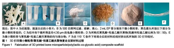
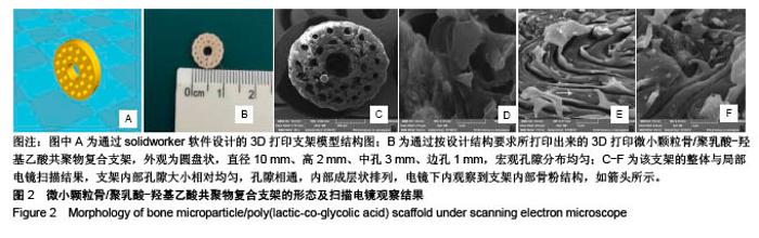
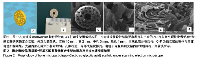
2.1 复合支架的制备过程、形态及电镜扫描结果 通过取兔四肢各管状骨,并经脱脂、脱蛋白处理后制得小片状骨块,再经球磨仪制研磨、500目筛网过滤、溶解、离心、冷冻干燥制得骨粉颗粒,将所得微小颗粒骨与PLGA按1∶5比例混合后,通过线材挤出机制得直径0.75 cm的均匀3D打印线材,线材制备过程见图1。 将线材通过熔炉沉积型3D打印技术成功制备成直径为10 mm×高2 mm的圆柱形多孔兔同种异体微小颗粒骨/ PLGA复合生物支架,电镜扫描结果示3D打印复合支架呈多孔状,内部结构成均匀层状排列,孔隙均匀,相互连通,见图2。"
"
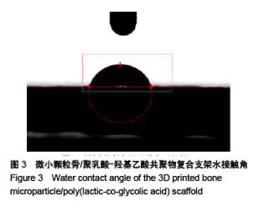
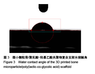
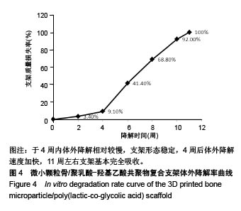
2.2 复合支架的孔隙率、吸水率、水接触角及体外降解率 该复合支架的孔隙率为(60.86±2.88)%,吸水率为(53.98± 2.04)%,水接触角为(76.27±0.34)°,见图3。体外测定该复合支架0,2,4,6,8,10,11周的质量损失率分别为0%,3.40%,9.10%,41.40%,68.80%,92.00%,100%,11周内该复合支架基本完全吸收,其降解率曲线,见图4,表明该支架材料于4周内体外降解相对较慢,支架形态稳定,4周后体外降解速度加快,11周左右支架基本完全吸收,是一种相对理想的可吸收支架材料。"

"
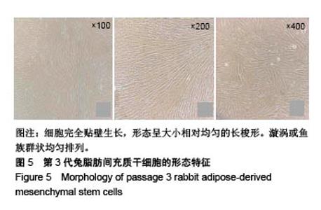
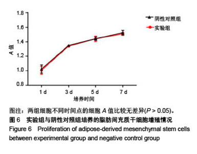
2.3 脂肪间充质干细胞的培养及CCK-8毒性实验结果 实验成功获得P3代兔脂肪间充质干细胞,见图5;CCK-8实验结果显示,实验组培养1,3,5,7 d的细胞A值与阴性对照组比较无差异(P > 0.05),见图6。实验组培养1,3,5,7 d的细胞相对增殖率分别为97.9%,99.1%,99.8%,98.9%,评价该支架材料细胞毒性1级,支架材料无明显细胞毒性,安全合格。"

"

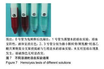
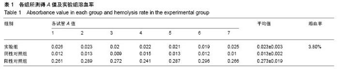
2.4 溶血实验结果 实验组和阴性对照组均无明显溶血现象发生,阳性对照组出现明显溶血现象,见图7。复合支架材料的溶血率为3.8%,小于5%,符合生物医用材料溶血率要求,见表1。"
"

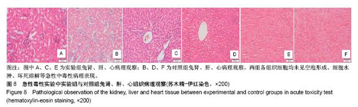
2.5 急性毒性实验 所有实验兔子腹腔注射复合支架浸提液后12,24,48,72 h后均未出现毒性反应表现,且注射后7 d内无惊厥、瘫痪、死亡等急性中毒表现,所有兔精神状态较好,饮食、睡眠、呼吸、尿便功能、体温均无异常表现。 注射浸提液7 d时处死所有实验兔,取心、肝、肾组织行苏木精-伊红病理染色切片,结果显示实验组心、肝、肾组织病理表现与对照组相同,各组织细胞结构无空泡、细胞水肿、坏死溶解等异常急性中毒性改变,见图8,证明该材料动物体内相容性良好,无明显体内毒性,为安全生物材料。"

| [1] 金灿,陈振琦.应用3D打印技术制作组织工程支架:修复骨缺损的研究回顾[J].中国组织工程研究,2017,21(10):1611-1616.[2] 周思佳,姜文学,尤佳.骨缺损修复材料:现状与需求和未来[J].中国组织工程研究, 2018, 22(14):2251-2258.[3] Lomas R,Chandrasekar A,Board TN.Bone allograft in the U.K.: perceptions andrealities.Hip Int. 2013;23(5):427-433.[4] Kuroda S,Katayama A,Takano -Yamamoto T.Severe anterior open-bite case treated using titanium screw anchorage.Angle Orthod.2004;74(4):558-567.[5] Yuan J,Zhen P,Zhao HB,et al.The preliminary performance study of the 3D printing of a tricalcium phosphate scaffold for the loading of sustained release anti-tuberculosis drugs.Mater Sci.2015;50(5):2138-2147.[6] Zhang Z,Zhang R,Song Z.Skull defect reconstruction based on a new hybrid level set. Biomed Mater Eng. 2014;24(6): 3343-3351.[7] Guarino V,Ambrosio L.Temperature-driven processing techniques for manufacturing fully interconnected porous scaffolds in bone tissue engineering.Proc Inst Mech Eng H. 2010;224(12):1389-1400.[8] 谢光友,杨海涛,吕富荣,等.兔脂肪间充质干细胞的培养鉴定及体外磁标记MR成像[J].第三军医大学学报,2014,36(15):1567-1571.[9] 国家技术监督局发布.中华人民共和国国家标准GB/T 1568-1997[M].北京:中国标准出版社,1997.[10] Sears NA,Dhavalikar PS,Seshadri D,et al.A Review of Three-Dimensional Printing in Tissue Engineering.Tissue Eng Part B Rev.2016;22(4):298-310.[11] 周晓,盛小伍,曾勇.骨组织工程的研究进展和面临的问题[J].组织工程与重建外科杂志,2016,12(5):319-321.[12] Turnbull G,Clarke J,Picard F,et al.3D bioactive composite scaffolds for bone tissue engineering. Bioact Mater. 2018;3(3): 278-314.[13] Lee M,Wu BM.Recent advances in 3D printing of tissue engineering scaffolds.Methods Mol Biol.2012;868:257-267.[14] 林楷丰,何树,宋岳,等.无丝3D打印技术常温构建仿生人工骨支架的研究[J].中华创伤骨科杂志,2016, 18(5):421-427.[15] Akbarzadeh R,Yousefi AM.Effects of processing parameters in thermally induced phase separation technique on porous architecture of scaffolds for bone tissue engineering. J Biomed Mater Res B Appl Biomater.2014;102(6):1304-1135.[16] Murphy SV,Atala A.3D bioprinting of tissues and organs.Nat Biotechnol.2014;32(8):773-785.[17] Nguyen DT,Mccanless JD,Mecwan MM,et al.Balancing mechanical strength with bioactivity in chitosan-calcium phosphate 3D microsphere scaffolds for bone tissue engineering: air-vs. freeze-drying processes.J Biomater Sci Polym Ed.2013;24(9):1071-1083.[18] Wu C,Luo Y,Cuniberti G,et al.Three-dimensional printing of hierarchical and tough mesoporous bioactive glass scaffolds with a controllable pore architecture, excellent mechanical strength and mineralization ability.Acta Biomater. 2011;7(6): 2644-2650.[19] Nakayama Y,Takewa Y,Sumikura H,et al.In-body tissue - engineered aorticivalve(Biovalve type VII) architecture based on 3D printer molding.J Biomed mater Res B ApplBiomater. 2015;103(1):1-11.[20] 武成聪,王芳,荣树,等.3D打印应用在骨组织工程研究中的特点与进展[J].中国组织工程研究,2017, 21(15):2418-2423.[21] Fafenrot S,Grimmelsmann N,Wortmann M,et al. Three-Dimensional (3D) Printing of Polymer-Metal Hybrid Materials by Fused Deposition Modeling. Materials(Basel). 2017;10(10):1199.[22] Rosenzweig DH,Caredli E,Steffe T,et al.3D PrintedABS and PLA Scaffolds for Cartilage and NucleusPulposus Tissue Regeneration.Int J Mol Sci.2015;16(7):15118-15135.[23] Cao T,Ho KH,Teoh SH.Scaffold design and in vitro study of osteochondralcoculture in a three-dimensional porous polycaprolactone scaffold fabricated by fused deposition modeling. Tissue Eng.2003;9 Suppl 1(supplement 1):S103.[24] 彭齐峰.3D打印骨粉聚乳酸骨组织工程支架的实验研究[D].遵义:遵义医学院,2018.[25] 钱科宏.PLGA介导的多功能复合医用材料的制备及其性能研究[D].扬州:扬州大学,2016.[26] An G,Zhang WB,Ma DK,et al.Influence of VEGF/BMP-2 on the proliferation and osteogenetic differentiation of rat bone mesenchymal stem cells on PLGA/gelatin composite scaffold. Eur Rev Med Pharmacol Sci.2017;21(10):2316-2328.[27] Piao S, Kim IG,LeeJY,et al.Therapeutic effect of adipose-derived stem cells and BDNF-immobilized PLGA membrane in a rat model of cavernous nerve injury.J sex Med. 2012;9(8):1968-1979.[28] Blokhuis TJ,Lindner T.Allograft and bone morphogenetic proteins: an overview.Injury. 2008;39(9):S33-S36.[29] 龙志成,宋兴华,龙仕杰,等.脂肪干细胞复合HA/β-TCP与同种异体骨修复兔脊柱骨缺损的比较[J].中国矫形外科杂志, 2018, 26(2): 164-169.[30] 瞿卫,丁铃,顾龙殿,等.小颗粒同种异体骨微创植骨治疗四肢骨缺损[J].中国骨与关节损伤杂志,2017, 32(9):1000-1001.[31] Zolnik BS,Burgess DJ.Effect of acidic pH on PLGA microsphere degradation and release.J Control Release. 2007;122(3):338-344.[32] Yang Y,Yang S,Wang Y,et al.Anti-infective efficacy, cytocompatibility and biocompatibility of a 3D-printed osteoconductive composite scaffold functionalized with quaternized chitosan.Acta Biomater.2016;46:112-128.[33] Liu C,Li Y,Wang J,et al.Improving Hydrophilicity and Inducing Bone-Like Apatite Formation on PPBES by Polydopamine Coating for Biomedical Application.Molecules. 2018;23(7): 1643.[34] Wilson CJ,Clegg RE,Leavesley DI,et al.Mediation of biomaterial-cell interactions by adsorbed proteins: a review. Tissue Eng.2005;11(2):1-18.[35] Hirasawa M,Tsutsumiarai C,Takakusaki K,et al. Superhydrophilic co-polymer coatings on denture surfaces reduce Candida albicans adhesion-An in vitro study.Arch Oral Biol.2017;87:143.[36] Zhang P, Hamamura K, Yokota H.A brief review of bone adaptation to unloading.Genomics Proteomics Bioinformatics. 2008;6(1):4-7.[37] 周晓,盛小伍,曾勇.骨组织工程的研究进展和面临的问题[J].组织工程与重建外科杂志,2016,12(5):319-321.[38] Rezwan K,Chen QZ,Blaker JJ,et al. Biodegradable and bioactive porous polymer/inorganic composite scaffolds for bone tissue engineering.Biomaterials.2006;27(18):3413-3431.[39] Liu Z,Ji J,Tang S,et al.Biocompatibility, degradability, bioactivity and osteogenesis of mesoporous/macroporous scaffolds of mesoporous diopside/poly(L-lactide) composite.J R Soc Interface.2015;12(111):20150507.[40] Watson BM, Kasper FK, Engel PS,et al.Synthesis and characterization of injectable, biodegradable, phosphate- containing,chemically cross-linkable, thermoresponsivemacromers for bone tissue engineering. Biomacromolecules.2014;15(5):1788-1796.[41] Arrigoni E,de Girolamo L,Di Giancamillo A, et al. Adipose-derived stem cells and rabbit bone regeneration: histomorphometric, immunohistochemical and mechanical characterization. J Orthop Sci.2013;18(2):331-339.[42] Tevlin R,Walmsley GG,Marecic O,et al. Stem and progenitor cells: advancing bone tissue engineering.Drug Deliv Transl Res.2016;6(2):1-15. |
| [1] | Zhang Tongtong, Wang Zhonghua, Wen Jie, Song Yuxin, Liu Lin. Application of three-dimensional printing model in surgical resection and reconstruction of cervical tumor [J]. Chinese Journal of Tissue Engineering Research, 2021, 25(9): 1335-1339. |
| [2] | Zeng Yanhua, Hao Yanlei. In vitro culture and purification of Schwann cells: a systematic review [J]. Chinese Journal of Tissue Engineering Research, 2021, 25(7): 1135-1141. |
| [3] | Xu Dongzi, Zhang Ting, Ouyang Zhaolian. The global competitive situation of cardiac tissue engineering based on patent analysis [J]. Chinese Journal of Tissue Engineering Research, 2021, 25(5): 807-812. |
| [4] | Wu Zijian, Hu Zhaoduan, Xie Youqiong, Wang Feng, Li Jia, Li Bocun, Cai Guowei, Peng Rui. Three-dimensional printing technology and bone tissue engineering research: literature metrology and visual analysis of research hotspots [J]. Chinese Journal of Tissue Engineering Research, 2021, 25(4): 564-569. |
| [5] | Chang Wenliao, Zhao Jie, Sun Xiaoliang, Wang Kun, Wu Guofeng, Zhou Jian, Li Shuxiang, Sun Han. Material selection, theoretical design and biomimetic function of artificial periosteum [J]. Chinese Journal of Tissue Engineering Research, 2021, 25(4): 600-606. |
| [6] | Liu Fei, Cui Yutao, Liu He. Advantages and problems of local antibiotic delivery system in the treatment of osteomyelitis [J]. Chinese Journal of Tissue Engineering Research, 2021, 25(4): 614-620. |
| [7] | Li Xiaozhuang, Duan Hao, Wang Weizhou, Tang Zhihong, Wang Yanghao, He Fei. Application of bone tissue engineering materials in the treatment of bone defect diseases in vivo [J]. Chinese Journal of Tissue Engineering Research, 2021, 25(4): 626-631. |
| [8] | Zhang Zhenkun, Li Zhe, Li Ya, Wang Yingying, Wang Yaping, Zhou Xinkui, Ma Shanshan, Guan Fangxia. Application of alginate based hydrogels/dressings in wound healing: sustained, dynamic and sequential release [J]. Chinese Journal of Tissue Engineering Research, 2021, 25(4): 638-643. |
| [9] | Chen Jiana, Qiu Yanling, Nie Minhai, Liu Xuqian. Tissue engineering scaffolds in repairing oral and maxillofacial soft tissue defects [J]. Chinese Journal of Tissue Engineering Research, 2021, 25(4): 644-650. |
| [10] | Xing Hao, Zhang Yonghong, Wang Dong. Advantages and disadvantages of repairing large-segment bone defect [J]. Chinese Journal of Tissue Engineering Research, 2021, 25(3): 426-430. |
| [11] | Chen Siqi, Xian Debin, Xu Rongsheng, Qin Zhongjie, Zhang Lei, Xia Delin. Effects of bone marrow mesenchymal stem cells and human umbilical vein endothelial cells combined with hydroxyapatite-tricalcium phosphate scaffolds on early angiogenesis in skull defect repair in rats [J]. Chinese Journal of Tissue Engineering Research, 2021, 25(22): 3458-3465. |
| [12] | Wang Hao, Chen Mingxue, Li Junkang, Luo Xujiang, Peng Liqing, Li Huo, Huang Bo, Tian Guangzhao, Liu Shuyun, Sui Xiang, Huang Jingxiang, Guo Quanyi, Lu Xiaobo. Decellularized porcine skin matrix for tissue-engineered meniscus scaffold [J]. Chinese Journal of Tissue Engineering Research, 2021, 25(22): 3473-3478. |
| [13] | Mo Jianling, He Shaoru, Feng Bowen, Jian Minqiao, Zhang Xiaohui, Liu Caisheng, Liang Yijing, Liu Yumei, Chen Liang, Zhou Haiyu, Liu Yanhui. Forming prevascularized cell sheets and the expression of angiogenesis-related factors [J]. Chinese Journal of Tissue Engineering Research, 2021, 25(22): 3479-3486. |
| [14] | Liu Chang, Li Datong, Liu Yuan, Kong Lingbo, Guo Rui, Yang Lixue, Hao Dingjun, He Baorong. Poor efficacy after vertebral augmentation surgery of acute symptomatic thoracolumbar osteoporotic compression fracture: relationship with bone cement, bone mineral density, and adjacent fractures [J]. Chinese Journal of Tissue Engineering Research, 2021, 25(22): 3510-3516. |
| [15] | Liu Liyong, Zhou Lei. Research and development status and development trend of hydrogel in tissue engineering based on patent information [J]. Chinese Journal of Tissue Engineering Research, 2021, 25(22): 3527-3533. |
| Viewed | ||||||
|
Full text |
|
|||||
|
Abstract |
|
|||||