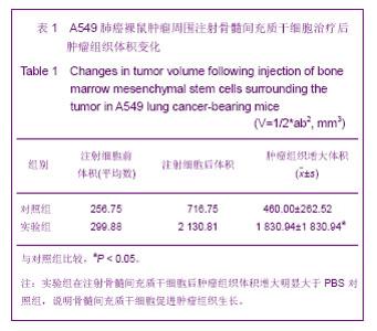
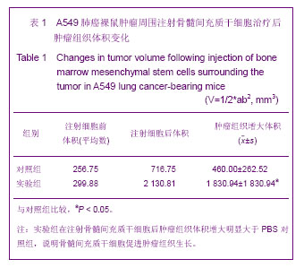
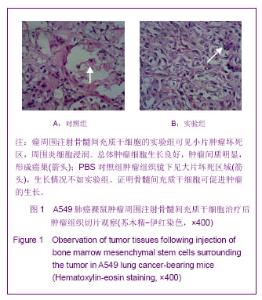
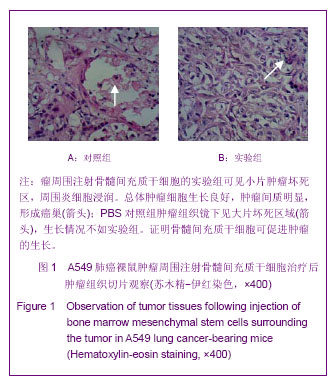
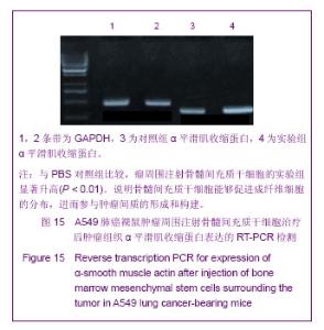
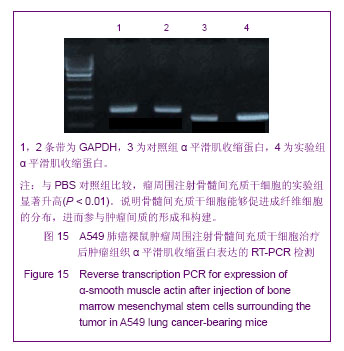
| [1] García-Castro J, Alemany R, Cascalló M,et al. Treatment of metastatic neuroblastoma with systemic oncolytic virotherapy delivered by autologous mesenchymal stem cells: an exploratory study. Cancer Gene Ther. 2010;17(7):476-483. [2] Rossignol J, Boyer C, Thinard R, et al. Mesenchymal stem cells induce a weak immune response in the rat striatum after allo or xenotransplantation. J Cell Mol Med. 2009;13(8B):2547-2558. [3] 封冰.间充质干细胞与肿瘤:抑制或促进?[J].中国肿瘤生物治疗杂志,2009,16(5):532-536.[4] 朱磊,姜晓峰.间充质干细胞与肿瘤关系的研究进展[J].中华肿瘤防治杂志,2011,18(4):317-320.[5] Bergfeld SA, DeClerck YA. Bone marrow-derived mesenchymal stem cells and the tumor microenvironment. Cancer Metastasis Rev. 2010;29(2):249-261. [6] Arnhold SJ, Goletz I, Klein H, et al. Isolation and characterization of bone marrow-derived equine mesenchymal stem cells. Am J Vet Res. 2007;68(10):1095- 10105. [7] Baddoo M, Hill K, Wilkinson R, et al. Characterization of mesenchymal stem cells isolated from murine bone marrow by negative selection. J Cell Biochem. 2003;89(6):1235-1249.[8] Deryugina EI, Müller-Sieburg CE. Stromal cells in long-term cultures: keys to the elucidation of hematopoietic development? Crit Rev Immunol. 1993;13(2):115-150. [9] Chamberlain G, Fox J, Ashton B, et al. Concise review: mesenchymal stem cells: their phenotype, differentiation capacity, immunological features, and potential for homing. Stem Cells. 2007;25(11):2739-2749. [10] Roni V, Habeler W, Parenti A,et al. Recruitment of human umbilical vein endothelial cells and human primary fibroblasts into experimental tumors growing in SCID mice. Exp Cell Res. 2003;287(1):28-38. [11] Studeny M, Marini FC, Champlin RE, et al. Bone marrow-derived mesenchymal stem cells as vehicles for interferon-beta delivery into tumors. Cancer Res. 2002;62(13): 3603-3608. [12] Shinojima N, Hossain A, Takezaki T, et al. TGF-β Mediates Homing of Bone Marrow-Derived Human Mesenchymal Stem Cells to Glioma Stem Cells. Cancer Res. 2013 Jan 30. [13] Condon MS. The role of the stromal microenvironment in prostate cancer. Semin Cancer Biol. 2005;15(2):132-137. [14] Ge S, Mao Y, Yi Y, et al. Comparative proteomic analysis of secreted proteins from nasopharyngeal carcinoma- associated stromal fibroblasts and normal fibroblasts. Exp Ther Med. 2012;3(5):857-860.[15] Micke P, Ostman A. Tumour-stroma interaction: cancer-associated fibroblasts as novel targets in anti-cancer therapy? Lung Cancer. 2004;45 Suppl 2:S163-175. [16] Nakayama H, Enzan H, Miyazaki E, et al. The role of myofibroblasts at the tumor border of invasive colorectal adenocarcinomas. Jpn J Clin Oncol. 1998;28(10):615-620. [17] 刘晗,陈军,徐祗顺.间充质干细胞与肿瘤生长发展关系的研究现状[J].中国组织工程研究与临床康复,2010,14(19):3565-3568. |