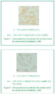
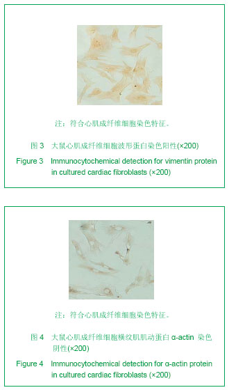
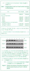
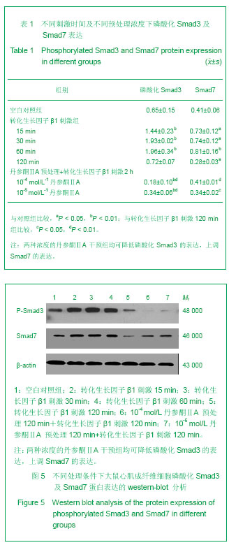
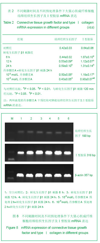
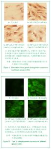
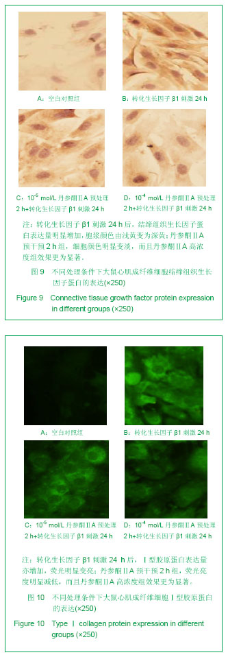
| [1] Baudino T A,Carver W, Giles W, et al. Cardiac ?broblasts: friend or foe?. Am J Physiol Heart Circ Physiol.2006; 291(3): H1015-H1026.[2] Bai D, Gao Q, Li C, et al.A conserved TGFβ1/HuR feedback circuit regulates the fibrogenic response in fibroblasts.Int J Biochem Cell Biol.2012;44(6):1031-1039.[3] Drobic V, Cunnington RH, Bedosky KM, et al. Differential and combined effects of cardiotrophin-1 and transforming growth factor-β1 on cardiac myo?broblast proliferation and contraction. Am J Physiol Heart Circ Physiol.2007;293(2): H1053-H1064.[4] Shyu KG, Wang BW, Wu GJ,et al.Mechanical stretch via transforming growth factor-β1 activates microRNA208a to regulate endoglin expression in cultured rat cardiac myoblasts. Chin Med J (Engl).2012;125(12):2205-2212.[5] Frank V, Alian M. Transforming growth factor-β1and fibrosis. World J Gastroenterol.2007;13(22): 3056-3062.[6] Leask A.Potential therapeutic targets for cardiac fibrosis: TGF-beta,angiotenangiotensin,endothelin,CCN2,and PDGF,partners in fibroblast activation.Circ Res.2010;106( 11): 1675-1680.[7] Kania G, Blyszczuk P, STEIN S, et al. Heart-Infiltrating Prominin-1(+)/CD133(+) Progenitor Cells Represent the Cellular Source of Transforming Growth Factor beta-Mediated Cardiac Fibrosis in Experimental Autoimmune Myocarditis. Circ Res.2009;105(5): 462,U151.[8] Glazer NL, Macy EM, Lumley T, et al.Transforming growth factor beta-1 and incidence of heart failure in older adults: The Cardiovascular Health Study[J].Circulation.2012;126(7):840-850.[9] Zhang DM,Qin Y,Niu FL,et al.Liaoning Zhongyi Zazhi. 2008; 35(12):1934-1936.张冬梅,秦英,牛福玲,等,丹参酮ⅡA对AngⅡ诱导的心肌成纤维细胞增殖及Ⅰ型胶原合成的影响[J]. 辽宁中医杂志, 2008, 35(12):1934-1936. [10] Sun XW,Cao L,Yu GH,et al.Disan Junyi Daxue Xuebao. 2007;29(7):585-587.孙兴旺,曹灵,于国华,等. 丹参酮Ⅱ-A磺酸钠对纤维化人肾间质成纤维细胞体外增殖及cyclin E蛋白表达的影响[J]. 第三军医大学学报, 2007, 29(7):585-587. [11] Dobaczewski M,Bujak M,Li N,et al.Smad3 signaling critically regulates fibroblast phenotype and function in healing myocardial infarction.Circ Res.2010;107( 3):418-428.[12] Moustaka SA, Souchelnytskyi S, Heldin CK. Smad regulation in transforming growth factor-βsignal transduction. Cell Science. 2001;114(6): 4359-4369.[13] Cunnington RH, Nazari M, Dixon MC. C-Ski, Smurf2, and Arkadia as regulators of transforming growth factor-β signaling: new targets for managing myofibroblast function and cardiac fibrosis. Can J Physiol and Pharma.2009;87(10):764-772.[14] He JH, Chen YL, Chen BL, et al. B-type natriuretic peptide attenuates cardiac hypertrophy via the transforming growth factor-beta1/smad7 pathway in vivo and in vitro. Clin and Exp Pharma and Physiol.2010;37(3):283-289. [15] Yang F, Huang XR, Chuang AC, et al. Essential role for Smad3 in angiotensin II-induced tubular epithelial-mesenchymal transition. Pathol. 2010;221(4): 390-401. |