Chinese Journal of Tissue Engineering Research ›› 2021, Vol. 25 ›› Issue (19): 3009-3015.doi: 10.3969/j.issn.2095-4344.3538
Previous Articles Next Articles
Radial extracorporeal shock wave therapy regulates the proliferation and differentiation of neural stem cells in the hippocampus via Notch1/Hes1 pathway after cerebral ischemia
Su Mingzhu1, Ma Yuewen1, 2
- 1The First Hospital of China Medical University, Shenyang 110001, Liaoning Province, China; 2Department of Rehabilitation Medicine, The First Hospital of China Medical University, Shenyang 110001, Liaoning Province, China
-
Received:2020-05-07Revised:2020-05-13Accepted:2020-07-17Online:2021-07-09Published:2021-01-13 -
Contact:Ma Yuewen, MD, Professor, Chief physician, The First Hospital of China Medical University, Shenyang 110001, Liaoning Province, China; Department of Rehabilitation Medicine, The First Hospital of China Medical University, Shenyang 110001, Liaoning Province, China -
About author:Su Mingzhu, Master, Physician, The First Hospital of China Medical University, Shenyang 110001, Liaoning Province, China -
Supported by:the Liaoning Provincial Key Research & Development Guidance Program, No. 2017225013 (to MYW)
CLC Number:
Cite this article
Su Mingzhu, Ma Yuewen. Radial extracorporeal shock wave therapy regulates the proliferation and differentiation of neural stem cells in the hippocampus via Notch1/Hes1 pathway after cerebral ischemia[J]. Chinese Journal of Tissue Engineering Research, 2021, 25(19): 3009-3015.
share this article
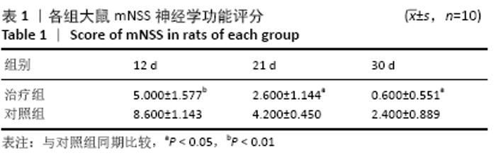
2.1 实验动物数量分析 清洁级健康雄性SD大鼠60只均进入结果分析。 2.2 大鼠神经功能评分 各组大鼠在造模成功后的12,21,30 d进行mNSS评分,见表1,mNSS评分越低,其神经功能恢复越好。治疗组大鼠的mNSS评分均小于对照组,提示体外放散式冲击波显著改善脑缺血大鼠的神经缺损症状。"

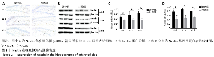
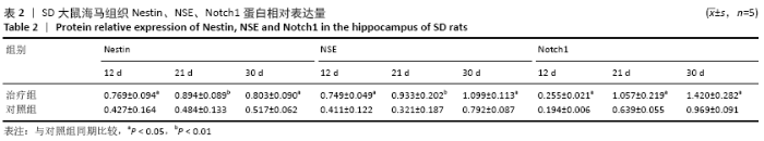
2.3 nestin在梗死侧海马区的表达 标本留取后,采用免疫组化、Western blot及Real time-PCR检测nestin蛋白及基因表达,见图2,表2,3。"

"


"
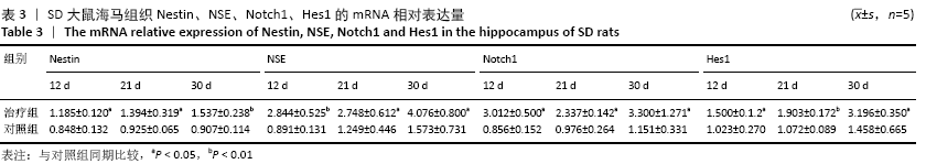
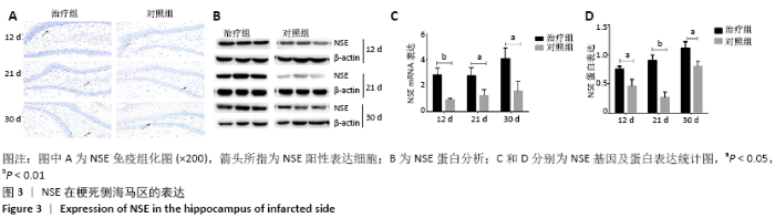
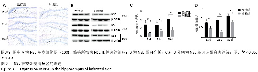
2.4 NSE在梗死侧海马区的表达 标本留取后,采用免疫组化、Western blot及Real time-PCR检测NSE蛋白及基因表达,见图3,表2,3。"
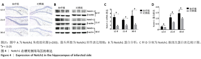
2.5 Notch1/Hes1通路在梗死侧海马区的表达 标本留取后,采用免疫组化、Western blot及Real time-PCR检测Notch1通路中的受体Notch1蛋白及基因表达,见图4;同时应用Real time-PCR检测Notch1信号通路下游基因Hes1的表达,见图5,表2,3。"

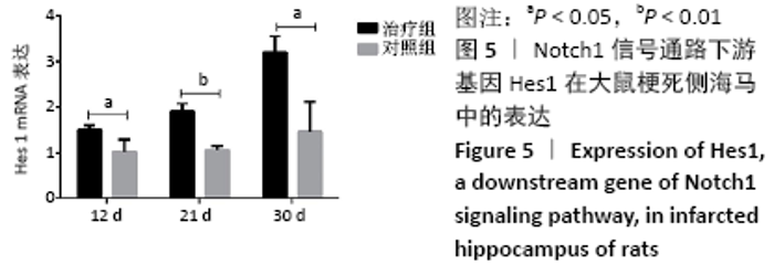
综合表1,图2,3可看出,体外放散式冲击波改善脑缺血后大鼠的神经功能与促进海马区神经干细胞的增殖与分化有关。结合图4,5可以看出,体外放散式冲击波促进海马区神经干细胞的增殖分化与Notch1信号通路有关。"

| [1] ZIAEE SM, TABESHMEHR P, HAIDER KH, et al. Optimization of time for neural stem cells transplantation for brain stroke in rats. Stem Cell Investig. 2017;4:29. [2] WRITING GROUP MEMBERS, MOZAFFARIAN D, BENJAMIN EJ, et al. Heart Disease and Stroke Statistics-2016 Update: A Report From the American Heart Association. Circulation. 2016;133(4):e38-360. [3] WARDLAW JM, MURRAY V, BERGE E, et al. Recombinant tissue plasminogen activator for acute ischaemic stroke: an updated systematic review and meta-analysis. Lancet. 2012;379(9834): 2364-2372. [4] 吴亚哲,陈伟伟.中国脑卒中流行概况[J].心脑血管病防治,2016, 16(6):410‐414. [5] TIBÆK M, DEHLENDORFF C, JØRGENSEN HS, et al. Increasing Incidence of Hospitalization for Stroke and Transient Ischemic Attack in Young Adults: A Registry-Based Study. J Am Heart Assoc. 2016;5(5):e003158. [6] YU H, CAO B, FENG M, et al. Combinated transplantation of neural stem cells and collagen type I promote functional recovery after cerebral ischemia in rats. Anat Rec (Hoboken). 2010;293(5):911-917. [7] WEI WJ, SHI B, GUAN X, et al. Mapping theme trends and knowledge structures for human neural stem cells: a quantitative and co-word biclustering analysis for the 2013-2018 period. Neural Regen Res. 2019;14(10):1823-1832. [8] ADACHI K, MIRZADEH Z, SAKAGUCHI M, et al. Beta-catenin signaling promotes proliferation of progenitor cells in the adult mouse subventricular zone. Stem Cells. 2007;25(11):2827-2836. [9] HAN X, JU JH, SHIN I. Glycogen synthase kinase 3-β phosphorylates novel S/T-P-S/T domains in Notch1 intracellular domain and induces its nuclear localization. Biochem Biophys Res Commun. 2012;423(2): 282-288. [10] BORGGREFE T, LAUTH M, ZWIJSEN A, et al. The Notch intracellular domain integrates signals from Wnt, Hedgehog, TGFβ/BMP and hypoxia pathways. Biochim Biophys Acta. 2016;1863(2):303-313. [11] JARGIN SV. Shock wave therapy of ischemic heart disease in the light of general pathology. Int J Cardiol. 2010;144(1):116-117. [12] QURESHI AA, ROSS KM, OGAWA R, et al. Shock wave therapy in wound healing. Plast Reconstr Surg. 2011;128(6):721e-727e. [13] LEE JH, CHO SH. Effect of extracorporeal shock wave therapy on denervation atrophy and function caused by sciatic nerve injury. J Phys Ther Sci. 2013;25(9):1067-1069. [14] ZHANG D, MENG Y, HAI H, et al. Radial Extracorporeal Shock Wave Therapy in an Individual With Primary Trigeminal Neuralgia: A Case Report and Literature Review. Am J Phys Med Rehabil. 2018; 97(5):e42-e45. [15] WERNER C, BYHAHN M, HESSE S. Non-invasive brain stimulation to promote alertness and awareness in chronic patients with disorders of consciousness: Low-level, near-infrared laser stimulation vs. focused shock wave therapy. Restor Neurol Neurosci. 2016;34(4):561-569. [16] KANG N, ZHANG J, YU X, et al. Radial extracorporeal shock wave therapy improves cerebral blood flow and neurological function in a rat model of cerebral ischemia. Am J Transl Res. 2017;9(4):2000-2012. [17] YUEN CM, CHUNG SY, TSAI TH, et al. Extracorporeal shock wave effectively attenuates brain infarct volume and improves neurological function in rat after acute ischemic stroke. Am J Transl Res. 2015;7(6): 976-994. [18] CHEN Y, XU J, HUANG Z, et al. An Innovative Approach for Enhancing Bone Defect Healing Using PLGA Scaffolds Seeded with Extracorporeal-shock-wave-treated Bone Marrow Mesenchymal Stem Cells (BMSCs). Sci Rep. 2017;7:44130. [19] LEONE L, RAFFA S, VETRANO M, et al. Extracorporeal Shock Wave Treatment (ESWT) enhances the in vitro-induced differentiation of human tendon-derived stem/progenitor cells (hTSPCs). Oncotarget. 2016;7(6):6410-6423. [20] XU L, ZHAO Y, WANG M, et al. Defocused low-energy shock wave activates adipose tissue-derived stem cells in vitro via multiple signaling pathways. Cytotherapy. 2016;18(12):1503-1514. [21] ZHANG J, KANG N, YU X, et al. Radial Extracorporeal Shock Wave Therapy Enhances the Proliferation and Differentiation of Neural Stem Cells by Notch, PI3K/AKT, and Wnt/β-catenin Signaling. Sci Rep. 2017;7(1):15321. [22] ZHANG R, ENGLER A, TAYLOR V. Notch: an interactive player in neurogenesis and disease. Cell Tissue Res. 2018;371(1):73-89. [23] KUME T. Ligand-dependent Notch signaling in vascular formation. Adv Exp Med Biol. 2012;727:210-222. [24] SUN F, MAO X, XIE L, et al. Notch1 signaling modulates neuronal progenitor activity in the subventricular zone in response to aging and focal ischemia. Aging Cell. 2013;12(6):978-987. [25] WANG JJ, ZHU JD, ZHANG XH, et al. Neuroprotective effect of Notch pathway inhibitor DAPT against focal cerebral ischemia/reperfusion 3 hours before model establishment. Neural Regen Res. 2019;14(3): 452-461. [26] IRVIN DK, ZURCHER SD, NGUYEN T, et al. Expression patterns of Notch1, Notch2, and Notch3 suggest multiple functional roles for the Notch-DSL signaling system during brain development. J Comp Neurol. 2001;436(2):167-181. [27] FELLING RJ, COVEY MV, WOLUJEWICZ P, et al. Astrocyte-produced leukemia inhibitory factor expands the neural stem/progenitor pool following perinatal hypoxia-ischemia. J Neurosci Res. 2016;94(12): 1531-1545. [28] CAI Z, ZHAO B, DENG Y, et al. Notch signaling in cerebrovascular diseases (Review). Mol Med Rep. 2016;14(4):2883-2898. [29] 赵俊红,林阳阳,何晓阔,等.电针对脑梗死大鼠海马齿状回Notch1调控作用的研究[J].中国康复医学杂志,2018,23(5):520-524. [30] 王丽娜,杨洋,王光,等.电针结合康复训练对缺血脑卒中大鼠Notch1与Notch4表达的影响[J].解剖科学进展,2018,24(2): 136-139. [31] 钟晴,童骄,钟欣池,等.脑泰方提取物对大鼠脑缺血再灌注损伤后海马神经干细胞增殖及Notch1蛋白表达的影响[J]. 中华中医药学刊,2016,34(8):1823-1826. [32] ZHAO JH, WANG B, WANG XH, et al. Effect of lncRNA GAS5 on the apoptosis of neurons via the notch1 signaling pathway in rats with cerebral infarction. Eur Rev Med Pharmacol Sci. 2019;23(22): 10083-10091. [33] SCHMIEDT E, CHAUSSY C. Extracorporeal shock-wave lithotripsy of kidney and ureteric stones. Urol Int. 1984;39(4):193-198. [34] MATTYASOVSZKY SG, LANGENDORF EK, RITZ U, et al. Exposure to radial extracorporeal shock waves modulates viability and gene expression of human skeletal muscle cells: a controlled in vitro study. J Orthop Surg Res. 2018;13(1):75. [35] NIKOLIKJ-DIMITROVA ED, GJERAKAROSKA-SAVEVSKA C, KOEVSKA V, et al. The Effectiveness of Radial Extracorporeal Shock Wave Therapy for Chronic Achilles Tendinopathy: A Case Report with 18 Months Follow-Up. Open Access Maced J Med Sci. 2018;6(3):523-527. [36] GERDESMEYER L, MAIER M, HAAKE M, et al. Physical-technical principles of extracorporeal shockwave therapy (ESWT). Orthopade. 2002;31(7):610-617. [37] FU M, SUN CK, LIN YC, et al. Extracorporeal shock wave therapy reverses ischemia-related left ventricular dysfunction and remodeling: molecular-cellular and functional assessment. PLoS One. 2011;6(9):e24342. [38] COOPER B, BACHOO P. Extracorporeal shock wave therapy for the healing and management of venous leg ulcers. Cochrane Database Syst Rev. 2018;6(6):CD011842. [39] OMAR MT, ALGHADIR A, AL-WAHHABI KK, et al. Efficacy of shock wave therapy on chronic diabetic foot ulcer: a single-blinded randomized controlled clinical trial. Diabetes Res Clin Pract. 2014;106(3):548-554. [40] TANG HY, ZHAO Y, LI YZ, et al. Effectiveness of extracorporeal shock wave monotherapy for avascular necrosis of femoral head: A systematic review protocol of randomized controlled trial. Medicine (Baltimore). 2019;98(14):e15119. [41] SCHMITZ C, CSÁSZÁR NB, MILZ S, et al. Efficacy and safety of extracorporeal shock wave therapy for orthopedic conditions: a systematic review on studies listed in the PEDro database. Br Med Bull. 2015;116(1):115-138. [42] LOHRER H, NAUCK T, KORAKAKIS V, et al. Historical ESWT Paradigms Are Overcome: A Narrative Review. Biomed Res Int. 2016;2016:3850461. [43] SEOL PH, HA KW, KIM YH, et al. Effect of Radial Extracorporeal Shock Wave Therapy in Patients With Fabella Syndrome. Ann Rehabil Med. 2016;40(6):1124-1128. [44] MIAO HC, WU F, DING J, et al. Effects of electroacupuncture intervention combined with polysaccharide of Gastrodia elata Blume on expression of nestin and cytokines of neural stem cells in the dentate gyrus of cerebral ischemia rats. Zhen Ci Yan Jiu. 2014;39(1): 40-45. [45] WAT JJ, WAT MJ. Sox7 in vascular development: review, insights and potential mechanisms. Int J Dev Biol. 2014;58(1):1-8. [46] HUANG L, LIU LF, LIU J, et al. Ginsenoside Rg1 protects against neurodegeneration by inducing neurite outgrowth in cultured hippocampal neurons. Neural Regen Res. 2016;11(2):319-325. [47] BRAI E, MARATHE S, ASTORI S, et al. Notch1 Regulates Hippocampal Plasticity Through Interaction with the Reelin Pathway, Glutamatergic Transmission and CREB Signaling. Front Cell Neurosci. 2015;9:447. [48] ZHAO J, SUI M, LÜ X, et al. Electroacupuncture promotes neural stem cell proliferation and neurogenesis in the dentate gyrus of rats following stroke via upregulation of Notch1 expression. Mol Med Rep. 2015;12(5):6911-6917. [49] ZHANG S, WANG P, REN L, et al. Protective effect of melatonin on soluble Aβ1-42-induced memory impairment, astrogliosis, and synaptic dysfunction via the Musashi1/Notch1/Hes1 signaling pathway in the rat hippocampus. Alzheimers Res Ther. 2016;8(1):40. |
| [1] | Lin Qingfan, Xie Yixin, Chen Wanqing, Ye Zhenzhong, Chen Youfang. Human placenta-derived mesenchymal stem cell conditioned medium can upregulate BeWo cell viability and zonula occludens expression under hypoxia [J]. Chinese Journal of Tissue Engineering Research, 2021, 25(在线): 4970-4975. |
| [2] | Pu Rui, Chen Ziyang, Yuan Lingyan. Characteristics and effects of exosomes from different cell sources in cardioprotection [J]. Chinese Journal of Tissue Engineering Research, 2021, 25(在线): 1-. |
| [3] | Zhang Chong, Liu Zhiang, Yao Shuaihui, Gao Junsheng, Jiang Yan, Zhang Lu. Safety and effectiveness of topical application of tranexamic acid to reduce drainage of elderly femoral neck fractures after total hip arthroplasty [J]. Chinese Journal of Tissue Engineering Research, 2021, 25(9): 1381-1386. |
| [4] | Wang Haiying, Lü Bing, Li Hui, Wang Shunyi. Posterior lumbar interbody fusion for degenerative lumbar spondylolisthesis: prediction of functional prognosis of patients based on spinopelvic parameters [J]. Chinese Journal of Tissue Engineering Research, 2021, 25(9): 1393-1397. |
| [5] | Chen Jinping, Li Kui, Chen Qian, Guo Haoran, Zhang Yingbo, Wei Peng. Meta-analysis of the efficacy and safety of tranexamic acid in open spinal surgery [J]. Chinese Journal of Tissue Engineering Research, 2021, 25(9): 1458-1464. |
| [6] | Wu Xun, Meng Juanhong, Zhang Jianyun, Wang Liang. Concentrated growth factors in the repair of a full-thickness condylar cartilage defect in a rabbit [J]. Chinese Journal of Tissue Engineering Research, 2021, 25(8): 1166-1171. |
| [7] | Shen Jinbo, Zhang Lin. Micro-injury of the Achilles tendon caused by acute exhaustive exercise in rats: ultrastructural changes and mechanism [J]. Chinese Journal of Tissue Engineering Research, 2021, 25(8): 1190-1195. |
| [8] | Zeng Zhen, Hu Jingwei, Li Xuan, Tang Linmei, Huang Zhiqiang, Li Mingxing. Quantitative analysis of renal blood flow perfusion using contrast-enhanced ultrasound in rats with hemorrhagic shock during resuscitation [J]. Chinese Journal of Tissue Engineering Research, 2021, 25(8): 1201-1206. |
| [9] | Tang Hui, Yao Zhihao, Luo Daowen, Peng Shuanglin, Yang Shuanglin, Wang Lang, Xiao Jingang. High fat and high sugar diet combined with streptozotocin to establish a rat model of type 2 diabetic osteoporosis [J]. Chinese Journal of Tissue Engineering Research, 2021, 25(8): 1207-1211. |
| [10] | Chai Le, Lü Jianlan, Hu Jintao, Hu Huahui, Xu Qingjun, Yu Jinwei, Quan Renfu. Signal pathway variation after induction of inflammatory response in rats with acute spinal cord injury [J]. Chinese Journal of Tissue Engineering Research, 2021, 25(8): 1218-1223. |
| [11] | Liu Xiangxiang, Huang Yunmei, Chen Wenlie, Lin Ruhui, Lu Xiaodong, Li Zuanfang, Xu Yaye, Huang Meiya, Li Xihai. Ultrastructural changes of the white zone cells of the meniscus in a rat model of early osteoarthritis [J]. Chinese Journal of Tissue Engineering Research, 2021, 25(8): 1237-1242. |
| [12] | Zhang Xiumei, Zhai Yunkai, Zhao Jie, Zhao Meng. Research hotspots of organoid models in recent 10 years: a search in domestic and foreign databases [J]. Chinese Journal of Tissue Engineering Research, 2021, 25(8): 1249-1255. |
| [13] | Shu Wenbo, Chen Mengchi, Li Hua, Huang Liqian, Huang Binbin, Zhang Wenhai, Wu Yachen, Wang Zefeng, Li Qiaoli, Liu Peng. Correlation between body fat distribution and characteristics of daily physical activity in college students [J]. Chinese Journal of Tissue Engineering Research, 2021, 25(8): 1277-1283. |
| [14] | Ji Zhixiang, Lan Changgong. Polymorphism of urate transporter in gout and its correlation with gout treatment [J]. Chinese Journal of Tissue Engineering Research, 2021, 25(8): 1290-1298. |
| [15] | Liu Cong, Liu Su. Molecular mechanism of miR-17-5p regulation of hypoxia inducible factor-1α mediated adipocyte differentiation and angiogenesis [J]. Chinese Journal of Tissue Engineering Research, 2021, 25(7): 1069-1074. |
| Viewed | ||||||
|
Full text |
|
|||||
|
Abstract |
|
|||||