Chinese Journal of Tissue Engineering Research ›› 2013, Vol. 17 ›› Issue (14): 2649-2655.doi: 10.3969/j.issn.2095-4344.2013.14.026
Previous Articles Next Articles
Isolation and culture of mesenchymal stem cells: From laboratory research to clinical application
Li Bing-yao1, Wu Xiao-yun2, Wu Yan1
- 1 Inner Mongolia Medical University, Hohhot 010059, Inner Mongolia Autonomous Region, China
2 Beijing Jingmeng Stem Cell High-Tech Co., Ltd., Beijing 100085, China
-
Received:2012-06-08Revised:2012-08-27Online:2013-04-02Published:2013-04-02 -
Contact:Wu Yan, M.D., Professor, Master’s supervisor, Inner Mongolia Medical University, Hohhot 010059, Inner Mongolia Autonomous Region, China yanw007@sina.com -
About author:Li Bing-yao★, Studying for master’s degree, Physician, Inner Mongolia Medical University, Hohhot 010059, Inner Mongolia Autonomous Region, China cell2112@sina.com -
Supported by:Stem Cell Innovation Team Program of Science and Technology Bureau of Inner Mongolia Autonomous Region of China, No. KJT2009
CLC Number:
Cite this article
Li Bing-yao, Wu Xiao-yun, Wu Yan1. Isolation and culture of mesenchymal stem cells: From laboratory research to clinical application [J]. Chinese Journal of Tissue Engineering Research, 2013, 17(14): 2649-2655.
share this article

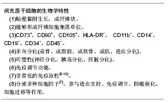
2.1 间充质干细胞的来源和生物学特性 间充质干细胞最早在骨髓中发现,因培养后细胞表型为成纤维细胞样,而被称为成纤维细胞集落形成单位。由于对骨髓血系细胞起支持诱导作用,可促进造血细胞克隆的形成,因而推测这种细胞可能是间质细胞的前体细胞,又因其来自于骨髓基质,将其称为“骨髓基质细胞”。早期实验研究用得最多的是骨髓来源的骨髓间充质干细胞。骨髓间充质干细胞占骨髓有核细胞的0.01%-0.000 1%。目前,越来越多的研究表明还能够从脂肪、滑膜、骨骼、肌肉、肺、肝、胰腺、羊水以及脐血等组织中分离和制备间充质干细胞[1]。但实验室和临床上用得最多的仍然还是骨髓来源的间充质干细胞。骨髓来源的间充质干细胞存在以下问题:随着年龄的老化,干细胞数目显著降低,增殖分化能力大幅度衰退;制备过程不容易质控;移植给异体可能引起免疫反应;取材时对患者有损伤,患者有骨髓疾病时不能采集。即使是健康供者,亦不能抽取太多的骨髓。这都限制了骨髓间充质干细胞临床应用,使得寻找骨髓以外其他可替代的间充质干细胞来源成为一个重要的问题。临床上作为脐血来源的间充质干细胞培养成功率很低,一般只有20%-30%的脐血样本培养后能分离出间充质干细胞。为了有效地提高脐血间充质干细胞原代培养成功率,应用正交实验设计法在间充质干细胞传统培养方法的基础上,通过添加细胞因子白细胞介素3和粒细胞-集落刺激因子的方法可以使脐血间充质干细胞的原代培养成功率由传统方法的25%左右提高到90%以上[2]。 脐带是孕妇与胎儿之间的物质交通支,也是分娩后的废弃物,收集脐带对产妇和胎儿均不造成伤害,取材也不受伦理学的困扰。另外从胚胎发育而言,脐带的免疫原性低,可以降低或避免免疫排斥。一些研究者比较了脐带间充质干细胞和骨髓间充质干细胞,发现前者比后者有更快的增殖能力和更好的成骨分化能力,因此脐带是临床上间充质干细胞的主要来源。也有一些研究者认为脂肪组织是间充质干细胞最理想的来源[3],因为其来源的间充质干细胞具有和骨髓源间充质干细胞相似的表型、增殖能力和分化能力,而且脂肪非常丰富且取材简单,捐献者可以避免痛苦[3]。 目前,国际干细胞治疗学会提出了鉴定人来源间充质干细胞的3条最低标准[4]:①在标准培养条件下,间充质干细胞具备对塑料底物的贴附性。②间充质干细胞群体表达CD105、CD73及CD90,而不表达造血标志物CD45和CD34。③经体外诱导,间充质干细胞能向成骨细胞、脂肪细胞及软骨细胞分化。这仅仅是间充质干细胞的体外鉴定,一些研究者一直寻求间充质干细胞的体内鉴定[5]。随着对间充质干细胞的深入研究,发现间充质干细胞可以分化成血细胞和肌细胞,还具有跨胚层分化的可塑性。间充质干细胞具有向外胚层神经细胞分化和内胚层肝细胞、胰岛细胞分化的能力[6]。而且间充质干细胞具有非常低得免疫原性。间充质干细胞分泌多种细胞因子发挥造血支持和免疫调节等功能[7-8]。间充质干细胞的这些生物学特性,显著推动了间充质干细胞的应用范围和应用价值。 间充质干细胞的标准定义:"
| [1] Camassola M, de Macedo Braga LM, Chagastelles PC,et al. Methodology, biology and clinical applications of human mesenchymal stem cells. Methods Mol Biol. 2012;879: 491-504.[2] Fan X, Liu T, Liu Y,et al. Optimization of primary culture condition for mesenchymal stem cells derived from umbilical cord blood with factorial design. Biotechnol Prog. 2009;25(2): 499-507.[3] Boquest AC, Collas P. Obtaining freshly isolated and cultured mesenchymal stem cells from human adipose tissue. Methods Mol Biol. 2012;879:269-278.[4] Dominici M, Le Blanc K, Mueller I, et al. Minimal criteria for defining multipotent mesenchymal stromal cells. The International Society for Cellular Therapy position statement. Cytotherapy. 2006;8(4):315-317.[5] da Silva Meirelles L, Caplan AI, Nardi NB. In search of the in vivo identity of mesenchymal stem cells. Stem Cells. 2008; 26(9):2287-2299.[6] Ngoc PK, Phuc PV, Nhung TH,et al. Improving the efficacy of type 1 diabetes therapy by transplantation of immunoisolated insulin-producing cells. Hum Cell. 2011;24(2):86-95.[7] Hwang JH, Lee MJ, Seok OS,et al. Cytokine expression in placenta-derived mesenchymal stem cells in patients with pre-eclampsia and normal pregnancies.Cytokine. 2010;49(1): 95-101.[8] Sumanasinghe RD, Pfeiler TW, Monteiro-Riviere NA,et al. Expression of proinflammatory cytokines by human mesenchymal stem cells in response to cyclic tensile strain. J Cell Physiol. 2009;219(1):77-83.[9] Huang XP, Sun Z, Miyagi Y,et al. Differentiation of allogeneic mesenchymal stem cells induces immunogenicity and limits their long-term benefits for myocardial repair. Circulation. 2010;122(23):2419-2429.[10] Schu S, Nosov M, O'Flynn L,et al. Immunogenicity of allogeneic mesenchymal stem cells. J Cell Mol Med. 2012; 16(9):2094-2103.[11] Liu L, Zhao X, Li P,et al. A novel way to isolate MSCs from umbilical cords. Eur J Immunol. 2012;42(8):2190-2193.[12] Pierini M, Dozza B, Lucarelli E,et al. Efficient isolation and enrichment of mesenchymal stem cells from bone marrow. Cytotherapy. 2012;14(6):686-693.[13] Chieregato K, Castegnaro S, Madeo D,et al. Epidermal growth factor, basic fibroblast growth factor and platelet-derived growth factor-bb can substitute for fetal bovine serum and compete with human platelet-rich plasma in the ex vivo expansion of mesenchymal stromal cells derived from adipose tissue. Cytotherapy. 2011;13(8):933-943.[14] Meyerrose T, Olson S, Pontow S,et al. Mesenchymal stem cells for the sustained in vivo delivery of bioactive factors. Adv Drug Deliv Rev. 2010;62(12):1167-1174.[15] Nekanti U, Mohanty L, Venugopal P,et al. Optimization and scale-up of Wharton's jelly-derived mesenchymal stem cells for clinical applications. Stem Cell Res. 2010;5(3):244-254.[16] Felka T, Schäfer R, De Zwart P,et al. Animal serum-free expansion and differentiation of human mesenchymal stromal cells. Cytotherapy. 2010;12(2):143-153.[17] Jung S, Sen A, Rosenberg L,et al. Identification of growth and attachment factors for the serum-free isolation and expansion of human mesenchymal stromal cells. Cytotherapy. 2010; 12(5):637-657.[18] Jung S, Panchalingam KM, Rosenberg L,et al. Ex vivo expansion of human mesenchymal stem cells in defined serum-free media. Stem Cells Int. 2012;2012:123030.[19] Hatlapatka T, Moretti P, Lavrentieva A,et al. Optimization of culture conditions for the expansion of umbilical cord-derived mesenchymal stem or stromal cell-like cells using xeno-free culture conditions.Tissue Eng Part C Methods. 2011;17(4): 485-493.[20] Poloni A, Maurizi G, Serrani F,et al. Human AB serum for generation of mesenchymal stem cells from human chorionic villi: comparison with other source and other media including platelet lysate. Cell Prolif. 2012;45(1):66-75.[21] Pérez-Simon JA, López-Villar O, Andreu EJ,et al. Mesenchymal stem cells expanded in vitro with human serum for the treatment of acute and chronic graft-versus-host disease: results of a phase I/II clinical trial. Haematologica. 2011;96(7):1072-1076.[22] Ben Azouna N, Jenhani F, Regaya Z,et al. Phenotypical and functional characteristics of mesenchymal stem cells from bone marrow: comparison of culture using different media supplemented with human platelet lysate or fetal bovine serum. Stem Cell Res Ther. 2012;3(1):6.[23] Tekkatte C, Vidyasekar P, Kapadia NK,et al. Enhancement of adipogenic and osteogenic differentiation of human bone-marrow-derived mesenchymal stem cells by supplementation with umbilical cord blood serum. Cell Tissue Res. 2012;347(2):383-395.[24] Phadnis SM, Joglekar MV, Venkateshan V,et al. Human umbilical cord blood serum promotes growth, proliferation, as well as differentiation of human bone marrow-derived progenitor cells.In Vitro Cell Dev Biol Anim. 2006;42(10): 283-286.[25] Duggal S, Brinchmann JE. Importance of serum source for the in vitro replicative senescence of human bone marrow derived mesenchymal stem cells. J Cell Physiol. 2011;226(11): 2908-2915.[26] Shetty P, Bharucha K, Tanavde V. Human umbilical cord blood serum can replace fetal bovine serum in the culture of mesenchymal stem cells. Cell Biol Int. 2007;31(3):293-298.[27] Ma HY, Yao L, Yu YQ,et al. An effective and safe supplement for stem cells expansion ex vivo: cord blood serum.Cell Transplant. 2012;21(5):857-869.[28] Jung J, Moon N, Ahn JY,et al. Mesenchymal stromal cells expanded in human allogenic cord blood serum display higher self-renewal and enhanced osteogenic potential.Stem Cells Dev. 2009;18(4):559-571. [29] Gottipamula S, Sharma A, Krishnamurthy S,et al. Human platelet lysate is an alternative to fetal bovine serum for large-scale expansion of bone marrow-derived mesenchymal stromal cells. Biotechnol Lett. 2012;34(7):1367-1374.[30] Xia W, Li H, Wang Z,et al. Human platelet lysate supports ex vivo expansion and enhances osteogenic differentiation of human bone marrow-derived mesenchymal stem cells. Cell Biol Int. 2011;35(6):639-643.[31] Abdelrazik H, Spaggiari GM, Chiossone L,et al. Mesenchymal stem cells expanded in human platelet lysate display a decreased inhibitory capacity on T- and NK-cell proliferation and function. Eur J Immunol. 2011;41(11):3281-3290.[32] Eibes G, dos Santos F, Andrade PZ,et al. Maximizing the ex vivo expansion of human mesenchymal stem cells using a microcarrier-based stirred culture system. J Biotechnol. 2010; 146(4):194-197.[33] dos Santos F, Andrade PZ, Eibes G,et al. Ex vivo expansion of human mesenchymal stem cells on microcarriers. Methods Mol Biol. 2011;698:189-198.[34] Dos Santos F, Andrade PZ, Boura JS,et al. Ex vivo expansion of human mesenchymal stem cells: a more effective cell proliferation kinetics and metabolism under hypoxia. J Cell Physiol. 2010;223(1):27-35.[35] de la Fuente R, Bernad A, Garcia-Castro J,et al. Retraction: Spontaneous human adult stem cell transformation. Cancer Res. 2010;70(16):6682.[36] Rubio D, Garcia-Castro J, Martín MC,et al. Spontaneous human adult stem cell transformation. Cancer Res. 2005; 65(8): 3035-3039.[37] Ramasamy R, Lam EW, Soeiro I,et al. Mesenchymal stem cells inhibit proliferation and apoptosis of tumor cells: impact on in vivo tumor growth. Leukemia. 2007;21(2):304-310.[38] Tian LL, Yue W, Zhu F,et al. Human mesenchymal stem cells play a dual role on tumor cell growth in vitro and in vivo. J Cell Physiol. 2011;226(7):1860-1867.[39] Kucerova L, Matuskova M, Hlubinova K,et al. Tumor cell behaviour modulation by mesenchymal stromal cells. Mol Cancer. 2010;9:129.[40] Kurtova AV, Balakrishnan K, Chen R,et al. Diverse marrow stromal cells protect CLL cells from spontaneous and drug-induced apoptosis: development of a reliable and reproducible system to assess stromal cell adhesion- mediated drug resistance. Blood. 2009;114(20): 4441-4450.[41] Vianello F, Villanova F, Tisato V,et al. Bone marrow mesenchymal stromal cells non-selectively protect chronic myeloid leukemia cells from imatinib-induced apoptosis via the CXCR4/CXCL12 axis. Haematologica. 2010;95(7): 1081-1089.[42] Spaeth EL, Dembinski JL, Sasser AK,et al. Mesenchymal stem cell transition to tumor-associated fibroblasts contributes to fibrovascular network expansion and tumor progression. PLoS One. 2009;4(4):e4992.[43] Takeuchi M, Takeuchi K, Kohara A,et al. Chromosomal instability in human mesenchymal stem cells immortalized with human papilloma virus E6, E7, and hTERT genes. In Vitro Cell Dev Biol Anim. 2007;43(3-4):129-138. |
| [1] | Pu Rui, Chen Ziyang, Yuan Lingyan. Characteristics and effects of exosomes from different cell sources in cardioprotection [J]. Chinese Journal of Tissue Engineering Research, 2021, 25(在线): 1-. |
| [2] | Zhang Xiumei, Zhai Yunkai, Zhao Jie, Zhao Meng. Research hotspots of organoid models in recent 10 years: a search in domestic and foreign databases [J]. Chinese Journal of Tissue Engineering Research, 2021, 25(8): 1249-1255. |
| [3] | Wang Zhengdong, Huang Na, Chen Jingxian, Zheng Zuobing, Hu Xinyu, Li Mei, Su Xiao, Su Xuesen, Yan Nan. Inhibitory effects of sodium butyrate on microglial activation and expression of inflammatory factors induced by fluorosis [J]. Chinese Journal of Tissue Engineering Research, 2021, 25(7): 1075-1080. |
| [4] | Wang Xianyao, Guan Yalin, Liu Zhongshan. Strategies for improving the therapeutic efficacy of mesenchymal stem cells in the treatment of nonhealing wounds [J]. Chinese Journal of Tissue Engineering Research, 2021, 25(7): 1081-1087. |
| [5] | Liao Chengcheng, An Jiaxing, Tan Zhangxue, Wang Qian, Liu Jianguo. Therapeutic target and application prospects of oral squamous cell carcinoma stem cells [J]. Chinese Journal of Tissue Engineering Research, 2021, 25(7): 1096-1103. |
| [6] | Xie Wenjia, Xia Tianjiao, Zhou Qingyun, Liu Yujia, Gu Xiaoping. Role of microglia-mediated neuronal injury in neurodegenerative diseases [J]. Chinese Journal of Tissue Engineering Research, 2021, 25(7): 1109-1115. |
| [7] | Li Shanshan, Guo Xiaoxiao, You Ran, Yang Xiufen, Zhao Lu, Chen Xi, Wang Yanling. Photoreceptor cell replacement therapy for retinal degeneration diseases [J]. Chinese Journal of Tissue Engineering Research, 2021, 25(7): 1116-1121. |
| [8] | Jiao Hui, Zhang Yining, Song Yuqing, Lin Yu, Wang Xiuli. Advances in research and application of breast cancer organoids [J]. Chinese Journal of Tissue Engineering Research, 2021, 25(7): 1122-1128. |
| [9] | Wang Shiqi, Zhang Jinsheng. Effects of Chinese medicine on proliferation, differentiation and aging of bone marrow mesenchymal stem cells regulating ischemia-hypoxia microenvironment [J]. Chinese Journal of Tissue Engineering Research, 2021, 25(7): 1129-1134. |
| [10] | Zeng Yanhua, Hao Yanlei. In vitro culture and purification of Schwann cells: a systematic review [J]. Chinese Journal of Tissue Engineering Research, 2021, 25(7): 1135-1141. |
| [11] | Kong Desheng, He Jingjing, Feng Baofeng, Guo Ruiyun, Asiamah Ernest Amponsah, Lü Fei, Zhang Shuhan, Zhang Xiaolin, Ma Jun, Cui Huixian. Efficacy of mesenchymal stem cells in the spinal cord injury of large animal models: a meta-analysis [J]. Chinese Journal of Tissue Engineering Research, 2021, 25(7): 1142-1148. |
| [12] | Hou Jingying, Yu Menglei, Guo Tianzhu, Long Huibao, Wu Hao. Hypoxia preconditioning promotes bone marrow mesenchymal stem cells survival and vascularization through the activation of HIF-1α/MALAT1/VEGFA pathway [J]. Chinese Journal of Tissue Engineering Research, 2021, 25(7): 985-990. |
| [13] | Shi Yangyang, Qin Yingfei, Wu Fuling, He Xiao, Zhang Xuejing. Pretreatment of placental mesenchymal stem cells to prevent bronchiolitis in mice [J]. Chinese Journal of Tissue Engineering Research, 2021, 25(7): 991-995. |
| [14] | Liang Xueqi, Guo Lijiao, Chen Hejie, Wu Jie, Sun Yaqi, Xing Zhikun, Zou Hailiang, Chen Xueling, Wu Xiangwei. Alveolar echinococcosis protoscolices inhibits the differentiation of bone marrow mesenchymal stem cells into fibroblasts [J]. Chinese Journal of Tissue Engineering Research, 2021, 25(7): 996-1001. |
| [15] | Fan Quanbao, Luo Huina, Wang Bingyun, Chen Shengfeng, Cui Lianxu, Jiang Wenkang, Zhao Mingming, Wang Jingjing, Luo Dongzhang, Chen Zhisheng, Bai Yinshan, Liu Canying, Zhang Hui. Biological characteristics of canine adipose-derived mesenchymal stem cells cultured in hypoxia [J]. Chinese Journal of Tissue Engineering Research, 2021, 25(7): 1002-1007. |
| Viewed | ||||||
|
Full text |
|
|||||
|
Abstract |
|
|||||