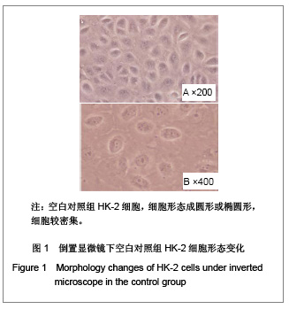
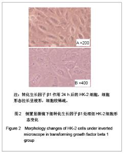
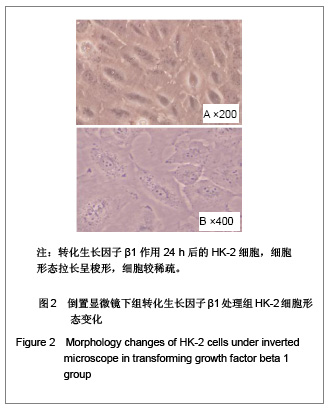
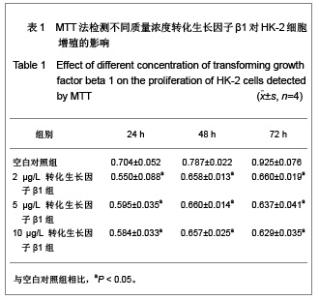
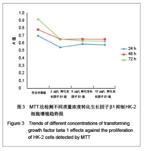
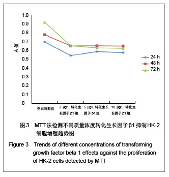
| [1] Bandara JM, Wijewardena HV, Liyanege J,et al. Chronic renal failure in Sri Lanka caused by elevated dietary cadmium: Trojan horse of the green revolution.Toxicol Lett. 2010;198(1): 33-39.[2] Feest T. Epidemiology and causes of chronic renal failure. Medicine. 2007;35(8):438-441.[3] Li DQ, Wang Y, Zhou GL,et al.Zhonghua Tangniaobing Zazhi. 2004;12(3): 219-224. 李丹清,王颖,周桂玲,等. 糖尿病肾病大鼠肾小管与肾小球病变的顺序及其意义[J].中华糖尿病杂志,2004,12(3): 219-224.[4] Hertig A.Epithelial-mesenchymal transition of the renal graft. Nephrol Ther. 2008;4 Suppl 1:S25-S28.[5] Ding Z, Chen Z, Chen X,et al. Adenovirus-mediated anti-sense ERK2 gene therapy inhibits tubular epithelial-mesenchymal transition and ameliorates renal allograft fibrosis.Transpl Immunol. 2011;25(1):34-41.[6] Yoshino J, Monkawa T, Tsuji M,et al. Snaill is involved in the renal epithelial-mesenchymal transition. Biochem Biophys Res Commun. 2007;362(1):63-68.[7] Margetts PJ, Bonniaud P, Liu L,et al.Transient overexpression of TGF-{beta}1 induces epithelial mesenchymal transition in the rodent peritoneum. J Am Soc Nephrol. 2005;16(2): 425-436.[8] Thiery JP, Acloque H, Huang RY,et al. Epithelial-mesenchymal transitions in development and disease. Cell. 2009;139(5):871-890.[9] Xu J, Lamouille S, Derynck R.TGF-beta-induced epithelial to mesenchymal transition.Cell Res. 2009;19(2):156-172.[10] Kalluri R, Weinberg RA.The basics of epithelial-mesenchymal transition. J Clin Invest. 2009;119(6):1420-1428.[11] Wang W, Koka V, Lan HY.Transforming growth factor-beta and Smad signalling in kidney diseases.Nephrology (Carlton). 2005;10(1):48-56.[12] Lü L, Zhang L, Wai MS,et al. Exocytosis of MTT formazan could exacerbate cell injury.Toxicol In Vitro. 2012;26(4): 636-644.[13] Kutz SM, Higgins CE, Samarakoon R,et al. TGF-beta 1-induced PAI-1 expression is E box/USF-dependent and requires EGFR signaling. Exp Cell Res. 2006;312(7): 1093-1105.[14] Yang LX,Li Y,Wang Q,et al. Zhongguo Zhongxiyi Jiehe Zazhi. 2008;9(12): 1040-1043. 杨丽霞,李彧,王谦,等. 姜黄素对TGF-β1诱导人肾小管上皮细胞转分化及分泌细胞外基质成分的影响[J]. 中国中西医结合肾病杂志,2008,9(12): 1040-1043.[15] Dudas PL, Argentieri RL, Farrell FX. BMP-7 fails to attenuate TGF-beta1-induced epithelial-to-mesenchymal transition in human proximal tubule epithelial cells. Nephrol Dial Transplant. 2009;24(5):1406-1416. |