Chinese Journal of Tissue Engineering Research ›› 2013, Vol. 17 ›› Issue (4): 625-632.doi: 10.3969/j.issn.2095-4344.2013.04.010
Previous Articles Next Articles
Comparison between cross-sectional anatomy and CT scanning for observing thoracolumbar vertebrae structure
Gong Teng, Wang Jing-gui
- Department of Orthopaedics, Affiliated Hospital of Medical College of Chinese People’s Armed Police Forces, Tianjin 300162, China
-
Received:2012-07-05Revised:2012-11-28Online:2013-01-22Published:2013-01-22 -
Contact:Wang Jing-gui, Chief physician, Professor, Department of Orthopedics, Affiliated Hospital of Medical College of Chinese People’s Armed Police Forces, Tianjin 300162, China -
About author:Gong Teng☆, Doctor, Attending physician, Department of Orthopedics, Affiliated Hospital of Medical College of Chinese People’s Armed Police Forces, Tianjin 300162, China gongtengwujin@126.com
CLC Number:
Cite this article
Gong Teng, Wang Jing-gui. Comparison between cross-sectional anatomy and CT scanning for observing thoracolumbar vertebrae structure[J]. Chinese Journal of Tissue Engineering Research, 2013, 17(4): 625-632.
share this article
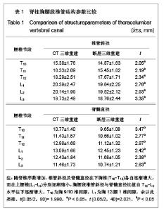
2.1 脊柱胸腰段椎管结构参数分析 随脊椎序数增加,椎管斜径及脊髓直径在下胸椎(T10-T12)各自逐渐增大,相互间改变幅度较大,而在上腰椎(L1-L3)分别逐渐缩小,上腰椎两指标总体均较下胸椎大,胸腰段椎管斜径与脊髓直径比值自T10-L3水平往下逐渐增大,见表1。"


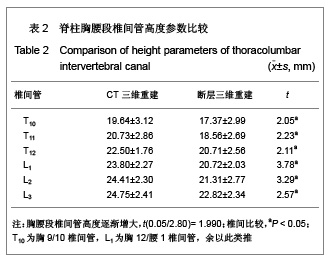
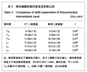
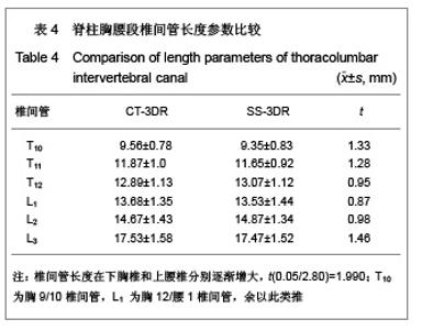
2.2 脊柱胸腰段椎间管径值参数比较 胸腰段椎间管高度逐渐增大,椎间管宽度在下胸椎依次增大,而于上腰椎又依次减小,椎间管高度明显大于相应椎间管宽度,椎间管长度在下胸椎和上腰椎分别逐渐增大,见表2-4。"

"
"

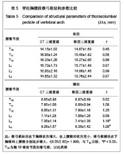
2.3 脊柱胸腰段椎弓根结构参数比较 椎弓根纵径在下胸椎依次增大,在上腰椎却依次变小,椎弓根横径在下胸椎和上腰椎分别逐步增大,见表5。CT薄扫和断层切片三维重建在观察各结构变化程度及规律时基本一致。"

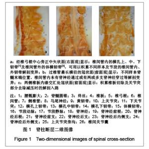
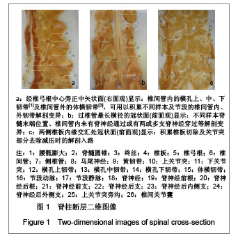
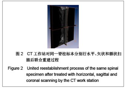
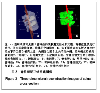
2.4 典型断层影像学分析 图1a可见椎间管内的横孔上、中、下韧带及椎间管外的体横韧带[1,3],可用以积累不同样本及节段的椎间管内、外韧带解剖变异资料;右侧腰1椎间管内的横孔中韧带上方有脊神经和节段动脉走行,其下方为节段静脉通过,右侧腰2脊神经则被横孔上、下韧带所包绕,横孔上韧带头侧通过节段动脉,横孔下韧带尾侧有节段静脉经过,在腰3椎体上有椎间管内韧带延伸至椎体的椎间管外(体横)韧带。图1b可用以积累不同样本脊髓末端位置、椎间管内未有脊神经通过或有两或多支脊神经穿过等解剖变异资料,可观察腰骶膨大、脊髓圆锥、马尾神经的构成及走行路径,记录脊神经前、后根自硬膜囊发出位置和椎间管、椎弓根断面形状。图1c可用以积累椎板切除及关节突部分去除减压时的解剖入路资料,从椎板至双侧关节突的上下嵴线有黄韧带附着,其于上关节突旁沟延伸为椎间关节囊,便于观察椎间管外口、根黄通道及盘黄间隙内相关结构。CT三维重建过程见图2。"
"

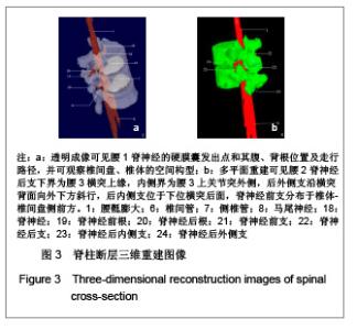
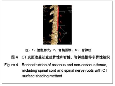
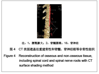
断层三维重建图像见图3,CT三维立体图像见图4。"
"
| [1] Xu SD,Guo SF. Beijing: The Publishing House of People’s Health. 2002:593-595. 胥少汀,郭世绂.脊髓损伤基础与临床[M].北京:人民卫生出版社, 2002:593-595.[2] Zheng YF, Zhang T, Wang ZG, et al. Zhonghua Guke Zazhi. 2007;27(1):26-29. 郑永发,张弢,王志钢,等.下胸椎椎管狭窄症的临床和治疗特点[J].中华骨科杂志,2007,27(1):26-29.[3] Guo SF. Tianji: Tianjing science & technology press. 1989: 129-136. 郭世绂.临床骨科解剖学[M].天津科学技术出版社,1989: 129-136.[4] Lou CF, Hu CF, Gao H, et al. Zhonghua Chuangshang Guke Zazhi. 2009;1l(3):201-205. 罗从风,胡承方,高洪,等.基于CT的胫骨平台骨折的三柱分型[J].中华创伤骨科杂志,2009,1l(3):201-205.[5] Liu JH, Shi XT. Jiepouxue Yonjiu.2010;06:457-458. 刘健华,石小田.前庭蜗器整体标本的设计与制作[J].解剖学研究, 2010,06:457-458.[6] Yuan P, Wang WC. Zhongnan Daxue Xuebao(Yixueban). 2010 ;35(1) :85-89. 袁平,王万春.膝关节三维有限元模型的建立及生物力学分析[J].中南大学学报(医学版),2010,35(1) :85-89.[7] Ding S, Jia KF, Zai LD, et al. Jiepouxue Yanjiu. 2010;06: 401-404, 407. 丁实,贾科锋,翟丽东,等.结节间沟及其毗邻结构断层解剖学研究[J].解剖学研究,.2010,06:401-404, 407.[8] Jia KF, Ding S, Zai LD, et al. Zhongguo Linchuang Jiepouxue Zazhi. 2011;29(2):140-144. 贾科锋,丁实,翟丽东,等.肘管与滑车上肘肌的解剖学研究及其临床意义[J].中国临床解剖学杂志,2011,29(2):140-144. [9] Gao W, Yuan W, Han Y, et al. Zhongguo Linchuang Jiepouxue Zazhi. 2011;29(4): 413-417. 高炜,袁武,韩悦,等.前列腺解剖带区的断层切片与MRI图像对照研究[J].中国临床解剖学杂志,2011,29(4): 413-417.[10] Lv YB, Zhao LW, Wu Y, et al. Zhongguo Linchuang Jiepouxue Zazhi. 2010;28(5):518-522. 吕杨波,赵立武,吴樾,等.视神经管手术入路断层与应用解剖学研究[J].中国临床解剖学杂志,2010,28(5):518-522.[11] Wang H, Miao YY, FU SQ. Jiepouxue Yanjiu. 2010;06: 405-408. 王华,苗莹莹,付升旗.翼腭间隙的断层影像解剖及临床意义[J].解剖学研究,2010,06:405-408.[12] Zai LD, Liu J, Yuan W, et al. Zhong guo Linchuang Jiepouxue Zazhi. 2009,27(04):405-407. 翟丽东,刘瑾,袁武,等.直肠阴道隔的解剖学研究及其临床意义[J].中国临床解剖学杂志,2009,27(04):405-407.[13] Liu J, Zai LD, Li YS. Zhongguo Linchuang Jiepouxue Zazhi. 2009;27(06):663-664. 刘瑾,翟丽东,李云生.直肠阴道隔(IWS)区域的薄层断面解剖学研究[J].中国临床解剖学杂志,2009,27(06):663-664.[14] Zhai LD, Liu J, Li YS, et al. Denonvilliers' fascia in women and its relationship with the fascia propria of the rectum examined by successive slices of celloidin-embedded pelvic viscera. Dis Colon Rectum. 2009;52(9):1564-571.[15] Zhai LD, Liu J, Li YS, et al. The male rectourethralis and deep transverse perineal muscles and their relationship to adjacent structures examined with successive slices of celloidin-embedded pelvic viscera. Eur Urol. 2011;59(3): 415-421.[16] Markaryan A, Nelson EG, Tretiakova M, et al. Technical report: laser microdissection of cochlear structures from celloidin embedded human temporal bone tissues and detection of the mitochondrial DNA common deletion using real time PCR. Hear Res. 2008;244(1-2):1-6.[17] Markaryan A, Nelson EG, Tretiakova M, et al. Technical report: immunofluorescence and TUNEL staining of celloidin embedded human temporal bone tissues. Hear Res. 2008;241(1-2):1-6.[18] Cai JM, Cheng LM, Jia YW. Zhongguo Jiaoxing Waike Zazhi. 2010;18(24):2042-2045. 蔡佳敏,程黎明,贾永伟.胸腰段椎体成形术单侧经椎弓根穿刺的数字化模拟研究[J].中国矫形外科杂志,2010,18(24):2042- 2045.[19] Ouyang ZH, Ouyang HJ. Zhongguo Yixue Yinxgiangxue Zazhi. 2011;19(01): 73-75. 欧阳志和,欧阳厚淦.胸腰结合段侧面血管的影像解剖学研究[J].中国医学影像学杂志,2011,19(01): 73-75.[20] Abbas J, Hamoud K, Masharawi YM, et al. Ligamentum flavum thickness in normal and stenotic spines. Spine. 2010; 35(12):1225-1230.[21] Masharawi Y, Dar G, Peleg S, et al. Lumbar facet anatomy changes in spondylolysis: a comparative skeletal study. Eur Spine J. 2007;16(7):993-999.[22] Dar G, Masharawi Y, Peleg S, et al. Schmorl's nodes distribution in the human spine and its possible etiology. Eur Spine J. Eur Spine J. 2010;19(4): 670-675. |
| [1] | Li Chenjie, Lü Linwei, Song Yang, Liu Jingna, Zhang Chunqiu. Measurement and statistical analysis of trabecular morphological parameters of titanium alloy peri-prosthesis under preload [J]. Chinese Journal of Tissue Engineering Research, 2021, 25(4): 516-520. |
| [2] | Han Shichong, Li Chang, Xing Haiyang, Ge Wenlong, Wang Gang . Finite element analysis of two internal fixation methods for treating extra-articular proximal tibial fractures [J]. Chinese Journal of Tissue Engineering Research, 2021, 25(15): 2329-2333. |
| [3] | Kang Peng, Zhang Fangxin, Maihemuti·Yakufu, Wei Weitao, Yilihamu·Toheti, Aierken·Amudong. True acetabulum morphology of Crowe type IV developmental dysplasia of the hip based on three-dimensional surgical simulation [J]. Chinese Journal of Tissue Engineering Research, 2020, 24(36): 5749-5754. |
| [4] | Chen Zegang, Ding Haili, Li Long, Wang Chun. Changes of bone metabolism after different intensity endurance exercises in growing rats [J]. Chinese Journal of Tissue Engineering Research, 2020, 24(35): 5582-5588. |
| [5] | Wu Libing, Xu Yangyang, He Yujie, Wang Haiyan, Gao Shang, Enhejirigala, Li Xiaohe, Li Zhijun. Anatomical etiology and clinical significance of three-dimensional digital measurement of kidney stones [J]. Chinese Journal of Tissue Engineering Research, 2020, 24(29): 4662-4666. |
| [6] |
Xu Yangyang, Gao Shang, Su Baoke, Wang Yidan, He Yujie, Li Kun, Wang Haiyan, Li Xiaohe.
Three-dimensional morphological characteristics of basal nuclei in children aged 3 or 4 [J]. Chinese Journal of Tissue Engineering Research, 2020, 24(17): 2724-2729. |
| [7] | Luo Xiaofei, Wei Xuan, Wang Jinliang, Wang Shaohua, Li Zhe, Bai Yu. Ultrastructural changes of tibial subchondral bone in patients with knee osteoarthritis: CT evaluation [J]. Chinese Journal of Tissue Engineering Research, 2020, 24(15): 2399-2404. |
| [8] | Wu Xiaoqi, Li Ding, Huang Xuecheng, Huang Wenhua. Establishing morphological database of lower limbs for providing appropriate clinical postoperative prosthesis socket [J]. Chinese Journal of Tissue Engineering Research, 2020, 24(15): 2297-2302. |
| [9] | Fu Yu, Zhang Yunfeng, Su Baoke, Zhao Yan, Xin Daqi, Wang Haiyan, Xu Yangyang, Zhang Cong, Wang Yidan, Wang Xing, Gao Shang, En He, Cai Yongqiang, Wang Jianzhong, Wang Zhiqiang, Gao Mingjie,Li Zhijun, Ma Jierong, Li Xiaohe. Application of three-dimensional digital operation planning to individualized adolescent idiopathic scoliosis surgery [J]. Chinese Journal of Tissue Engineering Research, 2019, 23(32): 5158-5163. |
| [10] | Jia Junfeng, Tang Chengjie, Yue Jintao, Li Feng. Finite element analysis of three different fixation methods for distal tibial fracture [J]. Chinese Journal of Tissue Engineering Research, 2019, 23(32): 5188-5194. |
| [11] | He Yujie, Zhang Shaojie, Li Zhijun, Li Xiaohe, Wang Haiyan, Wang Xing, Xu Yangyang, Gao Mingjie, Li Kun, Dai Lina. Three-dimensional digital morphological characteristics of the facet joint of the lower cervical spine in children aged 4-6 years [J]. Chinese Journal of Tissue Engineering Research, 2019, 23(28): 4558-4563. |
| [12] | Zhong Hua, Liu Jun, Cai Guoxiang, Duan Shaoyin. Individualized design and clinical application of metacarpophalangeal joint prosthesis: a one-case report [J]. Chinese Journal of Tissue Engineering Research, 2019, 23(20): 3164-3169. |
| [13] | Fang Xu, Dong Junfeng. Application of additive manufacturing technology in bone defects [J]. Chinese Journal of Tissue Engineering Research, 2019, 23(18): 2915-2920. |
| [14] | Luo Jian, Wang Lihua, Wang Tao, Wen Hui. Changes of stress and displacement of three-dimensional finite element model of ankle joint using different material assignment methods [J]. Chinese Journal of Tissue Engineering Research, 2019, 23(18): 2822-2826. |
| [15] | Wei Qiushi, Fang Bin, Chen Zhenqiu, He Mincong, Chen Xiaojun, Yang Fan, Zhang Qingwen, He Wei. Role of bone status in anterolateral portion of femoral head in the progression of osteonecrosis of the femoral head [J]. Chinese Journal of Tissue Engineering Research, 2019, 23(16): 2516-2522. |
| Viewed | ||||||
|
Full text |
|
|||||
|
Abstract |
|
|||||