Chinese Journal of Tissue Engineering Research ›› 2013, Vol. 17 ›› Issue (1): 23-30.doi: 10.3969/j.issn.2095-4344.2013.01.004
Previous Articles Next Articles
Treatment of traumatic brain injury in rats by RhoA gene silencing combinedwith umbilical cord mesenchymal stem cell transplantation
Feng Shi-jun, Han Jian-guo
- Department of Neurosurgery, First Affiliated Hospital of Baotou Medical College, Baotou 014010, Inner Mongolia Autonomous Region, China
-
Received:2012-10-05Revised:2012-12-05Online:2013-01-01Published:2013-01-01 -
About author:Feng Shi-jun★, Master, Associate chief physician, Department of Neurosurgery, First Affiliated Hospital of Baotou Medical College, Baotou 014010, Inner Mongolia Autonomous Region, China qxy20110824@163.com
CLC Number:
Cite this article
Feng Shi-jun, Han Jian-guo. Treatment of traumatic brain injury in rats by RhoA gene silencing combinedwith umbilical cord mesenchymal stem cell transplantation[J]. Chinese Journal of Tissue Engineering Research, 2013, 17(1): 23-30.
share this article
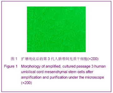
2.1 实验动物数量分析 实验选用Wistar大鼠84只,分为3组,实验过程无脱失,全部进入结果分析。 2.2 脐带间充质干细胞形态观察 见图1。"
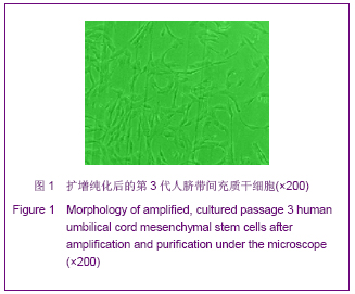
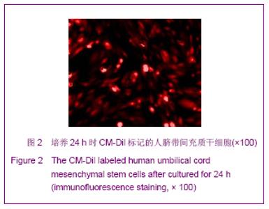
培养四五天后,贴壁细胞明显增多,细胞为长梭形或多角形,并可见有丝分裂,部分细胞克隆呈集落状生长,形态呈单一的长梭形,胞体中央膨大,培养八九天,细胞铺满培养瓶底面,成漩涡状生长,漩涡中心的细胞为复层生长,周围大部分细胞胞体呈单层的漩涡状或辐射状。 流式细胞仪检测结果显示CD34,CD45,CD29,CD90,CD105及CD49阳性,脐带间充质干细胞均一性好,纯度达98%以上。24 h时,CM-Dil标记的脐带间充质干细胞在荧光显微镜下呈红色荧光, FCM检测细胞标记率达100%,见图2。"
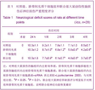
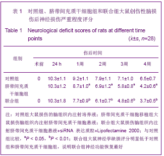
2.3 大鼠神经功能评价 三组大鼠神经功能评分与损伤前组比较,在伤后各时间点NSS评分增高(P < 0.01);伤后第1天3组NSS评分差异无显著性意义(P> 0.05);伤后1,2,3,4周,大鼠神经学缺损评分脐带间充质干细胞组低于对照组(P < 0.05),联合组明显低于对照组,差异有非常显著性意义(P < 0.01),见表1。"
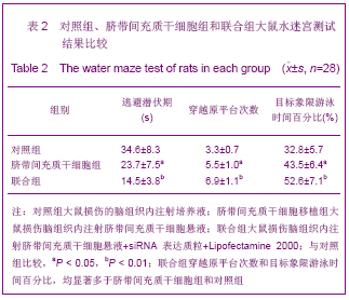
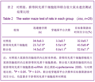
2.4 Morris水迷宫行为学检测结果 各组平均潜伏时间均逐渐缩短,1 d各组之间差异无显著性意义,2 d联合组逃避潜伏期较对照组、脐带间充质干细胞组明显缩短(P < 0.05),3-5 d联合组逃避潜伏期较对照组缩短更明显(P < 0.01)。空间探索试验穿越平台次数:与联合组相比, 脐带间充质干细胞组和对照组穿越原平台次数明显减少,差异有显著性意义(P < 0.05,P < 0.01);目标象限游泳时间占总时间的百分比:与联合组相比,脐带间充质干细胞组和对照组明显减少(P < 0.05,P < 0.01)。见表2。"
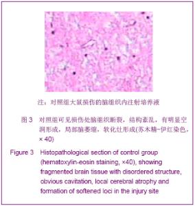
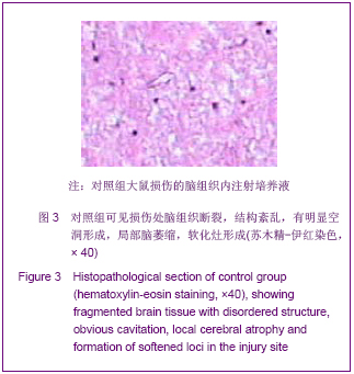
2.5 大鼠损伤处脑组织苏木精-伊红染色和荧光显微镜观察 移植4周后苏木精-伊红染色对照组可见损伤处脑组织被胶质细胞和胶质纤维充填,形成胶质瘢痕,软化灶形成,见图3。"
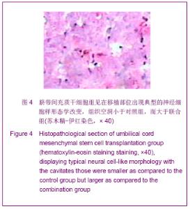
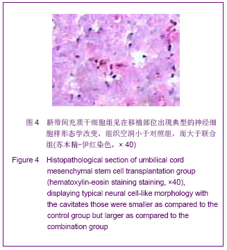
脐带间充质干细胞组见在移植部位出现典型的神经细胞样形态学改变,组织瘢痕和软化灶小于对照组,而大于联合组,见图4。"
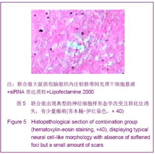
联合组出现典型的神经细胞样形态学改变且软化灶消失,有少量瘢痕,见图5。"

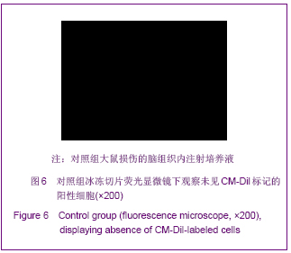
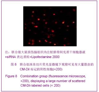
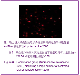
荧光显微镜观察结果显示大鼠损伤灶脑组织中的CM-Dil阳性细胞数:对照组为0,脐带间充质干细胞组为(28.42±6.47)个/高倍视野,联合组为(51.64±10.32)个/高倍视野,三者经过方差分析后两组之间的比较用Dunnett t 检验。各组之间差异有显著性意义(P< 0.01),见图6-8。"
"
"

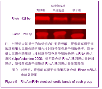
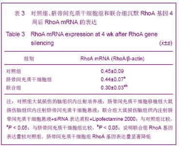
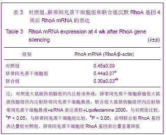
2.6 RT-PCR检测RhoA mRNA结果 经siRNA沉默RhoA基因4周后,联合组RhoA基因表达量较对照组、脐带间充质干细胞组RhoA基因表达量显著降低,差异有显著性意义(P < 0.05)。见图9,表3。"
"
| [1] Arabi YM, Haddad S, Tamim HM,et al. Mortality reduction after implementing a clinical practice guidelines–based management protocol for severe traumatic brain injury. J Crit Care.2010;25(2):190-195.[2] Lee HC, Chuang HC, Cho DY, et al. Applying Cerebral Hypothermia and Brain Oxygen Monitoring in Treating Severe Traumatic Brain Injury. World Neurosurg. 2010;74(6): 654-660.[3] Engelmann CM, Siert L. Cognitive disturbances following severe traumatic brain injury. Ugeskr Laege. 2007; 169(3): 217-219.[4] Liliang PC, Liang CL, Weng HC, et al. Proteins in Serum Predict Outcome After Severe Traumatic Brain Injury. J Surg Res. 2010;160(2): 302-3078.[5] Sekhon MS, Dhingra VK, Sekhon IS,et al.The safety of synthetic colloid in critically ill patients with severe traumatic brain injuries. J Crit Care.2011;26(4):357-362.[6] Yoo BY, Shin YH, Yoon HH,et al.Application of mesenchymal stem cells derived from bone marrow and umbilical cord in human hair multiplication.J Dermatol Sci. 2010, 60(2): 74-83.[7] Xu Y,Meng H,Li C,et alUmbilical cord-derived mesenchymal stem cells isolated by a novel explantation technique can differentiate into functional endothelial cells and promote revascularization. Stem Cells.2010;19(10):1511-1522. [8] Matsuse D,Kitada M,Kohama M,et al. Human umbilical cord-derived mesenchymal stromal cells differentiate into functional Schwann cells that sustain peripheral nerve regeneration.J Neuropathol Exp Neurol.2010;69(9):973-985.[9] Yuan GF, Li HX. Zhongguo Xiandai Yixue Zazhi. 2011;21(13): 1435-1438.苑国富,李红星.大鼠颅脑损伤后亚低温治疗对RhoA 及Nogo- A 表达的影响[J].中国现代医学杂志,2011, 21(13):1435-1438.[10] Wang QY, Liu WG, Wang ZY. Zhongguo Guke Linchuang yu Jichu Yanjiu Zazhi.2010;2(4):292-296.王求永,刘文革,王振宇.FTY720对大鼠急性脊髓损伤后RhoA 表达的影响[J].中国骨科临床与基础研究杂志,2010,2(4):292-296.[11] Ahmed Z, Dent RG, Suggate EL, et al. Disinhibition of neurotrophin-induced dorsal root ganglion cell neurite outgrowth on CNS myelin by siRNA-mediated knockdown of NgR, p75NTR and Rho-A. Molecular and Cellular Neuroscience.2005;28(3):509-523.[12] Chen L,Hui GZ,Zhang S,et al.Zhonghua Chuangshang Zazhi. 2009;25(6):498-502.陈镭,惠国桢,张赛,等.人脐血间充质干细胞移植促进大鼠颅脑损伤后神经功能的恢复[J].中华创伤杂志,2009,25(6):498- 502.[13] Horisawa T, Ishibashi T, Nishikawa H, et al. The effects of selective antagonists of serotonin 5-HT7 and 5-HT1A receptors on MK-801-induced impairment of learning and memory in the passive avoidance and Morris water maze tests in rats: Mechanistic implications for the beneficial effects of the novel atypical antipsychotic lurasidone . Behavioural Brain Res. 2011;220 (1): 83-90.[14] Mutlu O, Ulak G, Celikyurt IK et al, Effects of olanzapine, sertindole and clozapine on learning and memory in the Morris water maze test in naive and MK-801-treated mice.Pharmaco Biochem Behav. 2011;98(3): 398-404.[15] Li L, Ding J, Marshall C, et al.Pretraining affects Morris water maze performance with different patterns between control and ovariectomized plus d-galactose-injected mice. Behavioural Brain Research.2011;217(1): 244-247.[16] Hang RH, Xu YJ, Xie HF.Evaluating on recognition impairment after traumatic brain injury with WCST.Fa Yi Xue Za Zhi. 2011;27(5):346-349. [17] Han SR, Yee GT, Choi CY,et al.Cortical laminar necrosis in an infant with severe traumatic brain injury.J Korean Neurosurg Soc. 2011;50(5):472-474. [18] Shultz SR, Macfabe DF, Foley KA, et al.Sub-concussive brain injury in the Long-Evans rat induces acute neuroinflammation in the absence of behavioral impairments.Behav Brain Res. 2011;229(1):145-152. [19] Settervall CH, Sousa RM, Silva SC.In-hospital mortality and the Glasgow Coma Scale in the first 72 hours after traumatic brain injury.Rev Lat Am Enfermagem. 2011;19(6):1337-1343. [20] Yamashita S, Hasuo H, Tokutomi T,et al. Edaravone attenuates impairment of synaptic plasticity in granule cell layer of the dentate gyrus following traumatic brain injury. Kurume Med J. 2011;58(2):47-58.[21] Han SR, Yee GT, Choi CY, et al Cortical laminar necrosis in an infant with severe traumatic brain injury.J Korean Neurosurg Soc. 2011;50(5):472-474. [22] Hang RH, Xu YJ, Xie HF,et al. Evaluating on recognition impairment after traumatic brain injury with WCST.Fa Yi Xue Za Zhi. 2011;7(5):346-349. [23] He JT, Mang J, Mei CL,et al. Neuroprotective effects of exogenous activin a on oxygen-glucose deprivation in PC12 cells.Molecules. 2011;30(17):315-327.[24] Li GX,Wang CM,Gong LP,et al.Zhongguo Linchuang Shenjing Waike Zazhi. 2009,14(5):288-290.黎国雄,王传湄, 龚利平,等.神经干细胞立体定向脑内移植治疗大鼠重型颅脑损伤[J].中国临床神经外科杂志,2009,14(5): 288-290.[25] Zhao EY, Wang LD, Wen QQ, et al. Fasudil hydrochloridedifferentiates bone marrow mesenchymal stem cells into neurons via Notch signaling. Neural Regen Res. 2010;5(11):814-819.[26] Dergham P,Ellezam B,Essagian C,et al.Rho signaling pathwaytargeted to promote spinal cord repair. J Neurosci. 2002;22(15):6570-6577. [27] Govek EE,Newey SE,Aelstl V. The role of the Rho GTPases in neuronal development. Genes Dev.2005;19(10): 1-49.[28] Neuhof S,Moers J,Ricks M. Proliferation, diferentiation, and cytokine secretion of human umbilical cord blood-derivedmononuclear ceils in vitro.Exp Hematol.2007; 35(7):1119-1131.[29] Pang KM, Sung MA, Alrash-dan MS, et al. Trans-plantation of mesenchymal stem cells from human umbilical cord versus human umbilical cord blood for peripheral nerve regenera-tion. Neural Regen Res. 2010;5(11):838-845.[30] Ha Y,Choi JU,Yoon DH,et a1.Neural phenotype expression of cultured human cord blood cells in vitro. Neurureport. 2001; 12(16):3523-3530.[31] Buzahska L,Jurga M,Domafiska-Janik K.Neumnal differentiation of human umbilical cord blood neural stem- like cell line.Neurodegener Dis.2006;3(1-2):19-26.[32] Wang D,Zhang JJ,Mang JJ.Zhongguo Zuzhi Gongcheng Yanjiu yu Linchuang Kangfu. 2010,14(14): 2539-2544.王东,张建军,马景鑑.神经干细胞NgR基因沉默立体定向移植治疗大鼠脑损伤[J].中国组织工程研究与临床康复,2010, 14(14): 2539-2544.[33] Neuhof S,Moers J,Ricks M.Proliferation,diferentiation,and cytokine secretion of human umbilical cord blood-derived mononuclear ceils in vitro.Exp Hematol.2007;35(7): 1119-1131.[34] Zhou GQ,Jin Y,Zhang P.Zhongguo Zuzhi Gongcheng Yanjiu yu Linchuang Kangfu. 2010;14(45):8416-8420.周国庆,金怡,张鹏.沉默RhoA基因对骨髓间充质干细胞静脉移植治疗脑梗死大鼠的作用[J].中国组织工程研究与临床康复, 2010,14(45):8416-8420. |
| [1] | Jiang Tao, Ma Lei, Li Zhiqiang, Shou Xi, Duan Mingjun, Wu Shuo, Ma Chuang, Wei Qin. Platelet-derived growth factor BB induces bone marrow mesenchymal stem cells to differentiate into vascular endothelial cells [J]. Chinese Journal of Tissue Engineering Research, 2021, 25(25): 3937-3942. |
| [2] | Mo Jianling, He Shaoru, Feng Bowen, Jian Minqiao, Zhang Xiaohui, Liu Caisheng, Liang Yijing, Liu Yumei, Chen Liang, Zhou Haiyu, Liu Yanhui. Forming prevascularized cell sheets and the expression of angiogenesis-related factors [J]. Chinese Journal of Tissue Engineering Research, 2021, 25(22): 3479-3486. |
| [3] | Chen Lei, Zheng Rui, Jie Yongsheng, Qi Hui, Sun Lei, Shu Xiong. In vitro evaluation of adipose-derived stromal vascular fraction combined with osteochondral integrated scaffold [J]. Chinese Journal of Tissue Engineering Research, 2021, 25(22): 3487-3492. |
| [4] | Wei Qin, Zhang Xue, Ma Lei, Li Zhiqiang, Shou Xi, Duan Mingjun, Wu Shuo, Jia Qiyu, Ma Chuang. Platelet-derived growth factor-BB induces the differentiation of rat bone marrow mesenchymal stem cells into osteoblasts [J]. Chinese Journal of Tissue Engineering Research, 2021, 25(19): 2953-2957. |
| [5] | Chen Xiao, Guo Zhi, Chen Lina, Liu Xuanyong, Zhang Yihuizhi, Li Xumian, Wang Yueqiao, Wei Liya, Xie Jing, Lin Li. Factors affecting the mobilization and collection of autologous peripheral blood hematopoietic stem cells [J]. Chinese Journal of Tissue Engineering Research, 2021, 25(19): 2958-2962. |
| [6] | Guo Zhibin, Wu Chunfang, Liu Zihong, Zhang Yuying, Chi Bojing, Wang Bao, Ma Chao, Zhang Guobin, Tian Faming. Simvastatin stimulates osteogenic differentiation of bone marrow mesenchymal stem cells [J]. Chinese Journal of Tissue Engineering Research, 2021, 25(19): 2963-2968. |
| [7] | Li Congcong, Yao Nan, Huang Dane, Song Min, Peng Sha, Li Anan, Lu Chao, Liu Wengang. Identification and chondrogenic differentiation of human infrapatellar fat pad derived stem cells [J]. Chinese Journal of Tissue Engineering Research, 2021, 25(19): 2976-2981. |
| [8] | Gao Yuanhui, Xiang Yang, Cao Hui, Wang Shunlan, Zheng Linlin, He Haowei, Zhang Yingai, Zhang Shufang, Huang Denggao. Comparison of biological characteristics of adipose derived mesenchymal stem cells in Wuzhishan inbreed miniature pigs aged two different months [J]. Chinese Journal of Tissue Engineering Research, 2021, 25(19): 2988-2993. |
| [9] | Cao Yang, Zhang Junping, Peng Li, Ding Yi, Li Guanghui. Isolation and culture of rabbit aortic endothelial cells and biological characteristics [J]. Chinese Journal of Tissue Engineering Research, 2021, 25(19): 3000-3003. |
| [10] | Dai Min, Wang Shuai, Zhang Nini, Huang Guilin, Yu Limei, Hu Xiaohua, Yi Jie, Yao Li, Zhang Ligang. Biological characteristics of hypoxic preconditioned human amniotic mesenchymal stem cells [J]. Chinese Journal of Tissue Engineering Research, 2021, 25(19): 3004-3008. |
| [11] | Qin Yanchun, Rong Zhen, Jiang Ruiyuan, Fu Bin, Hong Xiaohua, Mo Chunmei. Chinese medicine compound preparation inhibits proliferation of CD133+ liver cancer stem cells and the expression of stemness transcription factors [J]. Chinese Journal of Tissue Engineering Research, 2021, 25(19): 3016-3023. |
| [12] | Dai Yaling, Chen Lewen, He Xiaojun, Lin Huawei, Jia Weiwei, Chen Lidian, Tao Jing, Liu Weilin. Construction of miR-146b overexpression lentiviral vector and the effect on the proliferation of hippocampal neural stem cells [J]. Chinese Journal of Tissue Engineering Research, 2021, 25(19): 3024-3030. |
| [13] | Chen Jie, Liao Chengcheng, Chen Zhiwei, Wang Yan. Bladder cancer stem cell markers and related signaling pathways: antibody targeted therapy [J]. Chinese Journal of Tissue Engineering Research, 2021, 25(19): 3090-3096. |
| [14] | Zhang Wei, Cui Shuaishuai, Zhou Zhichao, Hu Xiaohua, Yang Xiaohong. MicroRNA regulates histone deacetylase in the treatment of bone-related diseases [J]. Chinese Journal of Tissue Engineering Research, 2021, 25(17): 2767-2774. |
| [15] | Chen Liang, Meng Shu, Cheng Guoping, Ding Yi . Effects of fish scale collagen membrane on adhesion, proliferation and osteogenic differentiation of rat bone marrow mesenchymal stem cells [J]. Chinese Journal of Tissue Engineering Research, 2021, 25(16): 2494-2499. |
| Viewed | ||||||
|
Full text |
|
|||||
|
Abstract |
|
|||||