Chinese Journal of Tissue Engineering Research ›› 2019, Vol. 23 ›› Issue (9): 1384-1389.doi: 10.3969/j.issn.2095-4344.1607
Previous Articles Next Articles
Hepatocyte-like cells derived from induced pluripotent stem cells inhibit liver fibrosis in rats
Cheng Gang, Huang Denggao, Liang Ying
- Department of Radiation Oncology, Affiliated Haikou Hospital, Xiangya School of Medicine, Central South University, Haikou 570208, Hainan Province, China
-
Revised:2018-11-16Online:2019-03-28Published:2019-03-28 -
Contact:Liang Ying, Master candidate, Attending physician, Department of Radiation Oncology, Affiliated Haikou Hospital, Xiangya School of Medicine, Central South University, Haikou 570208, Hainan Province, China -
About author:Cheng Gang, Attending physician, Department of Radiation Oncology, Affiliated Haikou Hospital, Xiangya School of Medicine, Central South University, Haikou 570208, Hainan Province, China -
Supported by:the Natural Science Foundation of Hainan Province in 2016, No. 20168315 (to LY)
CLC Number:
Cite this article
Cheng Gang, Huang Denggao, Liang Ying. Hepatocyte-like cells derived from induced pluripotent stem cells inhibit liver fibrosis in rats[J]. Chinese Journal of Tissue Engineering Research, 2019, 23(9): 1384-1389.
share this article
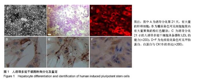
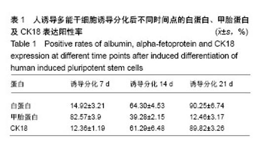
2.1 人诱导多能干细胞向肝样细胞诱导分化的形态变化 经肝向诱导分化至第21天,人诱导多能干细胞克隆团已经松散,以类圆形或多角形为主,呈铺路石样分布并密集排列,见图1A;糖原染色可见细胞胞浆内有大量聚集的粉红色糖原,见图1B;LDL摄取实验显示细胞胞质内有大量红色的荧光,见图1C;免疫组织化学法观察甲胎蛋白、白蛋白与CK18呈阳性表达,见图1D-F。人诱导多能干细胞向肝细胞诱导分化过程中的甲胎蛋白、白蛋白与CK18表达阳性率,见表1。"
"
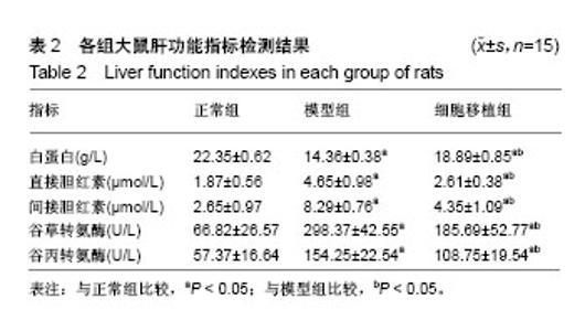
2.2 动物体内移植实验结果 入选45只SD大鼠,其中30只大鼠建立肝纤维化模型,3只造模失败,造模成功率为90%,随后采用相同方法造模,将实验动物补齐。 2.2.1 肝功能指标检测结果 与正常组比较,模型组、细胞移植组白蛋白显著降低,直接胆红素、间接胆红素、谷草转氨酶、谷丙转氨酶水平均显著升高,差异均有显著性意义(P < 0.05)。与模型组比较,细胞移植组白蛋白显著升高,直接胆红素、间接胆红素、谷草转氨酶、谷丙转氨酶水平均显著降低,差异均有显著性意义(P < 0.05),见表2。"


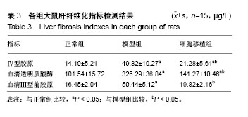
2.2.2 肝纤维化指标检测结果 与正常组比较,模型组Ⅳ型胶原、血清透明质酸酶、血清Ⅲ型前胶原水平均显著升高,差异均有显著性意义(P < 0.05)。与模型组比较,细胞移植组Ⅳ型胶原、血清透明质酸酶、血清Ⅲ型前胶原水平均显著降低,差异均有显著性意义(P < 0.05)。细胞移植组Ⅳ型胶原、血清透明质酸酶水平仍高于正常组,差异均有显著性意义(P < 0.05),见表3。"
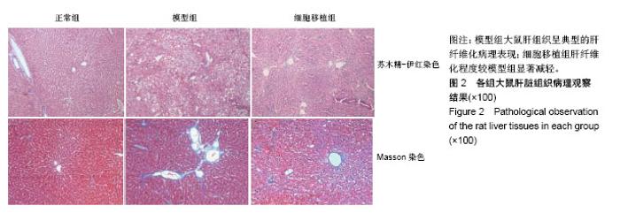
2.2.3 肝脏病理观察结果 细胞移植后4周苏木精-伊红染色、Masson染色显示,正常组肝组织肝小叶轮廓清晰,未见炎性细胞浸润,肝细胞形态正常,肝细胞索排列规则,肝窦无扩张;模型组肝小叶轮廓模糊不清,肝细胞疏松、肿胀,可见大量炎性细胞浸润,大量脂肪细胞呈空泡状改变,呈典型的肝纤维化改变;细胞移植组可见炎性细胞浸润、肝细胞变性与坏死,但程度较对照组明显减轻,病理改变介于正常组与对照组之间,见图2。"
| [1] Zoubek ME, Trautwein C, Strnad P. Reversal of liver fibrosis: From fiction to reality. Best Pract Res Clin Gastroenterol. 2017;31(2):129-141.[2] Pellicoro A, Ramachandran P, Iredale JP. Reversibility of liver fibrosis. Fibrogenesis Tissue Repair. 2012;5(Suppl 1):S26.[3] Pinzani M, Romanelli RG, Magli S. Progression of fibrosis in chronic liver diseases: time to tally the score. J Hepatol. 2001; 34(5):764-767.[4] 唐卫平,胡琦,戴小明,等.脂肪间充质干细胞移植对肝纤维化的治疗作用及其机制[J].中华肝脏病杂志,2018,26(1):28-33.[5] 薛改.脐带间充质干细胞对肝损伤的治疗作用及其机制研究[D].石家庄:河北医科大学,2017.[6] 刘卫,余森源,严和中,等. 骨髓间质干细胞移植对肝纤维化模型大鼠治疗效果[J].临床军医杂志,2017,45(9):903-907.[7] Kim JH, Jang YJ, An SY, et al. Enhanced Metabolizing Activity of Human ES Cell-Derived Hepatocytes Using a 3D Culture System with Repeated Exposures to Xenobiotics. Toxicol Sci. 2015;147(1):190-206.[8] Imai K,Tanoue A,Nakamura K,et al.An attempt to in vitro embryotoxicity test using mouse ES cells and human hepatocytes. Aatex.2013;18(1):33-38.[9] 杜盼,余江南,徐希明.人源多能干细胞体外定向诱导分化为肝细胞[J]. 江苏大学学报(医学版),2018,28(1):39-46.[10] 史德宝,吕礼应. 血清白蛋白检测方法对糖化白蛋白的影响[J].安徽医科大学学报,2015,50(12):1805-1808.[11] 王杨,谷冬梅,陈京山,等. 三种常规方法检测血清胆红素结果的可比性和偏倚评估[J]. 中国实验诊断学, 2017,21(10): 1744-1747.[12] 饶雅琪,张丽,曾金祥,等. 禾叶风毛菊醇提物对四氯化碳致小鼠肝损伤的保护作用[J]. 中国临床药理学杂志, 2017,33(20): 2058-2061.[13] 易梦秋,余旻,王鹏.多器官功能障碍综合征大鼠肺组织MMP-9、TIMP-1和Ⅳ型胶原的表达[J]. 中国病理生理杂志, 2018,34(1): 123-129.[14] Takahashi K, Yamanaka S. Induction of pluripotent stem cells from mouse embryonic and adult fibroblast cultures by defined factors. Cell. 2006;126(4):663-676.[15] Takahashi K, Tanabe K, Ohnuki M, et al. Induction of pluripotent stem cells from adult human fibroblasts by defined factors. Cell. 2007;131(5):861-872.[16] Song Z, Cai J, Liu Y, et al. Efficient generation of hepatocyte-like cells from human induced pluripotent stem cells. Cell Res. 2009;19(11):1233-1242.[17] Twaroski K, Mallanna SK, Jing R, et al. FGF2 mediates hepatic progenitor cell formation during human pluripotent stem cell differentiation by inducing the WNT antagonist NKD1. Genes Dev. 2015;29(23):2463-2474.[18] Takagi C, Yagi H, Hieda M, et al. Mesenchymal Stem Cells Contribute to Hepatic Maturation of Human Induced Pluripotent Stem Cells. Eur Surg Res. 2017;58(1-2):27-39.[19] Mallanna SK, Cayo MA, Twaroski K, et al. Mapping the Cell-Surface N-Glycoproteome of Human Hepatocytes Reveals Markers for Selecting a Homogeneous Population of iPSC-Derived Hepatocytes. Stem Cell Reports. 2016;7(3): 543-556.[20] 刘国菊,李丛丛,李睿坤,等.转化生长因子β1在纤维化疾病发病中作用的研究进展[J].山东医药, 2018,58(30):106-109.[21] 李梓豪,尹大龙,崔逸峰,等.肝硬化肝癌肝部分切除术后影响肝再生的抗纤维化因子研究[J].国际外科学杂志, 2018,45(2): 134-137.[22] 段佳悦,史清峰,王明光,等. 血管内皮生长因子、碱性成纤维细胞生长因子和内皮抑素在肝硬化中的表达及意义[J].临床荟萃, 2018,33(5):427-430.[23] Sulzbacher S, Schroeder IS, Truong TT, et al. Activin A-induced differentiation of embryonic stem cells into endoderm and pancreatic progenitors-the influence of differentiation factors and culture conditions. Stem Cell Rev. 2009;5(2):159-173.[24] 李毅,李欣,孙涛. CDX2基因修饰的脐带间充质干细胞向杯状细胞分化的可行性研究[J].生物技术通讯,2014,25(4):515-519.[25] Kang SJ, Lee HM, Park YI, et al. Chemically induced hepatotoxicity in human stem cell-induced hepatocytes compared with primary hepatocytes and HepG2. Cell Biol Toxicol. 2016;32(5):403-417.[26] 张文成.细胞重编程与诱导内胚层多能干细胞的获取与鉴定[D].北京:中国人民解放军军事医学科学院,解放军军事医学科学院, 2012.[27] Boulter L, Govaere O, Bird TG, et al. Macrophage-derived Wnt opposes Notch signaling to specify hepatic progenitor cell fate in chronic liver disease. Nat Med. 2012;18(4): 572-579.[28] Gieseck RL 3rd, Hannan NR, Bort R, et al. Maturation of induced pluripotent stem cell derived hepatocytes by 3D-culture. PLoS One. 2014;9(1):e86372.[29] Chen Y, Li Y, Wang X, et al. Amelioration of Hyperbilirubinemia in Gunn Rats after Transplantation of Human Induced Pluripotent Stem Cell-Derived Hepatocytes. Stem Cell Reports. 2015;5(1):22-30.[30] Roy-Chowdhury N, Wang X, Guha C, et al. Hepatocyte-like cells derived from induced pluripotent stem cells. Hepatol Int. 2017;11(1):54-69.[31] Sakurai F, Mitani S, Yamamoto T, et al. Human induced-pluripotent stem cell-derived hepatocyte-like cells as an in vitro model of human hepatitis B virus infection. Sci Rep. 2017;7:45698.[32] Huang CS, Lin HC, Lu KH, et al. Generation of high quality of hepatocyte-like cells from induced pluripotent stem cells with Parp1 but lacking c-Myc. J Chin Med Assoc. 2018;81(10): 871-877.[33] Cao YY, Zeng X. Research advances in hepatocyte-like cells from human induced pluripotent stem cells and their application. Zhonghua Gan Zang Bing Za Zhi. 2018;26(1):69-72.[34] Starokozhko V, Hemmingsen M, Larsen L, et al. Differentiation of human-induced pluripotent stem cell under flow conditions to mature hepatocytes for liver tissue engineering. J Tissue Eng Regen Med. 2018;12(5): 1273-1284.[35] Takayama K, Akita N, Mimura N, et al. Generation of safe and therapeutically effective human induced pluripotent stem cell-derived hepatocyte-like cells for regenerative medicine. Hepatol Commun. 2017;1(10):1058-1069.[36] Lorvellec M, Scottoni F, Crowley C, et al. Mouse decellularised liver scaffold improves human embryonic and induced pluripotent stem cells differentiation into hepatocyte-like cells. PLoS One. 2017;12(12):e0189586.[37] Kotaka M, Toyoda T, Yasuda K, et al. Adrenergic receptor agonists induce the differentiation of pluripotent stem cell-derived hepatoblasts into hepatocyte-like cells. Sci Rep. 2017;7(1):16734.[38] Asgari S, Moslem M, Bagheri-Lankarani K, et al. Differentiation and transplantation of human induced pluripotent stem cell-derived hepatocyte-like cells. Stem Cell Rev. 2013;9(4):493-504. |
| [1] | Zhang Tongtong, Wang Zhonghua, Wen Jie, Song Yuxin, Liu Lin. Application of three-dimensional printing model in surgical resection and reconstruction of cervical tumor [J]. Chinese Journal of Tissue Engineering Research, 2021, 25(9): 1335-1339. |
| [2] | Zeng Yanhua, Hao Yanlei. In vitro culture and purification of Schwann cells: a systematic review [J]. Chinese Journal of Tissue Engineering Research, 2021, 25(7): 1135-1141. |
| [3] | Xu Dongzi, Zhang Ting, Ouyang Zhaolian. The global competitive situation of cardiac tissue engineering based on patent analysis [J]. Chinese Journal of Tissue Engineering Research, 2021, 25(5): 807-812. |
| [4] | Wu Zijian, Hu Zhaoduan, Xie Youqiong, Wang Feng, Li Jia, Li Bocun, Cai Guowei, Peng Rui. Three-dimensional printing technology and bone tissue engineering research: literature metrology and visual analysis of research hotspots [J]. Chinese Journal of Tissue Engineering Research, 2021, 25(4): 564-569. |
| [5] | Chang Wenliao, Zhao Jie, Sun Xiaoliang, Wang Kun, Wu Guofeng, Zhou Jian, Li Shuxiang, Sun Han. Material selection, theoretical design and biomimetic function of artificial periosteum [J]. Chinese Journal of Tissue Engineering Research, 2021, 25(4): 600-606. |
| [6] | Liu Fei, Cui Yutao, Liu He. Advantages and problems of local antibiotic delivery system in the treatment of osteomyelitis [J]. Chinese Journal of Tissue Engineering Research, 2021, 25(4): 614-620. |
| [7] | Li Xiaozhuang, Duan Hao, Wang Weizhou, Tang Zhihong, Wang Yanghao, He Fei. Application of bone tissue engineering materials in the treatment of bone defect diseases in vivo [J]. Chinese Journal of Tissue Engineering Research, 2021, 25(4): 626-631. |
| [8] | Zhang Zhenkun, Li Zhe, Li Ya, Wang Yingying, Wang Yaping, Zhou Xinkui, Ma Shanshan, Guan Fangxia. Application of alginate based hydrogels/dressings in wound healing: sustained, dynamic and sequential release [J]. Chinese Journal of Tissue Engineering Research, 2021, 25(4): 638-643. |
| [9] | Chen Jiana, Qiu Yanling, Nie Minhai, Liu Xuqian. Tissue engineering scaffolds in repairing oral and maxillofacial soft tissue defects [J]. Chinese Journal of Tissue Engineering Research, 2021, 25(4): 644-650. |
| [10] | Xing Hao, Zhang Yonghong, Wang Dong. Advantages and disadvantages of repairing large-segment bone defect [J]. Chinese Journal of Tissue Engineering Research, 2021, 25(3): 426-430. |
| [11] | Chen Siqi, Xian Debin, Xu Rongsheng, Qin Zhongjie, Zhang Lei, Xia Delin. Effects of bone marrow mesenchymal stem cells and human umbilical vein endothelial cells combined with hydroxyapatite-tricalcium phosphate scaffolds on early angiogenesis in skull defect repair in rats [J]. Chinese Journal of Tissue Engineering Research, 2021, 25(22): 3458-3465. |
| [12] | Wang Hao, Chen Mingxue, Li Junkang, Luo Xujiang, Peng Liqing, Li Huo, Huang Bo, Tian Guangzhao, Liu Shuyun, Sui Xiang, Huang Jingxiang, Guo Quanyi, Lu Xiaobo. Decellularized porcine skin matrix for tissue-engineered meniscus scaffold [J]. Chinese Journal of Tissue Engineering Research, 2021, 25(22): 3473-3478. |
| [13] | Mo Jianling, He Shaoru, Feng Bowen, Jian Minqiao, Zhang Xiaohui, Liu Caisheng, Liang Yijing, Liu Yumei, Chen Liang, Zhou Haiyu, Liu Yanhui. Forming prevascularized cell sheets and the expression of angiogenesis-related factors [J]. Chinese Journal of Tissue Engineering Research, 2021, 25(22): 3479-3486. |
| [14] | Liu Chang, Li Datong, Liu Yuan, Kong Lingbo, Guo Rui, Yang Lixue, Hao Dingjun, He Baorong. Poor efficacy after vertebral augmentation surgery of acute symptomatic thoracolumbar osteoporotic compression fracture: relationship with bone cement, bone mineral density, and adjacent fractures [J]. Chinese Journal of Tissue Engineering Research, 2021, 25(22): 3510-3516. |
| [15] | Liu Liyong, Zhou Lei. Research and development status and development trend of hydrogel in tissue engineering based on patent information [J]. Chinese Journal of Tissue Engineering Research, 2021, 25(22): 3527-3533. |
| Viewed | ||||||
|
Full text |
|
|||||
|
Abstract |
|
|||||