Chinese Journal of Tissue Engineering Research ›› 2019, Vol. 23 ›› Issue (1): 41-46.doi: 10.3969/j.issn.2095-4344.1524
Previous Articles Next Articles
Differentiation of human umbilical cord mesenchymal stem cells into neuron-like cells: a comparison of two induction methods
Wang Wu1, Qi Baojun2, Wu Zhongyan1, Cui Yong1
- 1First Department Orthopedics, the Fifth Affiliated Hospital of Xinjiang Medical University, Urumqi 830011, Xinjiang Uygur Autonomous Region, China; 2Department of Orthopedics, Kaifeng Central Hospital, Kaifeng 475000, Henan Province, China
-
Revised:2018-09-05Online:2019-01-08Published:2018-11-28 -
Contact:Cui Yong, Associate chief physician, Associate professor, Master’s supervisor, First Department Orthopedics, the Fifth Affiliated Hospital of Xinjiang Medical University, Urumqi 830011, Xinjiang Uygur Autonomous Region, China -
About author:Wang Wu, Doctoral candidate, Attending physician, First Department Orthopedics, the Fifth Affiliated Hospital of Xinjiang Medical University, Urumqi 830011, Xinjiang Uygur Autonomous Region, China. Qi Baojun, Master, Physician, Department of Orthopedics, Kaifeng Central Hospital, Kaifeng 475000, Henan Province, China. Wang Wu and Qi Baojun contributed equally to this work. -
Supported by:the Natural Science Foundation of Xinjiang Uygur Autonomous Region, No. 2016D01C239 (to WW)
CLC Number:
Cite this article
Wang Wu, Qi Baojun, Wu Zhongyan, Cui Yong. Differentiation of human umbilical cord mesenchymal stem cells into neuron-like cells: a comparison of two induction methods[J]. Chinese Journal of Tissue Engineering Research, 2019, 23(1): 41-46.
share this article
Add to citation manager EndNote|Reference Manager|ProCite|BibTeX|RefWorks
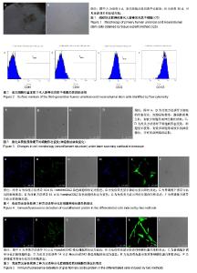
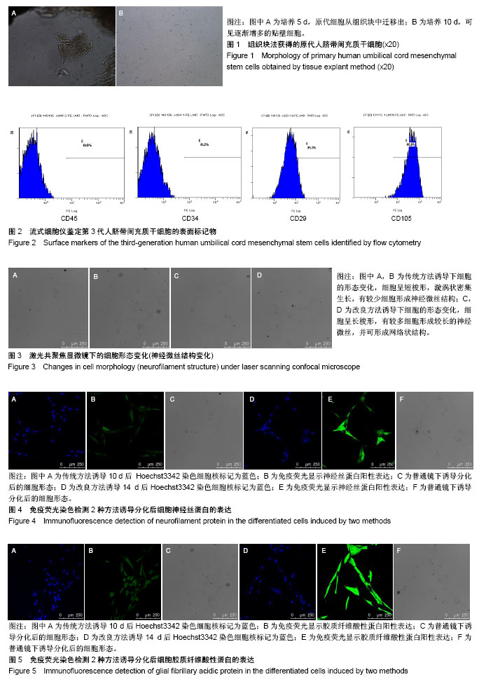
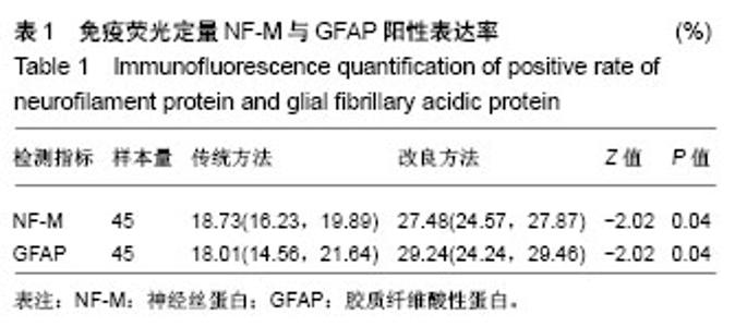
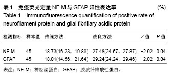
2.1 HUMSCs形态 原代培养第3天,倒置荧光显微镜观察可见组织块外周有散在的细胞游出。培养第5-7天,大量细胞由组织块中爬出,贴在皿底,呈长梭状,类似于成纤维细胞,汇聚后细胞排列紧密,呈群落样生长,聚合的细胞呈漩涡状。组织块法培养原代细胞达90%-95%融合的时间为14-16 d,可进行传代培养,见图1。传代数小时后HUMSCs快速贴壁,3-5 d后细胞可再次达到80%-90%的汇合度。经过数代培养后,细胞形状无明显变化,增殖能力无明显变化。 2.2 HUMSCs表型鉴定结果 流式细胞仪检测结果显示CD34、CD45表达为阴性,而CD29和CD105表达为阳性(阳性率分别为35.3%和95.9%),符合HUMSCs的特征性标记物表达,见图2,提示组织块法培养可获得HUMSCs。 2.3 诱导HUMSCs分化为神经元样细胞 HUMSCs通过2种方法诱导后均可分化成为神经元样细胞,部分细胞形态发生变化、胞体收缩形成突起呈星形,细胞之间网状连接,形态与神经元或神经胶质细胞近似。细胞生长状态良好,可存活时间较长,改良方案诱导分化后的细胞形态学变化尤为明显,细胞之间相互联系的神经微丝结构明显增多,明显优于前期实验的诱导方法,见图3。 2.4 免疫荧光染色检测诱导分化后细胞NF-M、GFAP的表达 免疫荧光染色提示HUMSCs用2种方法诱导后均可见NF-M和GFAP呈阳性表达,表明诱导分化后的细胞具有神经细胞的功能活性,见图4,5。 2.5 免疫荧光定量NF-M与GFAP阳性表达 传统方法组NF-M和GFAP的阳性表达率分别为18.73%和18.01%;改良方法组NF-M和GFAP的阳性表达率分别为27.48%和29.24%,后者明显优于前者,差异有显著性意义(P < 0.05),见表1。"
"
| [1] Pop DM, Sori??u O, ?u?man S, et al. Potential of placental-derived human mesenchymal stem cells for osteogenesis and neurogenesis. Rom J Morphol Embryol. 2015;56(3):989-996. [2] Nitkin CR, Bonfield TL. Concise Review: Mesenchymal Stem Cell Therapy for Pediatric Disease: Perspectives on Success and Potential Improvements. Stem Cells Transl Med. 2017;6(2):539-565. [3] Nekanti U, Mohanty L, Venugopal P, et al. Optimization and scale-up of Wharton's jelly-derived mesenchymal stem cells for clinical applications. Stem Cell Res. 2010;5(3):244-254. [4] Gao F, Chiu SM, Motan DA, et al. Mesenchymal stem cells and immunomodulation: current status and future prospects. Cell Death Dis. 2016;7:e2062. [5] Du WJ, Chi Y, Yang ZX, et al. Heterogeneity of proangiogenic features in mesenchymal stem cells derived from bone marrow, adipose tissue, umbilical cord, and placenta. Stem Cell Res Ther. 2016;7(1):163. [6] Antunes MA, Laffey JG, Pelosi P, et al. Mesenchymal stem cell trials for pulmonary diseases. J Cell Biochem. 2014;115(6):1023-1032. [7] Latifpour M, Shakiba Y, Amidi F, et al. Differentiation of human umbilical cord matrix-derived mesenchymal stem cells into germ-like cells. Avicenna J Med Biotechnol. 2014;6(4):218-227. [8] Tao R, Sun TJ, Han YQ, et al. Epimorphin-induced differentiation of human umbilical cord mesenchymal stem cells into sweat gland cells. Eur Rev Med Pharmacol Sci. 2014;18(9):1404-1410. [9] Li W, Ye B, Cai XY, et al. Differentiation of human umbilical cord mesenchymal stem cells into prostate-like epithelial cells in vivo. PLoS One. 2014;9(7):e102657. [10] Bai J, Hu Y, Wang YR, et al. Comparison of human amniotic fluid-derived and umbilical cord Wharton's Jelly-derived mesenchymal stromal cells: Characterization and myocardial differentiation capacity. J Geriatr Cardiol. 2012;9(2):166-171. [11] Hu Y, Liang J, Cui H, et al. Wharton's jelly mesenchymal stem cells differentiate into retinal progenitor cells. Neural Regen Res. 2013; 8(19):1783-1792. [12] Hang H, Yu Y, Wu N, et al. Induction of highly functional hepatocytes from human umbilical cord mesenchymal stem cells by HNF4α transduction. PLoS One. 2014;9(8):e104133. [13] Qu H, Liu X, Ni Y, et al. Laminin 411 acts as a potent inducer of umbilical cord mesenchymal stem cell differentiation into insulin-producing cells. J Transl Med. 2014;12:135. [14] Doan CC, Le TL, Hoang NS, et al. Differentiation of umbilical cord lining membrane-derived mesenchymal stem cells into endothelial-like cells. Iran Biomed J. 2014;18(2):67-75. [15] Paldino E, Cenciarelli C, Giampaolo A, et al. Induction of dopaminergic neurons from human Wharton's jelly mesenchymal stem cell by forskolin. J Cell Physiol. 2014;229(2):232-244. [16] Alves da Silva ML, Costa-Pinto AR, Martins A, et al. Conditioned medium as a strategy for human stem cells chondrogenic differentiation. J Tissue Eng Regen Med. 2015;9(6):714-723. [17] Ali H, Al-Yatama MK, Abu-Farha M, et al. Multi-lineage differentiation of human umbilical cord Wharton's Jelly Mesenchymal Stromal Cells mediates changes in the expression profile of stemness markers. PLoS One. 2015;10(4):e0122465. [18] Bárcia RN, Santos JM, Filipe M, et al. What Makes Umbilical Cord Tissue-Derived Mesenchymal Stromal Cells Superior Immunomodulators When Compared to Bone Marrow Derived Mesenchymal Stromal Cells. Stem Cells Int. 2015;2015:583984. [19] Hu SL, Luo HS, Li JT, et al. Functional recovery in acute traumatic spinal cord injury after transplantation of human umbilical cord mesenchymal stem cells. Crit Care Med. 2010;38(11):2181-2189. [20] Pang KM, Sung MA, Alrash-dan MS, et al. Tans-plantation of mesenchymal stem cells from human umbilical cord versus human umbilical cord blood for peripheral nerve regeneration. Neural Regen Res. 2010;5(11):838-845. [21] Cheng H, Liu X, Hua R, et al. Clinical observation of umbilical cord mesenchymal stem cell transplantation in treatment for sequelae of thoracolumbar spinal cord injury. J Transl Med. 2014;12:253. [22] Chen J, Venkat P, Zacharek A, et al. Neurorestorative therapy for stroke. Front Hum Neurosci. 2014;8:382. [23] 张飞,王一雄,武忠炎,等.人脐带间充质干细胞生物特性比较:胰酶冷消化和组织块法体外培养[J].中国组织工程研究, 2014,18(41):6614-6619.[24] McElreavey KD, Irvine AI, Ennis KT, et al. Isolation, culture and characterisation of fibroblast-like cells derived from the Wharton's jelly portion of human umbilical cord. Biochem Soc Trans. 1991;19(1):29S. [25] 王武,张飞,李贵才,等.以 EdU 体外标记人脐带间充质干细胞:5,10 μmol/L 是其最适浓度[J].中国组织工程研究, 2015,19(32):5167-5171.[26] Ianus A, Holz GG, Theise ND, et al. In vivo derivation of glucose-competent pancreatic endocrine cells from bone marrow without evidence of cell fusion. J Clin Invest. 2003;111(6):843-850. [27] Ra JC, Shin IS, Kim SH, et al. Safety of intravenous infusion of human adipose tissue-derived mesenchymal stem cells in animals and humans. Stem Cells Dev. 2011;20(8):1297-1308. [28] Nazarov I, Lee JW, Soupene E, et al. Multipotent stromal stem cells from human placenta demonstrate high therapeutic potential. Stem Cells Transl Med. 2012;1(5):359-372. [29] Rohban R, Pieber TR. Mesenchymal Stem and Progenitor Cells in Regeneration: Tissue Specificity and Regenerative Potential. Stem Cells Int. 2017;2017:5173732. [30] Guan LX, Guan H, Li HB, et al. Therapeutic efficacy of umbilical cord-derived mesenchymal stem cells in patients with type 2 diabetes. Exp Ther Med. 2015;9(5):1623-1630. [31] Zhao XF, Xu Y, Zhu ZY, et al. Clinical observation of umbilical cord mesenchymal stem cell treatment of severe systolic heart failure. Genet Mol Res. 2015;14(2):3010-3017. [32] D'souza N, Rossignoli F, Golinelli G, et al. Mesenchymal stem/stromal cells as a delivery platform in cell and gene therapies. BMC Med. 2015;13:186. [33] Hendijani F, Sadeghi-Aliabadi H, Haghjooy Javanmard S. Comparison of human mesenchymal stem cells isolated by explant culture method from entire umbilical cord and Wharton's jelly matrix. Cell Tissue Bank. 2014;15(4):555-565. [34] Zhou X, Gu J, Gu Y, et al. Human Umbilical Cord-Derived Mesenchymal Stem Cells Improve Learning and Memory Function in Hypoxic-Ischemic Brain-Damaged Rats via an IL-8-Mediated Secretion Mechanism Rather than Differentiation Pattern Induction. Cell Physiol Biochem. 2015;35(6):2383-2401. [35] Álvarez D, Levine M, Rojas M. Regenerative medicine in the treatment of idiopathic pulmonary fibrosis: current position. Stem Cells Cloning. 2015;8:61-65. [36] Rismanchi N, Floyd CL, Berman RF, et al. Cell death and long-term maintenance of neuron-like state after differentiation of rat bone marrow stromal cells: a comparison of protocols. Brain Res. 2003;991(1-2):46-55. [37] 孙丽,于丽,张华芳,等.人脐带间充质干细胞的富集及向神经细胞的诱导分化[J].神经解剖杂志,2011,27(2):185-190.[38] 郜元军,钱伟,汪涉海,等.大鼠骨髓基质干细胞诱导分化为肠神经细胞的体外研究[J].中华消化杂志,2006,26(10):661-665. |
| [1] | Pu Rui, Chen Ziyang, Yuan Lingyan. Characteristics and effects of exosomes from different cell sources in cardioprotection [J]. Chinese Journal of Tissue Engineering Research, 2021, 25(在线): 1-. |
| [2] | Lin Qingfan, Xie Yixin, Chen Wanqing, Ye Zhenzhong, Chen Youfang. Human placenta-derived mesenchymal stem cell conditioned medium can upregulate BeWo cell viability and zonula occludens expression under hypoxia [J]. Chinese Journal of Tissue Engineering Research, 2021, 25(在线): 4970-4975. |
| [3] | Zhang Tongtong, Wang Zhonghua, Wen Jie, Song Yuxin, Liu Lin. Application of three-dimensional printing model in surgical resection and reconstruction of cervical tumor [J]. Chinese Journal of Tissue Engineering Research, 2021, 25(9): 1335-1339. |
| [4] | Hou Jingying, Yu Menglei, Guo Tianzhu, Long Huibao, Wu Hao. Hypoxia preconditioning promotes bone marrow mesenchymal stem cells survival and vascularization through the activation of HIF-1α/MALAT1/VEGFA pathway [J]. Chinese Journal of Tissue Engineering Research, 2021, 25(7): 985-990. |
| [5] | Shi Yangyang, Qin Yingfei, Wu Fuling, He Xiao, Zhang Xuejing. Pretreatment of placental mesenchymal stem cells to prevent bronchiolitis in mice [J]. Chinese Journal of Tissue Engineering Research, 2021, 25(7): 991-995. |
| [6] | Liang Xueqi, Guo Lijiao, Chen Hejie, Wu Jie, Sun Yaqi, Xing Zhikun, Zou Hailiang, Chen Xueling, Wu Xiangwei. Alveolar echinococcosis protoscolices inhibits the differentiation of bone marrow mesenchymal stem cells into fibroblasts [J]. Chinese Journal of Tissue Engineering Research, 2021, 25(7): 996-1001. |
| [7] | Fan Quanbao, Luo Huina, Wang Bingyun, Chen Shengfeng, Cui Lianxu, Jiang Wenkang, Zhao Mingming, Wang Jingjing, Luo Dongzhang, Chen Zhisheng, Bai Yinshan, Liu Canying, Zhang Hui. Biological characteristics of canine adipose-derived mesenchymal stem cells cultured in hypoxia [J]. Chinese Journal of Tissue Engineering Research, 2021, 25(7): 1002-1007. |
| [8] | Geng Yao, Yin Zhiliang, Li Xingping, Xiao Dongqin, Hou Weiguang. Role of hsa-miRNA-223-3p in regulating osteogenic differentiation of human bone marrow mesenchymal stem cells [J]. Chinese Journal of Tissue Engineering Research, 2021, 25(7): 1008-1013. |
| [9] | Lun Zhigang, Jin Jing, Wang Tianyan, Li Aimin. Effect of peroxiredoxin 6 on proliferation and differentiation of bone marrow mesenchymal stem cells into neural lineage in vitro [J]. Chinese Journal of Tissue Engineering Research, 2021, 25(7): 1014-1018. |
| [10] | Zhu Xuefen, Huang Cheng, Ding Jian, Dai Yongping, Liu Yuanbing, Le Lixiang, Wang Liangliang, Yang Jiandong. Mechanism of bone marrow mesenchymal stem cells differentiation into functional neurons induced by glial cell line derived neurotrophic factor [J]. Chinese Journal of Tissue Engineering Research, 2021, 25(7): 1019-1025. |
| [11] | Duan Liyun, Cao Xiaocang. Human placenta mesenchymal stem cells-derived extracellular vesicles regulate collagen deposition in intestinal mucosa of mice with colitis [J]. Chinese Journal of Tissue Engineering Research, 2021, 25(7): 1026-1031. |
| [12] | Pei Lili, Sun Guicai, Wang Di. Salvianolic acid B inhibits oxidative damage of bone marrow mesenchymal stem cells and promotes differentiation into cardiomyocytes [J]. Chinese Journal of Tissue Engineering Research, 2021, 25(7): 1032-1036. |
| [13] | Li Cai, Zhao Ting, Tan Ge, Zheng Yulin, Zhang Ruonan, Wu Yan, Tang Junming. Platelet-derived growth factor-BB promotes proliferation, differentiation and migration of skeletal muscle myoblast [J]. Chinese Journal of Tissue Engineering Research, 2021, 25(7): 1050-1055. |
| [14] | Wang Feng, Zhou Liyu, Saijilafu, Qi Shibin, Ma Yanxia, Wei Shanwen. CaMKII-Smad1 promotes axonal regeneration of peripheral nerves [J]. Chinese Journal of Tissue Engineering Research, 2021, 25(7): 1064-1068. |
| [15] | Wang Xianyao, Guan Yalin, Liu Zhongshan. Strategies for improving the therapeutic efficacy of mesenchymal stem cells in the treatment of nonhealing wounds [J]. Chinese Journal of Tissue Engineering Research, 2021, 25(7): 1081-1087. |
| Viewed | ||||||
|
Full text |
|
|||||
|
Abstract |
|
|||||