Chinese Journal of Tissue Engineering Research ›› 2019, Vol. 23 ›› Issue (23): 3705-3709.doi: 10.3969/j.issn.2095-4344.1317

Previous Articles     Next Articles

Role of myofibroblasts in the development of mouse intrahepatic bile ducts 

Yang Junjun1, Li Dewei2, Wang Wei2, Feng Yuan1   

  1. (1Department of Infections, the Second Clinical College of North Sichuan Medical College, Nanchong Central Hospital, Nanchong 637000, Sichuan Province, China; 2Department of Hepatobiliary Surgery, the First Affiliated Hospital of Chongqing Medical University, Chongqing 400016, China)
  • Received:2019-03-08 Online:2019-08-18 Published:2019-08-18
  • Contact: Li Dewei, MD, Professor, Department of Hepatobiliary Surgery, the First Affiliated Hospital of Chongqing Medical University, Chongqing 400016, China
  • About author:Yang Junjun, Master, Attending physician, Department of Infections, the Second Clinical College of North Sichuan Medical College, Nanchong Central Hospital, Nanchong 637000, Sichuan Province, China
  • Supported by:

    the National Natural Science Foundation of China (General Program), No. 81470898 (to LDW)

Abstract:

BACKGROUND: Existing evidence has shown that myofibroblasts can promote hepatic progenitor cells to differentiate into bile duct cells. But, the role of myofibroblasts in the development of bile duct in vivo remains unclear.
OBJECTIVE: To investigate the developmental features of intrahepatic bile duct in mice and role of myofibroblasts in the development of bile duct.
METHODS: C57 mice, SPF grade were proved by the Laboratory Animal Center of Chongqing Medical University, and the study was approved by the Ethics Committee of Experimental Animal Ethics of Chongqing Medical University, approval number: 2017-182. The mice in gestational day 14.5 and 16.5, postnatal 0 and 10 days (P0, P10) mice were selected. After anesthesia, the liver was removed from fetal mice of embryonic day 14.5 and 16.5 (E14.5, E16.5) and P0 and P10 mice. The expression levels of α smooth muscle actin, Sox9 and cytokeratin 19 in liver were examined by immunocytochemical method.
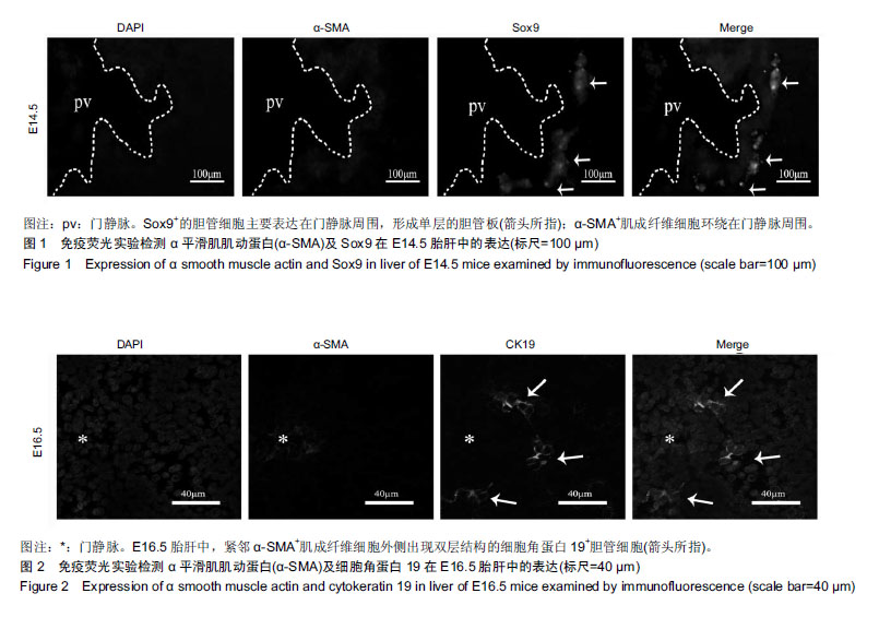
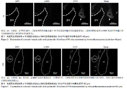
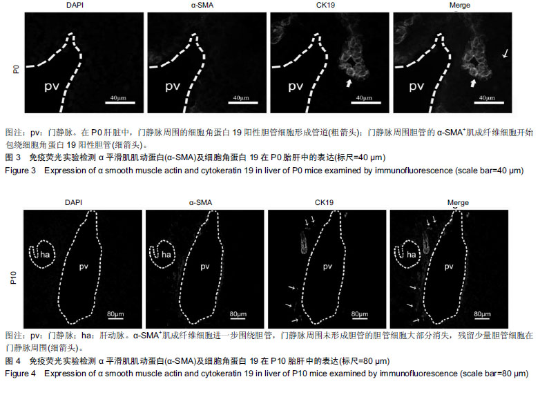
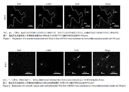
RESULTS AND CONCLUSION: (1) In the fetal liver of E14.5 mice, myofibroblasts positive for α smooth muscle actin were main located around the portal vein, which was adjacent to the bile duct cells positive for Sox9, and single layered ring called ductal plate formed. (2) In the fetal liver of E16.5 mice, the ductal plate positive for cytokeratin 19, which neared the myofibroblasts positive for α smooth muscle actin, became focally bilayered. (3) In the liver of P0 mice, the cholangiocytes positive for cytokeratin 19 formed bile ducts, which were surrounded by myofibroblasts positive for α smooth muscle actin. (4) In the liver of P10 mice, the bile duct expanded further, and cholangiocytes that could not form bile duct surrounding the portal vein disappeared. The bile duct was encircled by myofibroblasts positive for α smooth muscle actin, and a well-formed hepatic artery was found near the bile duct. (5) The developmental characteristics of intrahepatic bile ducts are systematic displayed. Myofibroblasts positive for α smooth muscle actin are closely related to intrahepatic bile duct development, and play critical role.

Key words: myofibroblasts, bile duct, development, liver, mice, cytokeratin 19, the National Natural Science Foundation of China

CLC Number: