Chinese Journal of Tissue Engineering Research ›› 2018, Vol. 22 ›› Issue (26): 4116-4122.doi: 10.3969/j.issn.2095-4344.0913
Previous Articles Next Articles
Properties of glass ionomer cements after addition of organic dye solutions
Wang Ping-ting, Bao Yi-jun, Liu Ying
- Tianjin Medical University School & Hospital of Stomatology, Tianjin 300070, China
-
Received:2018-05-30 -
About author:Wang Ping-ting, Master, Attending physician, Tianjin Medical University School & Hospital of Stomatology, Tianjin 300070, China -
Supported by:the Science & Technology Development Fund of Tianjin Education Commission for Higher Education, No. 2016YD19; and the Science Foundation of Tianjin Medical University, No. 2015KYZM11
CLC Number:
Cite this article
Wang Ping-ting, Bao Yi-jun, Liu Ying . Properties of glass ionomer cements after addition of organic dye solutions[J]. Chinese Journal of Tissue Engineering Research, 2018, 22(26): 4116-4122.
share this article
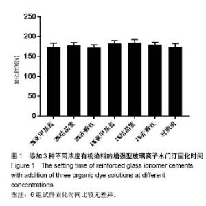
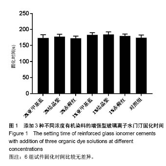
2.1 各组试件固化时间 与对照组比较,添加1%、2%亚甲基蓝、结晶紫、赤藓红染液后的玻璃离子水门汀材料固化时间无明显延长或缩短(P > 0.05)(图1),且与加入染料的浓度无相关性,固化时间均在150-200 s范围之内,符合临床操作需要。"
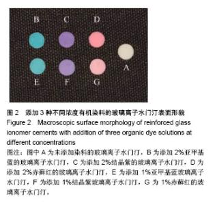
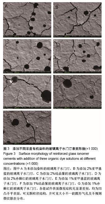
2.2 各组试件宏观及微观表面形貌 固化后的玻璃离子水门汀试样宏观表面形貌如图2所示,可观察到添加1%及2%亚甲基蓝、结晶紫、赤藓红染料后的玻璃离子水门汀与对照组具有明显的颜色差异,各组试件表面光滑平整,无明显裂隙、孔洞,且添加2%浓度染液的玻璃离子水门汀试件的着色较添加1%浓度染液的玻璃离子水门汀试件更深。 扫描电镜下试件微观表面形貌如图3所示,染色玻璃离子水门汀和对照组玻璃离子水门汀的表面微观结构无显著差别,均为凹凸不平表面,可见颗粒状结构,并可见大小不一的圆形气孔及不规则裂纹散在分布(裂纹由喷金过程中电子束打击及时间制作过程中脱水导致)。"
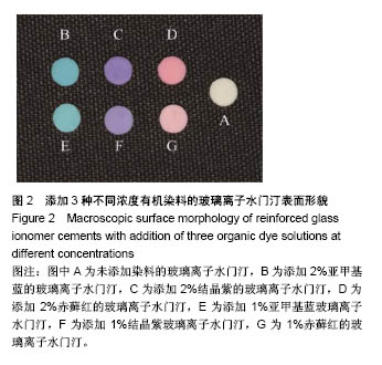
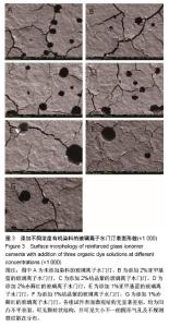
"
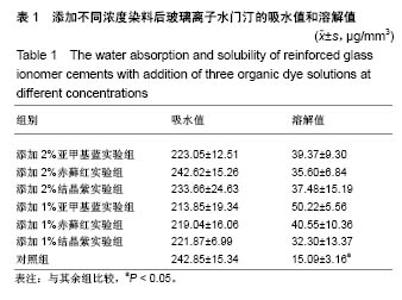
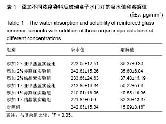
2.3 各组试件吸水值和溶解值 各组玻璃离子水门汀的吸水值及溶解值如表1所示,浸泡在人工唾液中7 d后,各实验组与对照组吸水值无显著差异(P > 0.05),对照组吸水值为(242.85±15.34) μg/mm3,染色组吸水值变化范围为(213.85±19.34) μg/mm3至(242.62±15.26) μg/mm3;各实验组溶解值明显高于对照组(P < 0.05),各染色组间溶解值比较无明显差异(P > 0.05),其中添加1%亚甲基蓝实验组溶解值最高,其余组别的溶解值范围为(32.30± 13.37) μg/mm3至(40.55±10.36) μg/mm3。"
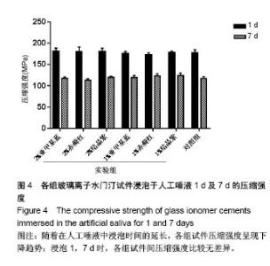
2.4 各组试件压缩强度 各组玻璃离子水门汀试件的压缩强度如图4所示,对照组储存1 d及7 d的压缩强度分别为(177.03±6.97) MPa和(117.26±4.26) MPa,各组染色玻璃离子水门汀储存1 d压缩强度数值范围为(172.67± 4.31) MPa至(181±6.77) MPa,储存7 d的压缩强度数值范围为(113.16±3.07) MPa至(124.31±5.06) MPa;随着在人工唾液中浸泡时间的延长,各组玻璃离子水门汀试件浸泡 7 d后的压缩强度呈现下降趋势,均显著低于本组浸泡1 d后的压缩强度(P < 0.05),而在1 d和7 d两个测试时间点各组之间压缩强度比较均无明显差异(P > 0.05)。"

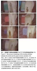
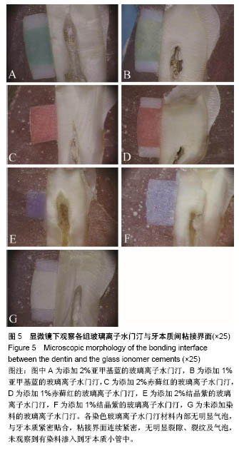
2.5 各组试件粘接界面形貌 各组玻璃离子水门汀材料与牙本质的粘接界面形貌如图5所示,可见各组染色玻璃离子水门汀材料内部无明显气泡(部分区域因材料脱水产生皲裂纹),与牙本质紧密贴合,粘接界面连续紧密,无明显裂隙、裂纹及气泡,未观察到有染料渗入到牙本质小管中。"
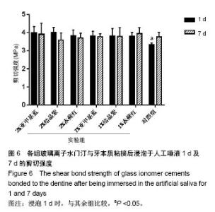
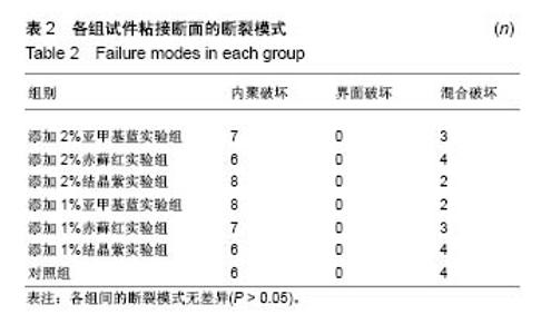
2.6 各组试件剪切粘接强度及断裂模式 各组玻璃离子水门汀与牙本质粘接试件浸泡于人工唾液1 d和7 d后的剪切粘接强度如图6所示。浸泡1 d后,对照组剪切粘接强度为(3.34±0.07) MPa,其余染色玻璃离子水门汀组剪切粘接强度数值范围为(3.80±0.19) MPa至(4.02±0.25) MPa,各实验组剪切粘接强度均显著高于对照组(P < 0.05);浸泡 7 d后,对照组剪切粘接强度达(3.76±0.23) MPa,但各组间剪切粘接强度不再有明显统计学差异(P > 0.05), 随着在人工唾液中储存时间的延长,对照组剪切粘接强度显著升高,各染色玻璃离子水门汀组剪切粘接强度并未随储存时间延长发生显著变化。 各组试件断裂模式结果如表2所示,各组的断裂模式主要为内聚破坏及混合破坏模式,未观察到单一的界面破坏模式。经卡方检验,各组间的断裂模式无统计学差异(P > 0.05)。"

"

| [1] Carrotte P.Endodontics: Part 1.The modern concept of root canal treatment. Br Dent J.2004; 197(4): 181-183.[2] Malmberg L,Bjorkner AE,Bergenholtz G.Establishment and maintenance of asepsis in endodontics - a review of the literature.Acta Odontol Scand.2016;74(6):431-435.[3] Rambabu T.Canal projection using gutta-percha points: A novel technique for pre-endodontic buildup of grossly destructed tooth.J Conserv Dent.2016;19(2):194-197.[4] Swanljung O,Vehkalahti MM.Root Canal Irrigants and Medicaments in Endodontic Malpractice Cases: A Nationwide Longitudinal Observation. J Endod.2018;44(4):559-564.[5] Heydrich R.Pre-endodontic treatment restorations. A modification of the 'donut' technique.J Am Dent Assoc.2005;136(5):641-642.[6] Holliday R.Cohen's pathways of the pulp, 10th edition.Br Dent J. 2011; 210(5):242.[7] Naoum H,Chandler N.Temporization for endodontics.Int Endod J. 2002;35(12):964-978.[8] Tanikonda R.Canal projection using gutta-percha points: A novel technique for pre-endodontic buildup of grossly destructed tooth.J Conserv Dent.2016;19(2):194-197.[9] Jensen A,Abbott P,Castro Salgado J.Interim and temporary restoration of teeth during endodontic treatment.Aust Dent J.2007;52(1 Suppl): S83-99.[10] Jensen AL,Abbott PV.Experimental model: dye penetration of extensive interim restorations used during endodontic treatment while under load in a multiple axis chewing simulator.J Endod.2007; 33(10): 1243-1246.[11] Castellucci A.Endodontics.Firenze:II Tridente (Firenze),2004:330-337.[12] Baig MS,Fleming GJ.Conventional glass-ionomer materials: A review of the developments in glass powder, polyacid liquid and the strategies of reinforcement.J Dent.2015;43(8):897-912.[13] Pegoraro TA,Da Silva NR,Carvalho RM.Cements for use in esthetic dentistry.Dent Clin North Am. 2007;51(2):453-471.[14] Cambruzzi JV,Marshall FJ,Pappin JB.Methylene blue dye: an aid to endodontic surgery.J Endod.1985;11(7):311-314.[15] Gutmann JL.Finding Small Root Canals.J Hist Dent.2016;64(1):14.[16] Scotti N,Comba A,Gambino A,et al.Microleakage at enamel and dentin margins with a bulk fills flowable resin.Eur J Dent.2014;8(1):1.[17] Chiniforush N,Pourhajibagher M,Shahabi S,et al.Clinical Approach of High Technology Techniques for Control and Elimination of Endodontic Microbiota.J Lasers Med Sci.2015;6(4):139-150.[18] Pourhajibagher M,Chiniforush N,Raoofian R,et al.Evaluation of photo-activated disinfection effectiveness with methylene blue against Porphyromonas gingivalis involved in endodontic infection: An in vitro study.Photodiagnosis Photodyn Ther.2016;16:132-135.[19] 苏英.甲紫对白念珠菌毒力因子作用的实验研究[J].中国麻风皮肤病杂志, 2009,25(9):648-650.[20] 李钧,王松灵.甲紫注入小型猪正常腮腺后超微病理学观察[J].现代口腔医学杂志,2000,14(6):411-411.[21] Ganesan L,Margolles-Clark E,Song Y,et al.The food colorant erythrosine is a promiscuous protein-protein interaction inhibitor. Biochem Pharmacol.2011;81(6):810-818.[22] Tahmassebi JF,Drogkari E,Wood SR.A study of the control of oral plaque biofilms via antibacterial photodynamic therapy.Eur Arch Paediatr Dent.2015;16(6):433-440.[23] Costa SB,De Oliveira RVD,Viégas Montenegro R,et al.Bond strength evaluation of composite resin bonded to glass ionomer cements after different periods of setting.Int J Adhes Adhes.2013;47:146-150.[24] 李若珍,吕永昌,毕良佳.四碘荧光素钠对口腔龈上菌斑抑制效果研究[J].中国实用口腔科杂志,2016,9(1): 27-30.[25] De Caluwe T,Vercruysse CW,Ladik I,et al.Addition of bioactive glass to glass ionomer cements: Effect on the physico-chemical properties and biocompatibility.Dent Mater.2017;33(4):e186-e203.[26] Ferreira JMS,Pinheiro SL,Sampaio FC,et al.Use of glass ionomer cement containing antibiotics to seal off infected dentin: a randomized clinical trial.Brazilian Dent J.2013;24(1):68-73.[27] Garcia IM,Leitune VCB,Balbinot GS,et al.Influence of niobium pentoxide addition on the properties of glass ionomer cements.Acta Biomater Odontol Scand.2016;2(1):138-143.[28] Algera TJ,Kleverlaan CJ,Prahl-Andersen B,et al.The influence of environmental conditions on the material properties of setting glass-ionomer cements.Dent Mater.2006;22(9):852-856.[29] Bhatia HP,Singh S,Sood S,et al.A Comparative Evaluation of Sorption, Solubility, and Compressive Strength of Three Different Glass Ionomer Cements in Artificial Saliva: An in vitro Study.Int J Clin Pediatr Dent. 2017;10(1):49-54.[30] 崔恺,陈吉华,赵三军.不同剂型玻璃离子水门汀溶解性比较研究[J].牙体牙髓牙周病学杂志,2008,18(2): 87-89.[31] Troca V,Fernandes K,Terrile A,et al.Effect of green propolis addition to physical mechanical properties of glass ionomer cements.J Appl Oral Sci.2011;19(2):100-105.[32] Bhatia H,Singh S,Sood S,et al.A Comparative Evaluation of Sorption, Solubility, and Compressive Strength of Three Different Glass Ionomer Cements in Artificial Saliva: An Study.Int J Clin Pediatr Dent. 2017; 10(1):49-54.[33] Hengtrakool C,Kukiattrakoon B,Kedjarune-Leggat U.Effect of naturally acidic agents on microhardness and surface micromorphology of restorative materials.Eur J Dent.2011;5(1):89-100.[34] Hewlett E,Caputo A,Wrobel D.Glass ionomer bond strength and treatment of dentin with polyacrylic acid.J Prosthet Dent.1991;66(6): 767-772.[35] Tedesco TK,Calvo AFB,Domingues GG,et al.Bond Strength of High-Viscosity Glass Ionomer Cements is Affected by Tubular Density and Location in Dentin?Microsc Microanal.2015;21(4):849-854.[36] Abo Al-Hana DA,El-Messairy AA,Shohayb FH,et al.Micro-shear bond strength of different composites and glass-ionomers used to reinforce root dentin.Tanta Dent J.2013;10(2):58-66.[37] Sidhu SK,Nicholson JW.A Review of Glass-Ionomer Cements for Clinical Dentistry.J Funct Biomater. 2016;7(3).pii: E16.doi: 10.3390/jfb7030016.[38] Cheetham JJ,Palamara JE,Tyas MJ,et al.Evaluation of the interfacial work of fracture of glass-ionomer cements bonded to dentin.J Mech Behav Biomed Mater.2014;29:427-437. |
| [1] | Zhang Tongtong, Wang Zhonghua, Wen Jie, Song Yuxin, Liu Lin. Application of three-dimensional printing model in surgical resection and reconstruction of cervical tumor [J]. Chinese Journal of Tissue Engineering Research, 2021, 25(9): 1335-1339. |
| [2] | Zeng Yanhua, Hao Yanlei. In vitro culture and purification of Schwann cells: a systematic review [J]. Chinese Journal of Tissue Engineering Research, 2021, 25(7): 1135-1141. |
| [3] | Xu Dongzi, Zhang Ting, Ouyang Zhaolian. The global competitive situation of cardiac tissue engineering based on patent analysis [J]. Chinese Journal of Tissue Engineering Research, 2021, 25(5): 807-812. |
| [4] | Wu Zijian, Hu Zhaoduan, Xie Youqiong, Wang Feng, Li Jia, Li Bocun, Cai Guowei, Peng Rui. Three-dimensional printing technology and bone tissue engineering research: literature metrology and visual analysis of research hotspots [J]. Chinese Journal of Tissue Engineering Research, 2021, 25(4): 564-569. |
| [5] | Chang Wenliao, Zhao Jie, Sun Xiaoliang, Wang Kun, Wu Guofeng, Zhou Jian, Li Shuxiang, Sun Han. Material selection, theoretical design and biomimetic function of artificial periosteum [J]. Chinese Journal of Tissue Engineering Research, 2021, 25(4): 600-606. |
| [6] | Liu Fei, Cui Yutao, Liu He. Advantages and problems of local antibiotic delivery system in the treatment of osteomyelitis [J]. Chinese Journal of Tissue Engineering Research, 2021, 25(4): 614-620. |
| [7] | Li Xiaozhuang, Duan Hao, Wang Weizhou, Tang Zhihong, Wang Yanghao, He Fei. Application of bone tissue engineering materials in the treatment of bone defect diseases in vivo [J]. Chinese Journal of Tissue Engineering Research, 2021, 25(4): 626-631. |
| [8] | Zhang Zhenkun, Li Zhe, Li Ya, Wang Yingying, Wang Yaping, Zhou Xinkui, Ma Shanshan, Guan Fangxia. Application of alginate based hydrogels/dressings in wound healing: sustained, dynamic and sequential release [J]. Chinese Journal of Tissue Engineering Research, 2021, 25(4): 638-643. |
| [9] | Chen Jiana, Qiu Yanling, Nie Minhai, Liu Xuqian. Tissue engineering scaffolds in repairing oral and maxillofacial soft tissue defects [J]. Chinese Journal of Tissue Engineering Research, 2021, 25(4): 644-650. |
| [10] | Xing Hao, Zhang Yonghong, Wang Dong. Advantages and disadvantages of repairing large-segment bone defect [J]. Chinese Journal of Tissue Engineering Research, 2021, 25(3): 426-430. |
| [11] | Chen Siqi, Xian Debin, Xu Rongsheng, Qin Zhongjie, Zhang Lei, Xia Delin. Effects of bone marrow mesenchymal stem cells and human umbilical vein endothelial cells combined with hydroxyapatite-tricalcium phosphate scaffolds on early angiogenesis in skull defect repair in rats [J]. Chinese Journal of Tissue Engineering Research, 2021, 25(22): 3458-3465. |
| [12] | Wang Hao, Chen Mingxue, Li Junkang, Luo Xujiang, Peng Liqing, Li Huo, Huang Bo, Tian Guangzhao, Liu Shuyun, Sui Xiang, Huang Jingxiang, Guo Quanyi, Lu Xiaobo. Decellularized porcine skin matrix for tissue-engineered meniscus scaffold [J]. Chinese Journal of Tissue Engineering Research, 2021, 25(22): 3473-3478. |
| [13] | Mo Jianling, He Shaoru, Feng Bowen, Jian Minqiao, Zhang Xiaohui, Liu Caisheng, Liang Yijing, Liu Yumei, Chen Liang, Zhou Haiyu, Liu Yanhui. Forming prevascularized cell sheets and the expression of angiogenesis-related factors [J]. Chinese Journal of Tissue Engineering Research, 2021, 25(22): 3479-3486. |
| [14] | Liu Chang, Li Datong, Liu Yuan, Kong Lingbo, Guo Rui, Yang Lixue, Hao Dingjun, He Baorong. Poor efficacy after vertebral augmentation surgery of acute symptomatic thoracolumbar osteoporotic compression fracture: relationship with bone cement, bone mineral density, and adjacent fractures [J]. Chinese Journal of Tissue Engineering Research, 2021, 25(22): 3510-3516. |
| [15] | Liu Liyong, Zhou Lei. Research and development status and development trend of hydrogel in tissue engineering based on patent information [J]. Chinese Journal of Tissue Engineering Research, 2021, 25(22): 3527-3533. |
| Viewed | ||||||
|
Full text |
|
|||||
|
Abstract |
|
|||||