Chinese Journal of Tissue Engineering Research ›› 2018, Vol. 22 ›› Issue (28): 4501-4506.doi: 10.3969/j.issn.2095-4344.0852
Previous Articles Next Articles
Establishment of a rabbit model of early femoroacetabular impingement and its morphological characteristics
Xiong Ao, Luan Xin-yu, Lin Jian-jing, Kang Bin, Zeng Hui
- Department of Bone and Joint, Peking University Shenzhen Hospital, Shenzhen 518036, Guangdong Province, China
-
Received:2018-01-31Online:2018-10-08Published:2018-10-08 -
About author:Xiong Ao, M.D., Associate chief physician, Department of Bone and Joint, Peking University Shenzhen Hospital, Shenzhen 518036, Guangdong Province, China -
Supported by:the Scientific Research Project of Health and Family Planning Commission of Shenzhen Municipality, No. 201501024
CLC Number:
Cite this article
Xiong Ao, Luan Xin-yu, Lin Jian-jing, Kang Bin, Zeng Hui. Establishment of a rabbit model of early femoroacetabular impingement and its morphological characteristics[J]. Chinese Journal of Tissue Engineering Research, 2018, 22(28): 4501-4506.
share this article

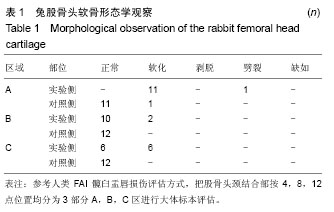
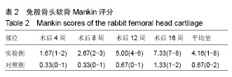
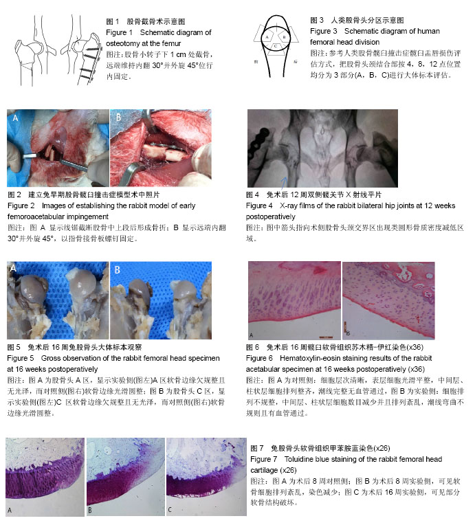
2.1 实验动物数量分析 实验选用新西兰兔12只,实验过程中无脱失,故进入结果分析的实验兔为12只。 2.2 影像学观察 部分新西兰兔术侧股骨头颈结合部出现直径约2 mm类圆形骨质密度减低区域,相邻的髋臼盂唇存在细微缺损(图4),其中术后12周2只,术后16周3只。其余新西兰兔实验侧髋关节与对照侧相比,未见明显变化。 2.3 大体标本观察 12只新西兰兔实验侧股骨头均出现不同程度软骨损伤(100%),对照侧仅出现1例股骨头软骨损伤(8%,P < 0.01,表1)。软骨损伤主要发生于股骨头颈结合部前上侧区域(A区),其中11例出现软骨软化,股骨头边缘粗糙,1例出现软骨劈裂。术侧股骨头C区共6例出现软骨软化,发生在术后12周和16周时,而B区仅在术后16周出现2例软骨软化(图5,表1)。"
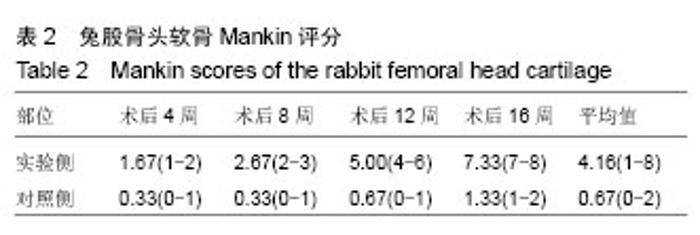
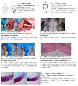
2.4 组织学观察 2.4.1 苏木精-伊红染色 显示对照侧软骨组织4层细胞清晰可辨,表层细胞光滑平整,中间层、柱状层细胞排列整齐,潮线完整无血管通过;而实验侧软骨表层细胞排列不规整,中间层、柱状层细胞数目减少并且排列紊乱,潮线弯曲不规则且有血管通过(图6),并且软骨细胞数目减少及排列紊乱的程度随术后时间的推移而加重。 2.4.2 甲苯胺蓝染色 显示术后8周实验侧可见软骨细胞排列紊乱,染色减少情况,术后12,6周实验侧可见部分软骨结构破坏(图7)。Mankin评分结果显示,总体样本实验侧分值明显高于对照侧(4.16[1-8],0.67[0-2],P < 0.01);术后不同时间节点,实验侧分值均高于对照侧(P < 0.05),并且实验侧的Mankin分值随术后时间的推移而升高(表2)。"
"
| [1] Myers SR, Eijer H, Ganz R.Anterior femoroacetabular impingement after periacetabular osteotomy. Clin Orthop Relat Res.1999;(363):93-99.[2] Eijer H, Myers SR, Ganz R.Anterior femoroacetabular impingement after femoral neck fractures.J Orthop Trauma. 2001;15(7): 475-481.[3] Ganz R,Parvizi J,Beck M,et al.Femoroacetabular impingement: a cause for osteoarthritis of the hip.Clin Orthop Relat Res.2003;(417):112-120.[4] Sangal RB, Waryasz GR, Schiller JR.Femoroacetabular impingement: a review of current concepts.R I Med J (2013). 2014;97(11):33-38.[5] Amanatullah DF, Antkowiak T, Pillay K, et al. Femoroacetabular impingement: current concepts in diagnosis and treatment. Orthopedics.2015l38(3):185-199.[6] Khan M, Bedi A, Fu F, Karlsson J, et al.New perspectives on femoroacetabular impingement syndrome.Nat Rev Rheumatol.2016;12(5):303-310.[7] Monazzam S, Bomar JD, Dwek JR,et al.Development and prevalence of femoroacetabular impingement-associated morphology in a paediatric and adolescent population: a CT study of 225 patients.Bone Joint J.2013;95-B(5): 598-604.[8] Ramachandran M, Azegami S, Hosalkar HS.Current concepts in the treatment of adolescent femoroacetabular impingement. J Child Orthop.2013;7(2):79-90.[9] Swenson KM, Erickson J, Peters C, et al.Hip pain in young adults: diagnosing femoroacetabular impingement. JAAPA. 2015;28(9):39-45.[10] Pathy R, Sink EL.Femoroacetabular impingement in children and adolescents. Curr Opin Pediatr.2016;28(1): 68-78.[11] Chaudhry H, Ayeni OR. The etiology of femoroacetabular impingement: what we know and what we don't. Sports Health.2014;6(2):157-161.[12] Leunig M, Beaule PE, Ganz R, et al. The concept of Femoroacetabular Impingement: Current status and future perspectives. Clin Orthop Relat Res.2009;467(3):616-622.[13] Hartofilakidis G, Bardakos NV, Babis GC, et al. An examination of the association between different morphotypes of femoroacetabular impingement in asymptomatic subjects and the development of osteoarthritis of the hip. J Bone Joint Surg Br. 2011;93(5):580-586.[14] Papalia R, Del Buono A, Franceschi F, et al. Femoroacetabular impingement syndrome management: arthroscopy or open surgery? Int Orthop.2012;36(5):903-914.[15] Collins JA, Ward JP, Youm T. Is prophylactic surgery for femoroacetabular impingement indicated? A systematic review.Am J Sports Med.2014;42(12):3009-3015.[16] Kuhns BD, Weber AE, Levy DM, et al.The Natural History of Femoroacetabular Impingement.Front Surg.2015;2:58.[17] Packer JD, Safran MR.The etiology of primary femoroacetabular impingement: genetics or acquired deformity? J Hip Preserv Surg.2015;2(3):249-257.[18] Zadpoor AA.Etiology of Femoroacetabular Impingement in Athletes: A Review of Recent Findings. Sports Med. 2015; 45(8):1097-1106.[19] Nepple JJ, Clohisy JC•ANCHOR Study Group Members. Evolution of Femoroacetabular Impingement Treatment: The ANCHOR Experience.Am J Orthop (Belle Mead NJ). 2017; 46(1): 28-34.[20] Siebenrock KA, Fiechter R, Tannast M, et al.Experimentally induced cam impingement in the sheep hip. J Orthop Res. 2013;31(4):580-587.[21] Adolphe M, Parodi AL Ethical issues in animal experimentation. Bull Acad Natl Med. 2009;193(8):1803-1804.[22] Beck M, Kalhor M, Leunig M, et al.Hip morphology influences the pattern of damage to the acetabular cartilage: femoroacetabular impingement as a cause of early osteoarthritis of the hip. J Bone Joint Surg Br. 2005;87(7): 1012-1018.[23] Mankin HJ.The reaction of articular cartilage to injury and osteoarthritis (first of two parts). N Engl J Med. 1974;291(24): 1285-1292.[24] Mankin HJ.The reaction of articular cartilage to injury and osteoarthritis (second of two parts). N Engl J Med. 1974;291 (25):1335-1340.[25] van der Sluijs JA, Geesink RG, van der Linden AJ, et al.The reliability of the Mankin score for osteoarthritis.J Orthop Res. 1992;10(1): 58-61.[26] Pollard TC, Villar RN, Norton MR, et al. Genetic influences in the aetiology of femoroacetabular impingement: a sibling study. J Bone Joint Surg Br.2010; 92(2):209-216.[27] Leunig M, Slongo T, Ganz R. Subcapital realignment in slipped capital femoral epiphysis: surgical hip dislocation and trimming of the stable trochanter to protect the perfusion of the epiphysis. Instr Course Lect.2008;57:499-507.[28] Banerjee P, McLean CR. Femoroacetabular impingement:a review of diagnosis and management. Curr Rev Musculosketlet Med. 2011;(1):23-32.[29] Fraitzl CR, Nelitz M, Cakir B, et al. Transfixation in slipped capital femoral epiphysis: long-term evidence for femoroacetabular impingement. Z Orthop Unfall. 2009;147(3): 334-340.[30] Laborie LB, Lehmann TG, Engesæter IØ, et al. Prevalence of radiographic findings thought to be associated with femoroacetabular impingement in a population-based cohort of 2081 healthy young adults. Radiology. 2011;260(2): 494-502.[31] Ramachandran M, Azegami S,Hosalkar HS. Current concepts in treatment of adolescent femoroacetabular impingement. J Child Orthop. 2013;7(2):79-90.[32] Tannast M, Goricki D, Beck M, et al.Hip damage occurs at the zone of femoroacetabular impingement. Clin Orthop Relat Res.2008;466(2): 273-280.[33] Crespo Rodríguez AM, de Lucas Villarrubia JC, Pastrana Ledesma MA, et al.Diagnosis of lesions of the acetabular labrum, of the labral-chondral transition zone, and of the cartilage in femoroacetabular impingement: Correlation between direct magnetic resonance arthrography and hip arthroscopy.Radiologia. 2015;57(2):131-141.[34] Meermans G, Konan S, Haddad FS, et al.Prevalence of acetabular cartilage lesions and labral tears in femoroacetabular impingement.Acta Orthop Belg. 2010;76(2): 181-188.[35] Flannelly J, Chambers MG, Dudhia J, et al.Metalloproteinase and tissue inhibitor of metalloproteinase expression in the murine STR/ort model of osteoarthritis. Osteoarthritis Cartilage.2002;10(9):722-733.[36] Staines KA, Madi K, Mirczuk SM,et al.Endochondral Growth Defect and Deployment of Transient Chondrocyte Behaviors Underlie Osteoarthritis Onset in a Natural Murine Model. Arthritis Rheumatol. 2016;68(4):880-891.[37] Akagi R, Sasho T, Saito M, Yet al.Effective knock down of matrix metalloproteinase-13 by an intra-articular injection of small interfering RNA (siRNA) in a murine surgically-induced osteoarthritis model.J Orthop Res. 2014;32(9):1175-1180.[38] Ferrell WR, Kelso EB, Lockhart JC, et al.Protease-activated receptor 2: a novel pathogenic pathway in a murine model of osteoarthritis. Ann Rheum Dis.2010;69(11):2051-2054.[39] Lim NH, Meinjohanns E, Meldal M, et al.In vivo imaging of MMP-13 activity in the murine destabilised medial meniscus surgical model of osteoarthritis. Osteoarthritis Cartilage. 2014;22(6):862-868.[40] Botter SM,van Osch GJ,Waarsing JH,et al.Quantification of subchondral bone changes in a murine osteoarthritis model using micro-CT. Biorheology.2006;43(3-4):379-388.[41] Haque Bhuyan MZ,Tamura Y,Sone E,et al.The intra-articular injection of RANKL-binding peptides inhibits cartilage degeneration in a murine model of osteoarthritis. J Pharmacol Sci.2017;134(2):124-130.[42] Leunig M, Beck M, Kalhor M, et al. Fibrocystic changes at anterosuperior femoral neck: prevalence in hips with femoroacetabular impingement. Radiology. 2005;236(1): 237-246.[43] Dimmick S, Stevens KJ, Brazier D, et al. Femoroacetabular impingement. Radiol Clin North Am. 2013;51(3):337-352. |
| [1] | Zhang Tongtong, Wang Zhonghua, Wen Jie, Song Yuxin, Liu Lin. Application of three-dimensional printing model in surgical resection and reconstruction of cervical tumor [J]. Chinese Journal of Tissue Engineering Research, 2021, 25(9): 1335-1339. |
| [2] | Peng Zhihao, Feng Zongquan, Zou Yonggen, Niu Guoqing, Wu Feng. Relationship of lower limb force line and the progression of lateral compartment arthritis after unicompartmental knee arthroplasty with mobile bearing [J]. Chinese Journal of Tissue Engineering Research, 2021, 25(9): 1368-1374. |
| [3] | Zhang Chao, Lü Xin. Heterotopic ossification after acetabular fracture fixation: risk factors, prevention and treatment progress [J]. Chinese Journal of Tissue Engineering Research, 2021, 25(9): 1434-1439. |
| [4] | Huang Dengcheng, Wang Zhike, Cao Xuewei. Comparison of the short-term efficacy of extracorporeal shock wave therapy for middle-aged and elderly knee osteoarthritis: a meta-analysis [J]. Chinese Journal of Tissue Engineering Research, 2021, 25(9): 1471-1476. |
| [5] | Liu Xiangxiang, Huang Yunmei, Chen Wenlie, Lin Ruhui, Lu Xiaodong, Li Zuanfang, Xu Yaye, Huang Meiya, Li Xihai. Ultrastructural changes of the white zone cells of the meniscus in a rat model of early osteoarthritis [J]. Chinese Journal of Tissue Engineering Research, 2021, 25(8): 1237-1242. |
| [6] | Zeng Yanhua, Hao Yanlei. In vitro culture and purification of Schwann cells: a systematic review [J]. Chinese Journal of Tissue Engineering Research, 2021, 25(7): 1135-1141. |
| [7] | Liu Xin, Yan Feihua, Hong Kunhao. Delaying cartilage degeneration by regulating the expression of aquaporins in rats with knee osteoarthritis [J]. Chinese Journal of Tissue Engineering Research, 2021, 25(5): 668-673. |
| [8] | Ma Zetao, Zeng Hui, Wang Deli, Weng Jian, Feng Song. MicroRNA-138-5p regulates chondrocyte proliferation and autophagy [J]. Chinese Journal of Tissue Engineering Research, 2021, 25(5): 674-678. |
| [9] | Cao Xuhan, Bai Zixing, Sun Chengyi, Yang Yanjun, Sun Weidong. Mechanism of “Ruxiang-Moyao” herbal pair in the treatment of knee osteoarthritis based on network pharmacology [J]. Chinese Journal of Tissue Engineering Research, 2021, 25(5): 746-753. |
| [10] | Li Yonghua, Feng Qiang, Tan Renting, Huang Shifu, Qiu Jinlong, Yin Heng. Molecular mechanism of Eucommia ulmoides active ingredients treating synovitis of knee osteoarthritis: an analysis based on network pharmacology [J]. Chinese Journal of Tissue Engineering Research, 2021, 25(5): 765-771. |
| [11] | Song Shan, Hu Fangyuan, Qiao Jun, Wang Jia, Zhang Shengxiao, Li Xiaofeng. An insight into biomarkers of osteoarthritis synovium based on bioinformatics [J]. Chinese Journal of Tissue Engineering Research, 2021, 25(5): 785-790. |
| [12] | Deng Zhenhan, Huang Yong, Xiao Lulu, Chen Yulin, Zhu Weimin, Lu Wei, Wang Daping. Role and application of bone morphogenetic proteins in articular cartilage regeneration [J]. Chinese Journal of Tissue Engineering Research, 2021, 25(5): 798-806. |
| [13] | Xu Dongzi, Zhang Ting, Ouyang Zhaolian. The global competitive situation of cardiac tissue engineering based on patent analysis [J]. Chinese Journal of Tissue Engineering Research, 2021, 25(5): 807-812. |
| [14] | Wu Zijian, Hu Zhaoduan, Xie Youqiong, Wang Feng, Li Jia, Li Bocun, Cai Guowei, Peng Rui. Three-dimensional printing technology and bone tissue engineering research: literature metrology and visual analysis of research hotspots [J]. Chinese Journal of Tissue Engineering Research, 2021, 25(4): 564-569. |
| [15] | Chang Wenliao, Zhao Jie, Sun Xiaoliang, Wang Kun, Wu Guofeng, Zhou Jian, Li Shuxiang, Sun Han. Material selection, theoretical design and biomimetic function of artificial periosteum [J]. Chinese Journal of Tissue Engineering Research, 2021, 25(4): 600-606. |
| Viewed | ||||||
|
Full text |
|
|||||
|
Abstract |
|
|||||