Chinese Journal of Tissue Engineering Research ›› 2018, Vol. 22 ›› Issue (34): 5484-5489.doi: 10.3969/j.issn.2095-4344.0684
Previous Articles Next Articles
Leukocyte- and platelet-rich fibrin promotes wound healing
Wu Yuxiang1, Si Shaoyan2, Xu Zhangrong3, Wang Aihong1, 3
- 1Department of Endocrinology, the 306th Hospital of PLA, Clinical Medicine College of Anhui Medical University, Beijing 100101, China; 2Special Medical Laboratory and Research Center, Beijing 100101, China; 3Department of Endocrinology, the 306th Hospital of PLA, Beijing 100101, China
-
Received:2018-06-29Online:2018-12-08Published:2018-12-08 -
Contact:Wang Aihong, MD, Master’s supervisor, Department of Endocrinology, the 306th Hospital of PLA, Clinical Medicine College of Anhui Medical University, Beijing 100101, China; Department of Endocrinology, the 306th Hospital of PLA, Beijing 100101, China -
About author:Wu Yuxiang, Master candidate, Department of Endocrinology, the 306th Hospital of PLA, Clinical Medicine College of Anhui Medical University, Beijing 100101, China -
Supported by:the PLA Medical Science and Technology Project for Youth Cultivation, No. 16QNP041; a grant from Beijing Municipal Science & Technology Commission, No. Z141107002514181; the Clinical Medical Research Special Project of Chinese Medical Association, No. 12020580358
CLC Number:
Cite this article
Wu Yuxiang, Si Shaoyan, Xu Zhangrong, Wang Aihong. Leukocyte- and platelet-rich fibrin promotes wound healing[J]. Chinese Journal of Tissue Engineering Research, 2018, 22(34): 5484-5489.
share this article

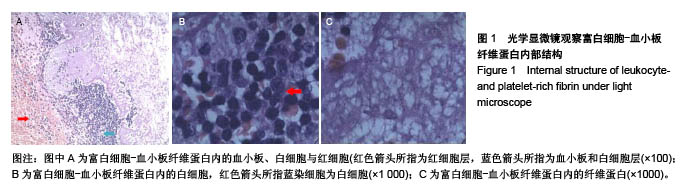
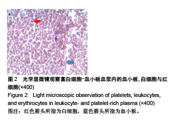
2.1 光镜下观察凝胶结构 富白细胞-血小板纤维蛋白内的血小板、白细胞与红细胞分界清晰(图1A),可见大量的白细胞及纤维蛋白簇集(图1B,C);富白细胞-血小板血浆的血小板、白细胞少,散在红细胞中(图2)?"

"
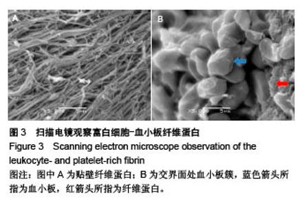
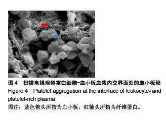
2.2 电镜下观察凝胶结构 电镜下观察发现富白细胞-血小板纤维蛋白中含有大量纤维蛋白,形成致密的特殊3D结构,交界处血小板叠加结构清晰(图3);富白细胞-血小板血浆交界处纤维较少夹杂大量血小板,部分血小板已 裂解(图4)。"


"
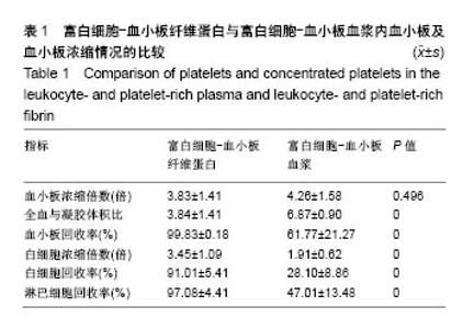
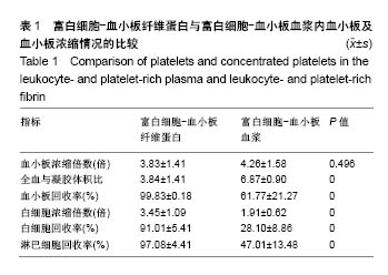
2.3 两种凝胶成分的比较 两种凝胶内血小板浓缩倍数比较无差异,但富白细胞-血小板纤维蛋白内有更高的血小板、白细胞及淋巴细胞回收率(P < 0.01),见表1。"

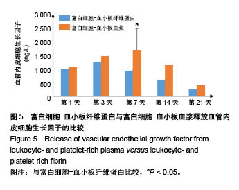
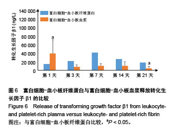
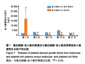
2.4 两种凝胶释放生长因子的比较 在富白细胞-血小板纤维蛋白中,血管内皮细胞生长因子质量浓度在第3天达高峰,转化生长因子β1质量浓度在第7天达高峰,血小板源性生长因子质量浓度在第14天达到高峰;在富白细胞-血小板血浆中,血管内皮细胞生长因子质量浓度在第7天达高峰,转化生长因子β1质量浓度在第1天达高峰,血小板源性生长因子质量浓度在第1天达高峰。 富白细胞-血小板血浆第7天释放的血管内皮细胞生长因子质量浓度显著高于富白细胞-血小板纤维蛋白[(947.79±303.18),(1 692.75±781.2) ng/L,P=0.008],两种凝胶第1,21天释放的转化生长因子ß1质量浓度比较差异有显著性意义[(14 369.77±12 489.34),(382 32.18± 314 65.05) ng/L,P=0.028;(19 295.26±22 477.88), (4 391.95± 4 113.73) ng/L,P=0.044],两种凝胶各个时间段释放血小板源性生长因子的质量浓度比较差异有显著性意义[(2 529.37±915.16),(16 835.82±9 523.03) ng/L,P < 0.001;(3 528.50±2 098.38),(1 271.38±382.94) ng/L,P=0.003;(3 220.56±1 417.73),(1 105.31±349.06) ng/L,P < 0.001;(5 393.52±3 480.30),(1 166.80±767.82) ng/L,P=0.001;(2 808.66±2 081.65),(726.12±259.29) ng/L,P=0.005]。两种凝胶中上述生长因子释放随时间变化的情况,见图5-7。"

"

"
| [1] Peice P,Krasner DL.Health-related quality of life and chronic wounds:evidence and implications for practice.A Clinical Source Book for Healthcare Professionals,2012:77-84.[2] Rahim K,Saleha S,Zhu X,et al.Bacterial Contribution in Chronicity of Wounds.Microb Ecol. 2017;73(3):710-721. [3] Budovsky A,Yarmolinsky L,Ben-Shabat S.Effect of medicinal plants on wound healing.Wound Repair Regen. 2015;23(2): 171-183.[4] 郝擎宇,葛乃航,宋德恒,等.慢性难愈性创面治疗方法的研究进展[J].感染、炎症、修复,2017,18(3):186-189.[5] 葛红.封闭负压引流技术在慢性难愈性创面中的应用[J].重庆医学,2017,46(A01):244-245.[6] 王彤华,周雄丽,谢利勤,等.湿性敷料在慢性伤口临床护理中的应用进展[J].中华现代护理杂志,2016, 22(15):2210-2212.[7] 田冰洁,王璐,王红红.慢性伤口清创术的研究进展[J].护理学杂志,2016,31(16):101-104.[8] Tambella AM,Attili AR,Dupré G,et al.Platelet-rich plasma to treat experimentally-induced skin wounds in animals: A systematic review and meta-analysis.PLoS One. 2018; 13(1):1-26.[9] Knafl D,Thalhammer F,Vossen MG.Vossen.In-vitro release pharmacokinetics of amikacin, teicoplanin and polyhexanide in a platelet rich fibrin—layer (PRF)—a laboratory evaluation of a modern, autologous wound treatment.PLoS One. 2017; 12(7):1-9.[10] Bielecki T,Dohan Ehrenfest DM.Platelet-rich plasma (PRP) and Platelet-Rich Fibrin (PRF): surgical adjuvants, preparations for in situ regenerative medicine and tools for tissue engineering.Curr Pharm Biotechnol. 2012;13(7): 1121-1130.[11] Suthar M,Gupta S,Bukhari S,et al.Treatment of chronic non-healing ulcers using autologous platelet rich plasma: a case series.J Biomed Sci.2017;24(1):16.[12] Chiaravalloti AJ,Zubkov B,Zubkov A.Treatment of a Chronic Cutaneous Surgical Wound With Platelet-Rich Fibrin. Dermatol Surg.2018;44(3):449-452.[13] Arshdeep,Kumaran MS.Platelet-rich plasma in dermatology: boon or a bane? Indian J Dermatol Venereol Lepro. 2014; 80(1):5-14.[14] Piccin A,Di Pierro AM,Corvetta D,et al.Severe skin radiodermatitis fully healed with the use of platelet gel and a hyperbaric chamber.Blood Transfus.2016;14(6):552-554.[15] McCarrel TM,Mall NA,Lee AS,et al.Considerations for the use of platelet-rich plasma in orthopedics.Sports Med. 2014;44(8): 1025-1036.[16] Kuffler DP.Platelet-rich plasma promotes axon regeneration, wound healing, and pain reduction: fact or fiction.Mol Neurobiol.2015;52(2):990-1014. [17] 任辉,岳彤,胡海燕,王婷,等.慢性创面患者生活质量及其影响因素的研究进展[J].现代临床护理,2018,17(1):47-53.[18] Bai MY,Wang CW,Wang JY,et al.Three-dimensional structure and cytokine distribution of platelet-rich fibrin.Clinics(Sao Paulo).2017;72(2):116-124.[19] 温天杨,王爱红,许樟荣.富白细胞-血小板血浆凝胶释放的炎性生长因子[J].中国组织工程研究,2013,17(16):2981-2988.[20] ?enses F,Önder ME,Koçyi?it ID,et al.Effect of Platelet-Rich Fibrin on Peripheral Nerve Regeneration.J Craniofac Surg. 2016;7(7):1759-1764.[21] Ding Y,Cui L,Zhao Q,et al.Platelet-Rich Fibrin Accelerates Skin Wound Healing in Diabetic Mice. Ann Plast Surg. 2017; 79(3):e15-e19.[22] Schär MO,Diaz-Romero J,Kohl S,et al. Platelet-rich concentrates differentially release growth factors and induce cell migration in vitro.Clin Orthop Relat Res. 2015;73(5): 1635-1643.[23] 康健,袁文.富血小板凝胶制备方法的比较与优选[J].中国组织工程研究,2014,18(3):476-481.[24] 戴静,陈建苏,招志毅.不同比例激活剂对富血小板凝胶的生物学影响[J].中国现代医学杂志,2017, 27(20):16-20.[25] 杨域,张卫,程飚.不同激活剂对人富血小板血浆形成凝胶及释放活性物质影响的实验研究[J].中华烧伤杂志,2017,33(1):12-17.[26] Clark JA,Humphries JE,Crean S,et al.Topical bovine thrombin: a 21-year review of topical bovine thrombin spontaneous case safety reports submitted to FDA's Adverse Event Reporting System. Pharmacoepidemiol Drug Saf.2010;19(2):107-114.[27] Schär MO,Diaz-Romero J,Kohl S,et al. Platelet-rich concentrates differentially release growth factors and induce cell migration in vitro.Clin Orthop Relat Res. 2015;473(5): 1635-1643.[28] De Pascale MR,Sommese L,Casamassimi A,et al.Platelet derivatives in regenerative medicine: an update.Transfus Med Rev.2015;29(1):52-61.[29] Bai MY,Wang CW,Wang JY,et al.Three-dimensional structure and cytokine distribution of platelet-rich fibrin.Clinics(Sao Paulo).2017;72(2):116-124.[30] Borzini P,Mazzucco I.Platelet-rich plasma(PRP) and platelet derivatives for topical therapyl what is true from the biologic view point[J].ISBT Science Series, 2007;2(1):272-281.[31] Bernuzzi G,Tardito S,Bussolati O,et al.Platelet gel in the treatment of cutaneous ulcers l the experience of the Immunohaematology and Transfusion Centre of Parma.Blood Transfus.2010;8(4): 237-247.[32] 孙斌,冯子阳,徐芳芳,等.富血小板血浆预防胸骨感染效果的Meta分析[J].中国输血杂志,2015, 28(3):295-298.[33] Cieslik-Bielecka A,Dohan Ehrenfest DM,Lubkowska A,et al. Microbicidal properties of Leukocyte- and Platelet-Rich Plasma/Fibrin (L-PRP/L-PRF): new perspectives.J Biol Regul Homeost Agents.2012;26(2 Suppl 1):43S-52S.[34] Klinger MH,Jelkmann W.Role of blood platelets in infection and inflammation.Interferon Cytokine Res. 2002;22(9): 913-922.[35] 练轶,毛俊丽,孙嵩,等.A-PRF与PRF的成分及超微结构观察[J].西南国防医药,2017,27(6):548-551. |
| [1] | Zhang Tongtong, Wang Zhonghua, Wen Jie, Song Yuxin, Liu Lin. Application of three-dimensional printing model in surgical resection and reconstruction of cervical tumor [J]. Chinese Journal of Tissue Engineering Research, 2021, 25(9): 1335-1339. |
| [2] | Jiang Hongying, Zhu Liang, Yu Xi, Huang Jing, Xiang Xiaona, Lan Zhengyan, He Hongchen. Effect of platelet-rich plasma on pressure ulcers after spinal cord injury [J]. Chinese Journal of Tissue Engineering Research, 2021, 25(8): 1149-1153. |
| [3] | Zeng Yanhua, Hao Yanlei. In vitro culture and purification of Schwann cells: a systematic review [J]. Chinese Journal of Tissue Engineering Research, 2021, 25(7): 1135-1141. |
| [4] | He Xiangzhong, Chen Haiyun, Liu Jun, Lü Yang, Pan Jianke, Yang Wenbin, He Jingwen, Huang Junhan. Platelet-rich plasma combined with microfracture versus microfracture in the treatment of knee cartilage lesions: a meta-analysis [J]. Chinese Journal of Tissue Engineering Research, 2021, 25(6): 964-969. |
| [5] | Xu Dongzi, Zhang Ting, Ouyang Zhaolian. The global competitive situation of cardiac tissue engineering based on patent analysis [J]. Chinese Journal of Tissue Engineering Research, 2021, 25(5): 807-812. |
| [6] | Wu Zijian, Hu Zhaoduan, Xie Youqiong, Wang Feng, Li Jia, Li Bocun, Cai Guowei, Peng Rui. Three-dimensional printing technology and bone tissue engineering research: literature metrology and visual analysis of research hotspots [J]. Chinese Journal of Tissue Engineering Research, 2021, 25(4): 564-569. |
| [7] | Cheng Jun, Tan Jun, Zhao Yun, Cheng Fangdong, Shi Guojia. Effect of thrombin concentration on the prevention of postoperative cerebrospinal leakage by fibrin glue [J]. Chinese Journal of Tissue Engineering Research, 2021, 25(4): 570-575. |
| [8] | Chang Wenliao, Zhao Jie, Sun Xiaoliang, Wang Kun, Wu Guofeng, Zhou Jian, Li Shuxiang, Sun Han. Material selection, theoretical design and biomimetic function of artificial periosteum [J]. Chinese Journal of Tissue Engineering Research, 2021, 25(4): 600-606. |
| [9] | Liu Fei, Cui Yutao, Liu He. Advantages and problems of local antibiotic delivery system in the treatment of osteomyelitis [J]. Chinese Journal of Tissue Engineering Research, 2021, 25(4): 614-620. |
| [10] | Li Xiaozhuang, Duan Hao, Wang Weizhou, Tang Zhihong, Wang Yanghao, He Fei. Application of bone tissue engineering materials in the treatment of bone defect diseases in vivo [J]. Chinese Journal of Tissue Engineering Research, 2021, 25(4): 626-631. |
| [11] | Zhang Zhenkun, Li Zhe, Li Ya, Wang Yingying, Wang Yaping, Zhou Xinkui, Ma Shanshan, Guan Fangxia. Application of alginate based hydrogels/dressings in wound healing: sustained, dynamic and sequential release [J]. Chinese Journal of Tissue Engineering Research, 2021, 25(4): 638-643. |
| [12] | Chen Jiana, Qiu Yanling, Nie Minhai, Liu Xuqian. Tissue engineering scaffolds in repairing oral and maxillofacial soft tissue defects [J]. Chinese Journal of Tissue Engineering Research, 2021, 25(4): 644-650. |
| [13] | Xing Hao, Zhang Yonghong, Wang Dong. Advantages and disadvantages of repairing large-segment bone defect [J]. Chinese Journal of Tissue Engineering Research, 2021, 25(3): 426-430. |
| [14] | Liu Fang, Shan Zhengming, Tang Yulei, Wu Xiaomin, Tian Weiqun. Effects of hemostasis and promoting wound healing of ozone sustained-release hydrogel [J]. Chinese Journal of Tissue Engineering Research, 2021, 25(22): 3445-3449. |
| [15] | Chen Siqi, Xian Debin, Xu Rongsheng, Qin Zhongjie, Zhang Lei, Xia Delin. Effects of bone marrow mesenchymal stem cells and human umbilical vein endothelial cells combined with hydroxyapatite-tricalcium phosphate scaffolds on early angiogenesis in skull defect repair in rats [J]. Chinese Journal of Tissue Engineering Research, 2021, 25(22): 3458-3465. |
| Viewed | ||||||
|
Full text |
|
|||||
|
Abstract |
|
|||||