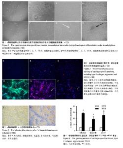
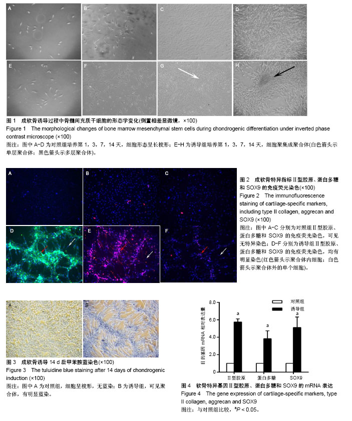
| [1] 黄弘轩,张姝江,白波.调控骨髓间充质干细胞成软骨分化的研究进展[J].中华关节外科杂志(电子版),2017,11(6):655-660.[2] Fahy N, Alini M, Stoddart MJ.Mechanical stimulation of mesenchymal stem cells: Implications for cartilage tissue engineering.J Orthop Res. 2018;36(1):52-63. [3] 汪泱,邓志锋,赖贤良,等.骨髓间充质干细胞的体外培养及定向诱导分化[J].中国临床医学,2004,11(2):202-204.[4] Li G, Yu F, Lei T,et al.Bone marrow mesenchymal stem cell therapy in ischemic stroke: mechanisms of action and treatment optimization strategies.Neural Regen Res. 2016; 11(6):1015-1024.[5] Wang Z, Li K, Sun H, et al. Icariin promotes stable chondrogenic differentiation of bone marrow mesenchymal stem cells in self?assembling peptide nanofiber hydrogel scaffolds.Mol Med Rep. 2018;17(6):8237-8243. [6] Bearden RN, Huggins SS, Cummings KJ, et al. In-vitro characterization of canine multipotent stromal cells isolated from synovium, bone marrow, and adipose tissue: a donor-matched comparative study.Stem Cell Res Ther. 2017; 8(1):218. [7] Olvera D, Sathy BN, Carroll SF, et al. Modulating microfibrillar alignment and growth factor stimulation to regulate mesenchymal stem cell differentiation.ActaBiomater. 2017; 64:148-160. [8] Glennon-Alty L, Williams R, Dixon S, et al. Induction of mesenchymal stem cell chondrogenesis by polyacrylate substrates.ActaBiomater. 2013;9(4):6041-6051. [9] Wang ZC, Sun HJ, Li KH, et al. Icariin promotes directed chondrogenic differentiation of bone marrow mesenchymal stem cells but not hypertrophy in vitro.ExpTher Med. 2014; 8(5):1528-1534. [10] Ray P, Hughes AJ, Sharif M, et al. Lectins selectively label cartilage condensations and the oticneuroepithelium within the embryonic chicken head.J Anat. 2017;230(3):424-434. [11] Ray P, Chapman SC.Cytoskeletal Reorganization Drives Mesenchymal Condensation and Regulates Downstream Molecular Signaling.PLoS One. 2015;10(8):e0134702. [12] Shimizu H, Yokoyama S, Asahara H.Growth and differentiation of the developing limb bud from the perspective of chondrogenesis.Dev Growth Differ. 2007;49(6):449-454. [13] Lubis AM, Lubis VK.Adult bone marrow stem cells in cartilage therapy.Acta Med Indones. 2012;44(1):62-68. [14] 杨强,丁晓明,徐宝山,等.含软骨钙化层的仿生支架体内构建组织工程骨软骨[J].中华骨科杂志,2018,38(6):321-329.[15] Mohamed-Ahmed S, Fristad I, Lie SA, et al. Adipose-derived and bone marrow mesenchymal stem cells: a donor-matched comparison.Stem Cell Res Ther. 2018;9(1):168. [16] 孙皓,左健.组织工程化软骨细胞和骨髓间充质干细胞用于修复同种异体关节软骨缺损[J].中国组织工程研究, 2012,16(19): 3602-3605.[17] Ruan SQ, Yan L, Deng J, et al. Preparation of a biphase composite scaffold and its application in tissue engineering for femoral osteochondral defects in rabbits.IntOrthop. 2017;41(9): 1899-1908. [18] 王奇,唐洪,曾伟南,等.骨髓间充质干细胞复合仿生凝胶修复猪关节软骨缺损[J].中华创伤杂志,2017,33(7):658-664.[19] Jiang X, Huang X, Jiang T, et al. The role of Sox9 in collagen hydrogel-mediated chondrogenic differentiation of adult mesenchymal stem cells (MSCs).Biomater Sci. 2018;6(6): 1556-1568. [20] 宋长志,徐小卒,吴亚,等.慢病毒介导的 Sox-9基因转染对大鼠关节软骨细胞炎性反应的影响[J].中华医院感染学杂志, 2016, 26(23):5342-5345.[21] 刘庆珍,吴尽,邳晓盟,等.软骨发育过程中负调控Sox9基因表达机制研究进展[J].农业科学与技术(英文版), 2017,18(6): 1038-1041.[22] Mao AS, Shin JW, Mooney DJ.Effects of substrate stiffness and cell-cell contact on mesenchymal stem cell differentiation. Biomaterials. 2016;98:184-191. [23] Cao B, Li Z, Peng R, et al. Effects of cell-cell contact and oxygen tension on chondrogenic differentiation of stem cells.Biomaterials. 2015;64:21-32. [24] de Windt TS, Saris DB, Slaper-Cortenbach IC, et al. Direct Cell-Cell Contact with Chondrocytes Is a Key Mechanism in Multipotent Mesenchymal Stromal Cell-Mediated Chondrogenesis.Tissue Eng Part A. 2015;21(19-20): 2536-2547. [25] 李拉,陈文庆,朱敬先,等.直接接触共培养技术在体外探索细胞间相互作用的应用研究[J].中国运动医学杂志, 2013,32(4): 337-342,352.[26] 刘霞,周广东,刘伟,等.细胞间直接接触诱导BMSC软骨分化的初步研究[J].组织工程与重建外科杂志,2010,6(2):70-74.[27] 田力,梁晓鹏,汪卓,等.体外非接触性共培养对MSC向软骨细胞诱导分化的实验观察[J].中国医药导报,2008,5(19):16-18.[28] Levorson EJ, Santoro M, Kasper FK, et al. Direct and indirect co-culture of chondrocytes and mesenchymal stem cells for the generation of polymer/extracellular matrix hybrid constructs. Acta Biomater. 2014;10(5):1824-1835. [29] Xu L, Wang Q, Xu F, et al. Mesenchymal stem cells downregulate articular chondrocyte differentiation in noncontact coculture systems: implications in cartilage tissue regeneration.Stem Cells Dev. 2013;22(11):1657-1669. [30] 冯均伟,王跃,吕波,等.骨髓间充质干细胞体外诱导成软骨细胞的动态观察[J].中国组织工程研究,2013,17(36):6409-6416. |

