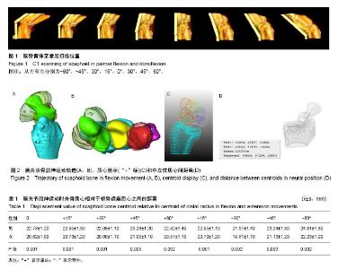
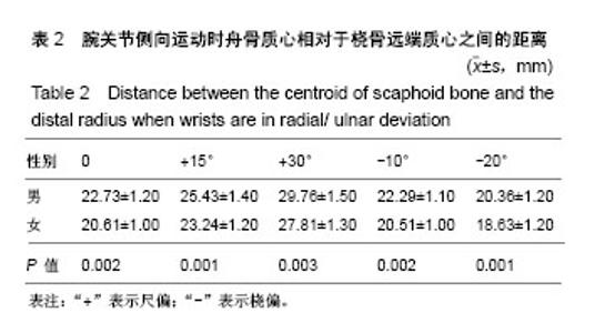
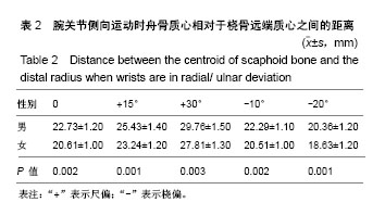
| [1] Wolfe SW, Crisco JJ, Katz LD. A non-invasive method for studying in vivo carpal kinematics. J Hand Surg Br.1997; 22(2):147-152. [2] Oura K, Moritomo H, Kataoka T, et al. Three-dimensional analysis of osteophyte formation on distal radius following scaphoid nonunion. J Orthop Sci. 2017;22(1):50-55. [3] 孟伟正,徐永清,李晶,等.正常人腕骨三维运动学研究方法[J].中华手外科杂志,2005,21(4):224-226.[4] 李俊.腕舟骨腰部骨折不同体位固定的生物力学研究[D]. 福州:福建中医学院 福建中医药大学,2006.[5] Hackney LA, Dodds SD. Assessment of scaphoid fracture healing. Curr Rev Musculoskelet Med.2011; 4 (1):16-22. [6] Pereira A, Hidalgo Diaz JJ, Saur M, et al. Carpal scaphoid non-union treatment: a retrospective trial comparing simple retrograde percutaneous screw fixation versus percutaneous screw fixation plus pulsed electromagnetic fields (Physiostim ((R))). Eur J Orthop Surg Traumatol.2017; 27(4): 521-525. [7] Reed DN, Fulcher SM, Harrison SJ. Unstable scaphoid nonunion treatment technique: use of a volar distal radius corticocancellous autograft. Tech Hand Up Extrem Surg.2012; 6 (2):91-94. [8] 刘俊健.石膏固定位置对腕舟骨骨折愈合和功能的影响[J].实用骨科杂志,2009,15(1):47-48.[9] Kaufmann R, Pfaeffle J, Blankenhorn B, et al. Kinematics of the midcarpal and radiocarpal joints in radioulnar deviation: an in vitro study. J Hand Surg Am.2005;30(5):937-942. [10] 樊志强.正常中腕关节运动学在体实验研究[D].昆明:昆明医学院, 2008.[11] 苏鸿博,刘德群,张文龙.正常男性腕关节主动极限位置稳定性分析[J].中国骨与关节损伤杂志,2015,30 (11):1174-1177.[12] Crisco JJ, Wolfe SW, Neu CP, et al. Advances in the in vivo measurement of normal and abnormal carpal kinematics. Orthop Clin North Am.2001;32(2):219-231, vii. [13] Omori S, Moritomo H, Omokawa S, et al. In vivo 3-dimensional analysis of dorsal intercalated segment instability deformity secondary to scapholunate dissociation: a preliminary report. J Hand Surg Am. 2013;38(7):1346-1355. [14] Bumbaširevi? M, Tomi? S, Leši? A, et al. The treatment of scaphoid nonunion using the Ilizarov fixator without bone graft, a study of 18 cases. J Orthop Surg Res. 2011;6(1):1-10. [15] Chu PJ, Shih JT. Arthroscopically Assisted Use of Injectable Bone Graft Substitutes for Management of Scaphoid Nonunions. Arthroscopy. 2011;27(1):31-37. [16] Hannemann P, Göttgens KW, Wely BJV, et al. Pulsed Electromagnetic Fields in the treatment of fresh scaphoid fractures. A multicenter, prospective, double blind, placebo controlled, randomized trial. BMC Musculoskelet Disord. 2011;12(1):90. [17] Biz C, Barison E, Ruggieri P, et al. Radiographic and functional outcomes after displaced intra-articular calcaneal fractures: a comparative cohort study among the traditional open technique (ORIF) and percutaneous surgical procedures (PS). J Orthop Surg Res. 2016;11(1):92. [18] Matsuki H, Ishikawa J, Iwasaki N, et al. Non-vascularized bone graft with Herbert-type screw fixation for proximal pole scaphoid nonunion. J Orthop Sci.2011;16(6):749-755. [19] 徐永清,钟世镇,朱青安,等.正常腕关节运动学的实验研究[J].中国临床解剖学杂志,2003,21(2):173-175.[20] Neogi T. Structural correlates of pain in osteoarthritis. Clin Exp Rheumatol. 2017;35 Suppl 107(5):75-78.[21] Canovas F, Roussanne Y, Captier G, et al. Study of carpal bone morphology and position in three dimensions by image analysis from computed tomography scans of the wrist. Surg Radiol Anat.2004; 26(3):186-190. |