Chinese Journal of Tissue Engineering Research ›› 2018, Vol. 22 ›› Issue (21): 3304-3309.doi: 10.3969/j.issn.2095-4344.0522
Previous Articles Next Articles
In vivo ectopic osteogenesis of adipose-derived mesenchymal stem cells/osteoblasts combined with hydroxyapatite/chitosan/polylactic acid
Tian Xue-tao1, Wang Xian-xun2, Wang Xiao-wei2
- 1Department of Traumatology, Affiliated Hospital of Jianghan University (Wuhan No. 6 Hospital), Wuhan 430015, Hubei Province, China; 2Department of Orthopedics, the Third People’s Hospital of Hubei Province, Wuhan 430033, Hubei Province, China
-
Revised:2018-03-04Online:2018-07-28Published:2018-07-28 -
Contact:Wang Xiao-wei, Master, Attending physician, Department of Orthopedics, the Third People’s Hospital of Hubei Province, Wuhan 430033, Hubei Province, China -
About author:Tian Xue-tao, Master, Associate chief physician, Department of Traumatology, Affiliated Hospital of Jianghan University (Wuhan No. 6 Hospital), Wuhan 430015, Hubei Province, China -
Supported by:the Research Project for Health and Family Plans in Hubei Province, No. WJ2015MB128
CLC Number:
Cite this article
Tian Xue-tao, Wang Xian-xun, Wang Xiao-wei. In vivo ectopic osteogenesis of adipose-derived mesenchymal stem cells/osteoblasts combined with hydroxyapatite/chitosan/polylactic acid[J]. Chinese Journal of Tissue Engineering Research, 2018, 22(21): 3304-3309.
share this article
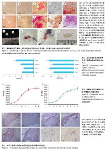
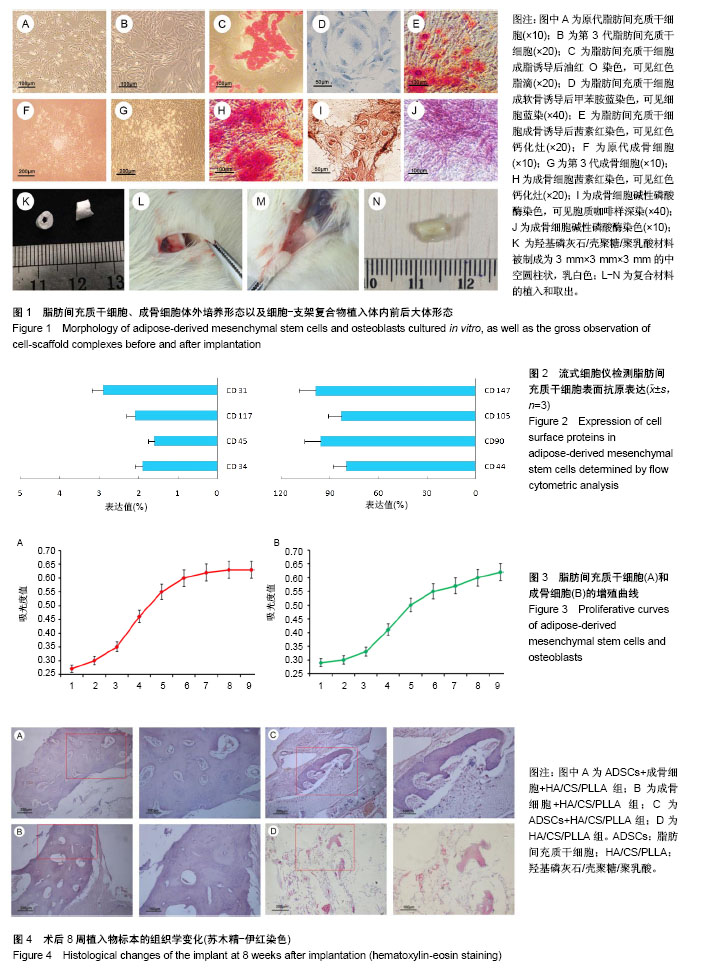
2.1 细胞形态学观察及染色鉴定结果 ADSCs原代培养4-6 h后开始贴壁,细胞大小不均匀,起初为多角形,梭形,3 d后类成纤维细胞明显增多,四五天时细胞进入对数生长期,六七天时细胞融合接近80%,可消化传代(图1A);传至第3代后细胞呈长梭形,形态均一,呈漩涡样生长(图1B),成脂诱导后油红O染色可见红染的脂滴(图1C),成软骨诱导后可见细胞质蓝染(图1D),成骨诱导后茜素红染色可见红染的钙化灶(图1E)。 骨组织块法获得的成骨细胞贴于细胞瓶底部,24 h后成骨细胞从骨块中爬出,葵花样生长,细胞呈三角形、多角形或梭形(图1F)。传至第3代后细胞呈鳞形,体积较大,多伪足,继续培养可见局部多层生长,并有钙化结节生成(图1G),培养14 d时茜素红染色可见红染的钙化灶(图1H),碱性磷酸酶染色可见胞质咖啡样深染(图1I,J)。 HA/CS/PLLA材料呈乳白色,被制成3 mm×3 mm×3 mm的圆柱状中空材料以便于植入体内(图1K-N)。 2.2 ADSCs表面抗原表达 CD147、CD90、CD105、CD44呈阳性表达(> 80%),而CD117、CD34、CD31、CD45则呈阴性表达(< 5%),见图2。 2.3 ADSCs和成骨细胞的增殖能力 随着培养时间的延长,细胞数逐渐增多,吸光度值增高;ADSCs呈“S”形增殖曲线,第1,2天为平台期,第3-5天为对数生长期,第6天以后细胞密度较大,细胞间接触抑制生长,进入平台期。而成骨细胞呈直线生长,第1,2天为平台期,第3-8天细胞基本呈线性生长,见图3。 2.4 大体观察结果 10只SD大鼠全部存活并进入结果分析,术后伤口愈合良好,无炎性反应,伤口一期愈合,期间进食正常。植入4周后可见材料有软组织长入,包裹,难以分离(图1L,M)。 2.5 苏木精-伊红染色观察组织学形态 ADSCs+成骨细胞+HA/CS/PLLA组:术后8周,移植物已完全被新生的编制骨替代,形成明显软骨陷窝,视野下可见大量钙化的骨组织,并形成骨髓腔,HA/CS/PLLA已被降解、吸收(图4A)。 成骨细胞+HA/CS/PLLA组:术后8周,移植物有新生编制骨及大量软骨形成,软骨基质较多,在钙化的骨组织中仍有未完全骨化的软骨。HA/CS/PLLA材料内有大量的纤维、血管、脂肪等结缔组织长入,支架开始降解(图4B)。 ADSCs+HA/CS/PLLA组:术后8周,支架材料表面及周边纤维组织内可见带状、条索状骨组织,内部可见软骨陷窝和软骨基质,周边有大量成骨细胞及未成熟的骨组织(图4C)。 HA/CS/PLLA组:术后8周,支架材料表面及周边纤维组织内可见岛状骨组织,支架材料降解较少,可见少量软骨细胞,周边可见少量成骨细胞和纤维组织(图4D)。 2.6 图像分析结果 各组术后8周植入物新骨形成差异明显,ADSCs+成骨细胞+HA/CS/PLLA组成骨分数为(33.8± 3.11)%,成骨细胞+HA/CS/PLLA组为(21.4±3.24)%,两组间比较差异有显著性意义(P < 0.05);HA/CS/PLLA组为(2.89±0.91)%,ADSCs+HA/CS/PLLA组成骨分数为(13.2± 1.59)%,与ADSCs+成骨细胞+HA/CS/PLLA组比较差异也有显著性意义(P < 0.05)。"
| [1] Roosens A, Asadian M, De Geyter N, et al. Complete Static Repopulation of Decellularized Porcine Tissues for Heart Valve Engineering: An in vitro Study. Cells Tissues Organs. 2017;204(5-6):270-282.[2] Yao W, Lay YE, Kot A, et al. Improved Mobilization of Exogenous Mesenchymal Stem Cells to Bone for Fracture Healing and Sex Difference. Stem Cells. 2016;34(10): 2587-2600.[3] Zhang H, Kot A, Lay YE, et al. Acceleration of Fracture Healing by Overexpression of Basic Fibroblast Growth Factor in the Mesenchymal Stromal Cells. Stem Cells Transl Med. 2017;6(10):1880-1893.[4] 陆钰,王鑫,甘朝兵,等. 多孔纳米羟基磷灰石/壳聚糖支架材料复合成骨细胞的异位成骨研究[J].生物骨科材料与临床研究,2012, 9(1):21-25.[5] Yuan H, Qin J, Xie J, et al. Highly aligned core-shell structured nanofibers for promoting phenotypic expression of vSMCs for vascular regeneration. Nanoscale. 2016;8(36): 16307-16322.[6] Zhang CY, Zhang CL, Wang JF, et al. Fabrication and in vitro investigation of nanohydroxyapatite, chitosan, poly(L‐lactic acid) ternary biocomposite. Journal of Applied Polymer Science. 2014;127(3):2152-2159.[7] Bodle JC, Hanson AD, Loboa EG. Adipose-derived stem cells in functional bone tissue engineering: lessons from bone mechanobiology. Tissue Eng Part B Rev. 2011;17(3):195-211.[8] Sakaguchi Y, Sekiya I, Yagishita K, et al. Comparison of human stem cells derived from various mesenchymal tissues: superiority of synovium as a cell source. Arthritis Rheum. 2005;52(8):2521-2529.[9] Feng W, Lv S, Cui J, et al. Histochemical examination of adipose derived stem cells combined with β-TCP for bone defects restoration under systemic administration of 1α,25(OH)2D3. Mater Sci Eng C Mater Biol Appl. 2015;54: 133-141.[10] Zhang H, Kot A, Lay YE, et al. Acceleration of Fracture Healing by Overexpression of Basic Fibroblast Growth Factor in the Mesenchymal Stromal Cells. Stem Cells Transl Med. 2017;6(10):1880-1893.[11] Vergroesen PP, Kroeze RJ, Helder MN, et al. The use of poly(L-lactide-co-caprolactone) as a scaffold for adipose stem cells in bone tissue engineering: application in a spinal fusion model. Macromol Biosci. 2011;11(6):722-730.[12] Roskies MG, Fang D, Abdallah MN, et al. Three-dimensionally printed polyetherketoneketone scaffolds with mesenchymal stem cells for the reconstruction of critical-sized mandibular defects. Laryngoscope. 2017;127(11):E392-E398.[13] Fang D, Roskies M, Abdallah MN, et al. Three-Dimensional Printed Scaffolds with Multipotent Mesenchymal Stromal Cells for Rabbit Mandibular Reconstruction and Engineering. Methods Mol Biol. 2017;1553:273-291.[14] Kang ML, Kim JE, Im GI. Vascular endothelial growth factor-transfected adipose-derived stromal cells enhance bone regeneration and neovascularization from bone marrow stromal cells. J Tissue Eng Regen Med. 2017;11(12): 3337-3348.[15] Godoy Zanicotti D, Coates DE, Duncan WJ. In vivo bone regeneration on titanium devices using serum-free grown adipose-derived stem cells, in a sheep femur model. Clin Oral Implants Res. 2017;28(1):64-75.[16] Lee SJ, Kim BJ, Kim YI, et al. Effect of Recombinant Human Bone Morphogenetic Protein-2 and Adipose Tissue-Derived Stem Cell on New Bone Formation in High-Speed Distraction Osteogenesis. Cleft Palate Craniofac J. 2016;53(1):84-92.[17] Tabatabaei Qomi R, Sheykhhasan M. Adipose-derived stromal cell in regenerative medicine: A review. World J Stem Cells. 2017;9(8):107-117.[18] Shakir M, Jolly R, Khan MS, et al. Nano-hydroxyapatite/ chitosan-starch nanocomposite as a novel bone construct: Synthesis and in vitro studies. Int J Biol Macromol. 2015;80: 282-292.[19] Godoy Zanicotti D, Coates DE, Duncan WJ. In vivo bone regeneration on titanium devices using serum-free grown adipose-derived stem cells, in a sheep femur model. Clin Oral Implants Res. 2017;28(1):64-75.[20] biocompatible evaluation of natural hydroxyapatite/chitosan composite for bone repair. J Appl Biomater Biomech. 2011; 9(1):11-18.[21] Park S, Heo HA, Lee KB, et al. Improved Bone Regeneration With Multiporous PLGA Scaffold and BMP-2-Transduced Human Adipose-Derived Stem Cells by Cell-Permeable Peptide. Implant Dent. 2017;26(1):4-11.[22] Zhang J, Zhou S, Zhou Y, et al. Adipose-Derived Mesenchymal Stem Cells (ADSCs) With the Potential to Ameliorate Platelet Recovery, Enhance Megakaryopoiesis, and Inhibit Apoptosis of Bone Marrow Cells in a Mouse Model of Radiation-Induced Thrombocytopenia. Cell Transplant. 2016;25(2):261-273.[23] Lai QG, Sun SL, Zhou XH, et al. Adipose-derived stem cells transfected with pEGFP-OSX enhance bone formation during distraction osteogenesis. J Zhejiang Univ Sci B. 2014;15(5): 482-490.[24] Zhang J, Zhou S, Zhou Y, et al. Adipose-Derived Mesenchymal Stem Cells (ADSCs) With the Potential to Ameliorate Platelet Recovery, Enhance Megakaryopoiesis, and Inhibit Apoptosis of Bone Marrow Cells in a Mouse Model of Radiation-Induced Thrombocytopenia. Cell Transplant. 2016;25(2):261-273.[25] Wan W, Zhang S, Ge L, et al. Layer-by-layer paper-stacking nanofibrous membranes to deliver adipose-derived stem cells for bone regeneration. Int J Nanomedicine. 2015;10: 1273-1290.[26] Zhang W, Zhang X, Wang S, et al. Comparison of the use of adipose tissue-derived and bone marrow-derived stem cells for rapid bone regeneration. J Dent Res. 2013;92(12): 1136-1141.[27] Lough DM, Chambers C, Germann G, et al. Regulation of ADSC Osteoinductive Potential Using Notch Pathway Inhibition and Gene Rescue: A Potential On/Off Switch for Clinical Applications in Bone Formation and Reconstructive Efforts. Plast Reconstr Surg. 2016;138(4):642e-652e.[28] 芮钢,胡宝山,郭元利,等.成骨细胞、血管内皮细胞复合组织工程骨修复骨缺损的实验研究[J]. 中华骨科杂志,2008, 28(2): 155-158.[29] Zhou X, Tao Y, Wang J, et al. Three-dimensional scaffold of type II collagen promote the differentiation of adipose-derived stem cells into a nucleus pulposus-like phenotype. J Biomed Mater Res A. 2016;104(7):1687-1693. |
| [1] | Pu Rui, Chen Ziyang, Yuan Lingyan. Characteristics and effects of exosomes from different cell sources in cardioprotection [J]. Chinese Journal of Tissue Engineering Research, 2021, 25(在线): 1-. |
| [2] | Lin Qingfan, Xie Yixin, Chen Wanqing, Ye Zhenzhong, Chen Youfang. Human placenta-derived mesenchymal stem cell conditioned medium can upregulate BeWo cell viability and zonula occludens expression under hypoxia [J]. Chinese Journal of Tissue Engineering Research, 2021, 25(在线): 4970-4975. |
| [3] | Zhang Tongtong, Wang Zhonghua, Wen Jie, Song Yuxin, Liu Lin. Application of three-dimensional printing model in surgical resection and reconstruction of cervical tumor [J]. Chinese Journal of Tissue Engineering Research, 2021, 25(9): 1335-1339. |
| [4] | Hou Jingying, Yu Menglei, Guo Tianzhu, Long Huibao, Wu Hao. Hypoxia preconditioning promotes bone marrow mesenchymal stem cells survival and vascularization through the activation of HIF-1α/MALAT1/VEGFA pathway [J]. Chinese Journal of Tissue Engineering Research, 2021, 25(7): 985-990. |
| [5] | Shi Yangyang, Qin Yingfei, Wu Fuling, He Xiao, Zhang Xuejing. Pretreatment of placental mesenchymal stem cells to prevent bronchiolitis in mice [J]. Chinese Journal of Tissue Engineering Research, 2021, 25(7): 991-995. |
| [6] | Liang Xueqi, Guo Lijiao, Chen Hejie, Wu Jie, Sun Yaqi, Xing Zhikun, Zou Hailiang, Chen Xueling, Wu Xiangwei. Alveolar echinococcosis protoscolices inhibits the differentiation of bone marrow mesenchymal stem cells into fibroblasts [J]. Chinese Journal of Tissue Engineering Research, 2021, 25(7): 996-1001. |
| [7] | Fan Quanbao, Luo Huina, Wang Bingyun, Chen Shengfeng, Cui Lianxu, Jiang Wenkang, Zhao Mingming, Wang Jingjing, Luo Dongzhang, Chen Zhisheng, Bai Yinshan, Liu Canying, Zhang Hui. Biological characteristics of canine adipose-derived mesenchymal stem cells cultured in hypoxia [J]. Chinese Journal of Tissue Engineering Research, 2021, 25(7): 1002-1007. |
| [8] | Geng Yao, Yin Zhiliang, Li Xingping, Xiao Dongqin, Hou Weiguang. Role of hsa-miRNA-223-3p in regulating osteogenic differentiation of human bone marrow mesenchymal stem cells [J]. Chinese Journal of Tissue Engineering Research, 2021, 25(7): 1008-1013. |
| [9] | Lun Zhigang, Jin Jing, Wang Tianyan, Li Aimin. Effect of peroxiredoxin 6 on proliferation and differentiation of bone marrow mesenchymal stem cells into neural lineage in vitro [J]. Chinese Journal of Tissue Engineering Research, 2021, 25(7): 1014-1018. |
| [10] | Zhu Xuefen, Huang Cheng, Ding Jian, Dai Yongping, Liu Yuanbing, Le Lixiang, Wang Liangliang, Yang Jiandong. Mechanism of bone marrow mesenchymal stem cells differentiation into functional neurons induced by glial cell line derived neurotrophic factor [J]. Chinese Journal of Tissue Engineering Research, 2021, 25(7): 1019-1025. |
| [11] | Duan Liyun, Cao Xiaocang. Human placenta mesenchymal stem cells-derived extracellular vesicles regulate collagen deposition in intestinal mucosa of mice with colitis [J]. Chinese Journal of Tissue Engineering Research, 2021, 25(7): 1026-1031. |
| [12] | Pei Lili, Sun Guicai, Wang Di. Salvianolic acid B inhibits oxidative damage of bone marrow mesenchymal stem cells and promotes differentiation into cardiomyocytes [J]. Chinese Journal of Tissue Engineering Research, 2021, 25(7): 1032-1036. |
| [13] | Wang Xianyao, Guan Yalin, Liu Zhongshan. Strategies for improving the therapeutic efficacy of mesenchymal stem cells in the treatment of nonhealing wounds [J]. Chinese Journal of Tissue Engineering Research, 2021, 25(7): 1081-1087. |
| [14] | Wang Shiqi, Zhang Jinsheng. Effects of Chinese medicine on proliferation, differentiation and aging of bone marrow mesenchymal stem cells regulating ischemia-hypoxia microenvironment [J]. Chinese Journal of Tissue Engineering Research, 2021, 25(7): 1129-1134. |
| [15] | Zeng Yanhua, Hao Yanlei. In vitro culture and purification of Schwann cells: a systematic review [J]. Chinese Journal of Tissue Engineering Research, 2021, 25(7): 1135-1141. |
| Viewed | ||||||
|
Full text |
|
|||||
|
Abstract |
|
|||||