Chinese Journal of Tissue Engineering Research ›› 2018, Vol. 22 ›› Issue (13): 2027-2032.doi: 10.3969/j.issn.2095-4344.0492
Previous Articles Next Articles
Human umbilical cord blood mesenchymal stem cells differentiate into neuron-like cells: in vitro induction by mouse nerve growth factor
Chen Jun, Yang Zi-jin
- Department of Pediatrics, Lianyungang Hospital Affiliated to Xuzhou Medical University (First People’s Hospital of Lianyungang), Lianyungang 222000, Jiangsu Province, China
-
Revised:2017-12-18Online:2018-05-08Published:2018-05-08 -
Contact:Yang Zi-jin, Chief physician, Master’s supervisor, Department of Pediatrics, Lianyungang Hospital Affiliated to Xuzhou Medical University (First People’s Hospital of Lianyungang), Lianyungang 222000, Jiangsu Province, China -
About author:Chen Jun, Master candidate, Department of Pediatrics, Lianyungang Hospital Affiliated to Xuzhou Medical University (First People’s Hospital of Lianyungang), Lianyungang 222000, Jiangsu Province, China
CLC Number:
Cite this article
Chen Jun, Yang Zi-jin. Human umbilical cord blood mesenchymal stem cells differentiate into neuron-like cells: in vitro induction by mouse nerve growth factor[J]. Chinese Journal of Tissue Engineering Research, 2018, 22(13): 2027-2032.
share this article
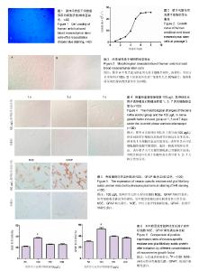
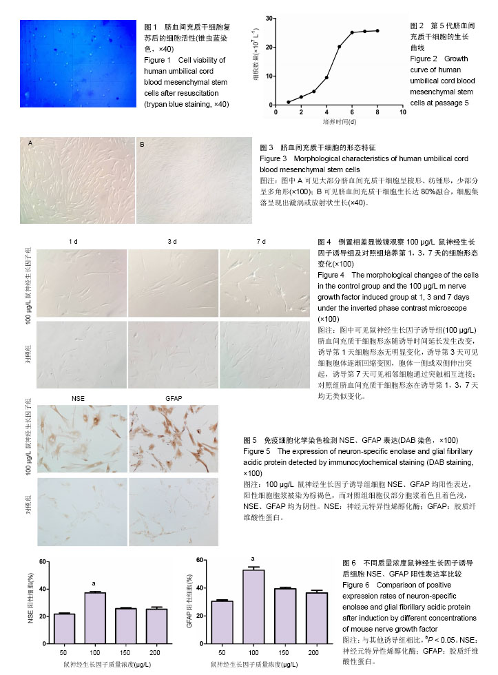
2.1 脐血间充质干细胞复苏后的细胞活性 用锥虫蓝染色检测复苏后细胞活性:正常活细胞的胞膜结构完整,锥虫蓝不能进入胞内,因此不能被染上颜色,此现象为“锥虫蓝拒染”;而死细胞或胞膜不完整的细胞,因胞膜破坏而可被锥虫蓝染成蓝色。实验中大部分活细胞为锥虫蓝拒染,少数死细胞被染成蓝色,计算细胞活性率达90%以上,表明细胞复苏后活性良好(图1)。 2.2 脐血间充质干细胞的生长曲线 最初1-3 d细胞生长处于延滞期,此段时间内细胞生长较为缓慢;从第3天开始到第6天止细胞进入对数生长期,此段时间内细胞生长极为迅速,细胞数量以几何级数增长;6 d后细胞生长进入平台期,相比对数生长期增速明显减缓,生长曲线总体呈“S”型(图2)。 2.3 脐血间充质干细胞的形态特征 倒置相差显微镜下观察可见脐血间充质干细胞形态为大小不一的长梭形、纺锤形 (图3A),折光性强。培养六七天后细胞基本达80%融合状态,此时可见细胞集落呈漩涡状或放射状生长(图3B)。 2.4 mNGF诱导过程中脐血间充质干细胞形态学变化 加入mNGF进行诱导分化培养后,每日在倒置相差显微镜下观察,诱导第1天各实验组脐血间充质干细胞形态均未见明显变化,诱导第 2,3天可见各实验组细胞形态发生改变——细胞胞体逐渐回缩变圆,胞体一侧或双侧伸出突起,呈现出单极、双极类神经元样改变,随着诱导时间的延长,可见细胞突起逐渐伸长并发出次级突起,诱导至第7天可见各实验组中相邻细胞通过突触相互连接,其中质量浓度为100 μg/L mNGF诱导组最为明显,而对照组脐血间充质干细胞形态则无明显改变,罕见类似细胞(图4)。7 d诱导时间内所有实验组细胞生长状态均良好,未见明显的脱落、死亡现象。 2.5 免疫细胞化学染色鉴定结果 对mNGF诱导第7天的脐血间充质干细胞进行免疫细胞化学染色,发现各mNGF诱导组细胞NSE、GFAP均阳性表达,阳性细胞胞浆被染为棕褐色,而对照组细胞仅部分胞浆着色且着色浅,为非特异性染色,故NSE、GFAP均不表达为阴性(图5)。 2.6 统计学分析 ①各mNGF诱导组细胞NSE阳性率差异有显著性意义(F=41.52,P < 0.01),50,100,150,200 μg/L mNGF诱导组NSE阳性率分别为(21.7±3.2)%,(37.2±3.4)%,(25.7±2.5)%,(25.3±4.9)%; ②各mNGF诱导组细胞GFAP阳性率差异也有显著性意义(F=37.01,P < 0.01),50,100,150,200 μg/L mNGF诱导组GFAP阳性率分别为(30.5±2.9)%,(52.7±5.2)%,(39.3±2.5)%,(36.5±4.2)%;③各mNGF诱导组组间比较,100 μg/L组NSE,GFAP细胞阳性率均高于50,150,200 μg/L组,差异有显著性意义(P < 0.05,图6)。"
| [1] Taran R, Mamidi MK, Singh G, et al. In vitro and in vivo neurogenic potential of mesenchymal stem cells isolated from different sources. J Biosci. 2014;39(1):157-169.[2] Wang L, Lu M. Regulation and direction of umbilical cord blood mesenchymal stem cells to adopt neuronal fate. Int J Neurosci. 2014;124(3):149-159.[3] Park HW, Moon HE, Kim HS, et al. Human umbilical cord blood-derived mesenchymal stem cells improve functional recovery through thrombospondin1, pantraxin3, and vascular endothelial growth factor in the ischemic rat brain. J Neurosci Res. 2015 93(12):1814-1825.[4] Zhang C, Huang L, Gu J, et al. Therapy for Cerebral Palsy by Human Umbilical Cord Blood Mesenchymal Stem Cells Transplantation Combined With Basic Rehabilitation Treatment: A Case Report. Glob Pediatr Health. 2015;2(1): 1-7.[5] Cui Y, Ma S, Zhang C, et al. Human umbilical cord mesenchymal stem cells transplantation improves cognitive function in Alzheimer's disease mice by decreasing oxidative stress and promoting hippocampal neurogenesis. Behav Brain Res. 2017;320:291-301.[6] Ning G, Tang L, Wu Q, et al. Human umbilical cord blood stem cells for spinal cord injury: early transplantation results in better local angiogenesis. Regen Med. 2013;8(3):271-281.[7] Zhang X, Zhang Q, Li W, et al. Therapeutic effect of human umbilical cord mesenchymal stem cells on neonatal rat hypoxic-ischemic encephalopathy. J Neurosci Res. 2014; 92(1):35-45.[8] Shahbazi A, Safa M, Alikarami F, et al. Rapid Induction of Neural Differentiation in Human Umbilical Cord Matrix Mesenchymal Stem Cells by cAMP-elevating Agents. Int J Mol Cell Med. 2016;5(3):167-177.[9] Rafieemehr H, Kheirandish M, Soleimani M. Improving the neuronal differentiation efficiency of umbilical cord blood-derived mesenchymal stem cells cultivated under appropriate conditions. Iran J Basic Med Sci. 2015;18(11): 1100-1106.[10] Zeng Y, Rong M, Liu Y, et al. Electrophysiological characterisation of human umbilical cord blood-derived mesenchymal stem cells induced by olfactory ensheathing cell-conditioned medium. Neurochem Res. 2013;38(12): 2483-2489.[11] Hei WH, Almansoori AA, Sung MA, et al. Adenovirus vector-mediated ex vivo gene transfer of brain-derived neurotrophic factor (BDNF) tohuman umbilical cord blood-derived mesenchymal stem cells (UCB-MSCs) promotescrush-injured rat sciatic nerve regeneration. Neurosci Lett. 2017;643:111-120.[12] Nan C, Guo L, Zhao Z, et al. Tetramethylpyrazine induces differentiation of human umbilical cord-derived mesenchymal stem cells into neuron-like cells in vitro. Int J Oncol. 2016; 48(6):2287-2294.[13] 李立军,时宇博,宗强,等.局部应用明胶海绵浸润鼠神经生长因子治疗周围神经损伤[J]. 中国组织工程研究,2015,19(30) :4827-4831.[14] 郝冬荣,厉红,庞保东.鼠神经生长因子治疗小儿面神经炎42例[J].中国药业,2014, 23(1):82-84.[15] 许马利,王杨. 鼠神经生长因子治疗新生儿缺氧缺血性脑病的Meta分析[J]. 中国临床药理学杂志,2016, 32(7):652-654.[16] 李巧秀,张俊清,翟红印,等. 鼠神经生长因子运动区注射治疗小儿脑瘫疗效观察[J]. 中国妇幼保健,2015, 30(28):4889-4891.[17] 方雅秀,谭燕,侯乐. 鼠神经生长因子治疗病毒性脑炎的疗效[J]. 实用医学杂志,2013, 29(11):1837-1838.[18] Liu F, Xuan A, Chen Y, et al. Combined effect of nerve growth factor and brain?derived neurotrophic factor on neuronal differentiation of neural stem cells and the potential molecular mechanisms. Mol Med Rep. 2014;10(4):1739-1745.[19] Han Z, Wang CP, Cong N, et al. Therapeutic value of nerve growth factor in promoting neural stem cell survival and differentiation and protecting against neuronal hearing loss. Mol Cell Biochem. 2017;428(1-2):149-159.[20] 胡海棠,林英勇,扈会平. 鼠神经生长因子随机双盲安慰剂对照ⅠⅡⅢ期临床试验中注射部位疼痛的综合分析[J]. 中国药物警戒, 2010, 7(7):397-400.[21] Tomioka T, Shimazaki T, Yamauchi T, et al. LIM homeobox 8 (Lhx8) is a key regulator of the cholinergic neuronal function via a tropomyosin receptor kinase A (TrkA)-mediated positive feedback loop. J Biol Chem. 2014;289(2):1000-1010.[22] Webber CA, Salame J, Luu GL, et al. Nerve growth factor acts through the TrkA receptor to protect sensory neurons from the damaging effects of the HIV-1 viral protein, Vpr. Neuroscience. 2013;252:512-525.[23] 肖力,何俐. 酪氨酸激酶A和酪氨酸激酶B与脑缺血缺氧性损伤[J]. 中国卒中杂志,2008,3(6):460-464.[24] Zhao J, Cheng YY, Fan W, et al. Botanical drug puerarin coordinates with nerve growth factor in the regulation of neuronal survival and neuritogenesis via activating ERK1/2 and PI3K/Akt signaling pathways in the neurite extension process.CNS Neurosci Ther. 2015;21(1):61-70.[25] Ahmad I, Yue WY, Fernando A, et al. p75NTR is highly expressed in vestibular schwannomas and promotes cell survival by activating nuclear transcription factor κB. Glia. 2014;62(10):1699-1712.[26] Zhang Y, Hu W. NFκB signaling regulates embryonic and adult neurogenesis. Front Biol (Beijing). 2012;7(4):277-291.[27] McKelvey L, Gutierrez H, Nocentini G, et al. The intracellular portion of GITR enhances NGF-promoted neurite growth through an inverse modulation of Erk and NF-κB signalling. Biol Open. 2012;1(10):1016-1023.[28] Muthaiah VPK, Michael FM, Palaniappan T, et al. JNK1 and JNK3 play a significant role in both neuronal apoptosis and necrosis. Evaluation based on in vitro approach using tert-butylhydroperoxide induced oxidative stress in neuro-2A cells and perturbation through 3-aminobenzamide. Toxicol In Vitro. 2017;41:168-178.[29] 苏肖英,汪海涛,孟茜,等. 神经生长因子诱导神经干细胞分化的作用机制探讨[J]. 广东医学,2012, 33(1):44-48.[30] Lim JY, Park SI, Oh JH, et al. Brain-derived neurotrophic factor stimulates the neural differentiation of human umbilical cord blood-derived mesenchymal stem cells and survival of differentiated cells through MAPK/ERK and PI3K/Akt-dependent signaling pathways. J Neurosci Res. 2008;86(10):2168-2178.[31] Zhang X, Zhang L, Cheng X, et al. IGF-1 promotes Brn-4 expression and neuronal differentiation of neural stem cells via the PI3K/Akt pathway. PLoS One. 2014;9(12):e113801.[32] Zhao L, Feng Y, Chen X, et al. Effects of IGF-1 on neural differentiation of human umbilical cord derived mesenchymal stem cells. Life Sci. 2016;151:93-101.[33] Lim JY, Park SI, Kim SM, et al. Neural differentiation of brain-derived neurotrophic factor-expressing human umbilical cord blood-derived mesenchymal stem cells in culture via TrkB-mediated ERK and β-catenin phosphorylation and following transplantation into the developing brain. Cell Transplant. 2011;20(11-12):1855-1866.[34] 帅春,汪灏. 激活PI3K/Akt信号通路抗少突胶质细胞凋亡的研究[J]. 广州医药, 2014,45(5):4-7. |
| [1] | Lin Qingfan, Xie Yixin, Chen Wanqing, Ye Zhenzhong, Chen Youfang. Human placenta-derived mesenchymal stem cell conditioned medium can upregulate BeWo cell viability and zonula occludens expression under hypoxia [J]. Chinese Journal of Tissue Engineering Research, 2021, 25(在线): 4970-4975. |
| [2] | Pu Rui, Chen Ziyang, Yuan Lingyan. Characteristics and effects of exosomes from different cell sources in cardioprotection [J]. Chinese Journal of Tissue Engineering Research, 2021, 25(在线): 1-. |
| [3] | Zhang Tongtong, Wang Zhonghua, Wen Jie, Song Yuxin, Liu Lin. Application of three-dimensional printing model in surgical resection and reconstruction of cervical tumor [J]. Chinese Journal of Tissue Engineering Research, 2021, 25(9): 1335-1339. |
| [4] | Wang Xianyao, Guan Yalin, Liu Zhongshan. Strategies for improving the therapeutic efficacy of mesenchymal stem cells in the treatment of nonhealing wounds [J]. Chinese Journal of Tissue Engineering Research, 2021, 25(7): 1081-1087. |
| [5] | Wang Shiqi, Zhang Jinsheng. Effects of Chinese medicine on proliferation, differentiation and aging of bone marrow mesenchymal stem cells regulating ischemia-hypoxia microenvironment [J]. Chinese Journal of Tissue Engineering Research, 2021, 25(7): 1129-1134. |
| [6] | Zeng Yanhua, Hao Yanlei. In vitro culture and purification of Schwann cells: a systematic review [J]. Chinese Journal of Tissue Engineering Research, 2021, 25(7): 1135-1141. |
| [7] | Kong Desheng, He Jingjing, Feng Baofeng, Guo Ruiyun, Asiamah Ernest Amponsah, Lü Fei, Zhang Shuhan, Zhang Xiaolin, Ma Jun, Cui Huixian. Efficacy of mesenchymal stem cells in the spinal cord injury of large animal models: a meta-analysis [J]. Chinese Journal of Tissue Engineering Research, 2021, 25(7): 1142-1148. |
| [8] | Hou Jingying, Yu Menglei, Guo Tianzhu, Long Huibao, Wu Hao. Hypoxia preconditioning promotes bone marrow mesenchymal stem cells survival and vascularization through the activation of HIF-1α/MALAT1/VEGFA pathway [J]. Chinese Journal of Tissue Engineering Research, 2021, 25(7): 985-990. |
| [9] | Shi Yangyang, Qin Yingfei, Wu Fuling, He Xiao, Zhang Xuejing. Pretreatment of placental mesenchymal stem cells to prevent bronchiolitis in mice [J]. Chinese Journal of Tissue Engineering Research, 2021, 25(7): 991-995. |
| [10] | Liang Xueqi, Guo Lijiao, Chen Hejie, Wu Jie, Sun Yaqi, Xing Zhikun, Zou Hailiang, Chen Xueling, Wu Xiangwei. Alveolar echinococcosis protoscolices inhibits the differentiation of bone marrow mesenchymal stem cells into fibroblasts [J]. Chinese Journal of Tissue Engineering Research, 2021, 25(7): 996-1001. |
| [11] | Fan Quanbao, Luo Huina, Wang Bingyun, Chen Shengfeng, Cui Lianxu, Jiang Wenkang, Zhao Mingming, Wang Jingjing, Luo Dongzhang, Chen Zhisheng, Bai Yinshan, Liu Canying, Zhang Hui. Biological characteristics of canine adipose-derived mesenchymal stem cells cultured in hypoxia [J]. Chinese Journal of Tissue Engineering Research, 2021, 25(7): 1002-1007. |
| [12] | Geng Yao, Yin Zhiliang, Li Xingping, Xiao Dongqin, Hou Weiguang. Role of hsa-miRNA-223-3p in regulating osteogenic differentiation of human bone marrow mesenchymal stem cells [J]. Chinese Journal of Tissue Engineering Research, 2021, 25(7): 1008-1013. |
| [13] | Lun Zhigang, Jin Jing, Wang Tianyan, Li Aimin. Effect of peroxiredoxin 6 on proliferation and differentiation of bone marrow mesenchymal stem cells into neural lineage in vitro [J]. Chinese Journal of Tissue Engineering Research, 2021, 25(7): 1014-1018. |
| [14] | Zhu Xuefen, Huang Cheng, Ding Jian, Dai Yongping, Liu Yuanbing, Le Lixiang, Wang Liangliang, Yang Jiandong. Mechanism of bone marrow mesenchymal stem cells differentiation into functional neurons induced by glial cell line derived neurotrophic factor [J]. Chinese Journal of Tissue Engineering Research, 2021, 25(7): 1019-1025. |
| [15] | Duan Liyun, Cao Xiaocang. Human placenta mesenchymal stem cells-derived extracellular vesicles regulate collagen deposition in intestinal mucosa of mice with colitis [J]. Chinese Journal of Tissue Engineering Research, 2021, 25(7): 1026-1031. |
| Viewed | ||||||
|
Full text |
|
|||||
|
Abstract |
|
|||||