Chinese Journal of Tissue Engineering Research ›› 2018, Vol. 22 ›› Issue (13): 2114-2119.doi: 10.3969/j.issn.2095-4344.0487
Previous Articles Next Articles
Superparamagnetic iron oxide labeling influences in vitro differentiation of induced pluripotent stem cells
Xie Qing-song, Shan Yu-dong, Fu Xiao-jun, Xu Xin-long, Hua Jie, Wen Lu-tong
- Department of Neurosurgery, Cixi People’s Hospital, Cixi Hospital of Wenzhou Medical University, Ningbo 315300, Zhejiang Province, China
-
Revised:2017-11-24Online:2018-05-08Published:2018-05-08 -
Contact:Xie Qing-song, Department of Neurosurgery, Cixi People’s Hospital, Cixi Hospital of Wenzhou Medical University, Ningbo 315300, Zhejiang Province, China -
About author:Xie Qing-song, Master, Associate chief physician, Department of Neurosurgery, Cixi People’s Hospital, Cixi Hospital of Wenzhou Medical University, Ningbo 315300, Zhejiang Province, China -
Supported by:the Natural Science Foundation of Zhejiang Province, No. LY15H090018; the Natural Science Foundation of Ningbo Municipality, No. 2014A610253
CLC Number:
Cite this article
Xie Qing-song, Shan Yu-dong, Fu Xiao-jun, Xu Xin-long, Hua Jie, Wen Lu-tong. Superparamagnetic iron oxide labeling influences in vitro differentiation of induced pluripotent stem cells[J]. Chinese Journal of Tissue Engineering Research, 2018, 22(13): 2114-2119.
share this article
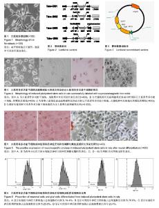
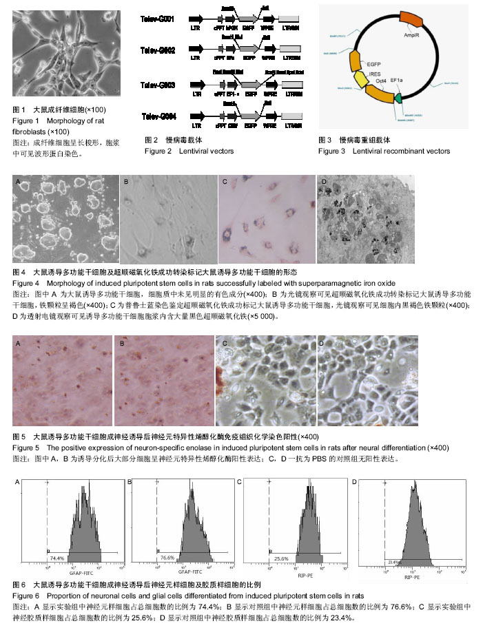
2.1 荧光显微镜观察分离培养获得的成纤维细胞 取传代后48 h的大鼠成纤维细胞,荧光显微镜观察可见细胞贴壁生长,呈梭形且生长速度快(图1),胞浆中可见波形蛋白染色,获得的成纤维细胞纯度约为96%。 2.2 慢病毒基因重组载体系统的构建 分别用EcoRⅠ酶切慢病毒载体(图2)和含有目的基因(Oct4,Sox2,Klf4和c-Myc)的质粒,割胶回收线性化的载体及含有目的基因的质粒片段。将包含有目的基因的质粒片段与该慢病毒载体连接,得到慢病毒基因重组载体(图3)。将含有目的基因的慢病毒基因重组载体转染到293T细胞中获得大量能够表达该重组载体的高质量病毒。 2.3 标记前后iPSCs的形态 取培养的iPSCs,吸掉细胞培养基,用PBS冲洗,胰酶消化,在荧光显微镜下观察,此时的iPSCs细胞质中未见明显的有色成分(图4A);在光学显微镜下观察SPIO转染标记的iPSCs,此时的iPSCs细胞质中局部染色,呈浅褐色(图4B)。SPIO标记细胞行普鲁士蓝染色后,光镜观察可见细胞质内大量蓝色致密颗粒(图4C),iPSCs的磁化标记有效率达95%以上(SPIO标记阳性率=阳性细胞数/细胞总数×100%)。透射电镜观察可见iPSCs胞浆内含大量黑色超顺磁氧化铁(图4D)。 2.4 免疫组化检测神经元细胞标记物神经元特异性烯醇化酶的阳性表达 诱导分化后大部分细胞呈神经元特异性烯醇化酶阳性表达,iPSCs诱导分化率为(82.28±17.72)%,见图5A,B,而使用一抗为PBS的对照组则无阳性表达,见图5C,D。 2.5 流式细胞仪检测分化为神经元样细胞及胶质样细胞的比例 实验组的神经元样细胞占总细胞的比例为74.4%,对照组的神经元样细胞占总细胞的比例为76.6%,两组差异无显著性意义(P > 0.05)。实验组的胶质样细胞和对照组的胶质样细胞占总细胞的比例分别为25.6%和23.4%,两组差异无显著性意义(P > 0.05)。见图6。"
| [1] Nakamura M, Okano H. Cell transplantation therapies for spinal cord injury focusing on induced pluripotent stem cells. Cell Res. 2013;23(1):70-80.[2] Malik N, Rao MS. A review of the methods for human iPSC derivation. Methods Mol Biol. 2013;997:23-33.[3] Ho MC, Huang CY, Lee JJ, et al. Generation of an induced pluripotent stem cell line, IBMS-iPSC-014-05, from a female autosomal dominant polycystic kidney disease patient carrying a common mutation of R803X in PKD2. Stem Cell Res. 2017;25:38-41.[4] Haq S, Suresh B, Ramakrishna S. Deubiquitylating enzymes as cancer stem cell therapeutics. Biochim Biophys Acta. 2017;1869(1):1-10.[5] Warren CR, Cowan CA. Humanity in a Dish: Population Genetics with iPSCs. Trends Cell Biol. 2017 Nov 23. [Epub ahead of print][6] Yoo M, Carromeu C, Kwon O, et al. The L1 adhesion molecule normalizes neuritogenesis in Rett syndrome-derived neural precursor cells. Biochem Biophys Res Commun. 2017; 494(3-4):504-510. [7] Geis FK, Galla M, Hoffmann D, et al. Potent and reversible lentiviral vector restriction in murine induced pluripotent stem cells. Retrovirology. 2017;14(1):48.[8] Barczewska M, Wojtkiewicz J, Habich A, et al. MR monitoring of minimally invasive delivery of mesenchymal stem cells into the porcine intervertebral disc. PLoS One. 2013;8(9):e74658.[9] Wang J, Xie J, Zhou X, et al. Ferritin enhances SPIO tracking of C6 rat glioma cells by MRI. Mol Imaging Biol. 2011;13(1): 87-93.[10] Cromer Berman SM, Kshitiz, Wang CJ, et al. Cell motility of neural stem cells is reduced after SPIO-labeling, which is mitigated after exocytosis. Magn Reson Med. 2013;69(1): 255-262.[11] 马艳,张德清,陈乐,等.超顺磁性氧化铁纳米粒子标记人骨髓和脐带间充质干细胞[J].中国组织工程研究,2012,16(45): 8398-8405.[12] Kim JB, Zaehres H, Wu G, et al. Pluripotent stem cells induced from adult neural stem cells by reprogramming with two factors. Nature. 2008;454(7204):646-650.[13] Liu H, Zhu F, Yong J, et al. Generation of induced pluripotent stem cells from adult rhesus monkey fibroblasts. Cell Stem Cell. 2008;3(6):587-590.[14] Li XX, Li KA, Qin JB, et al. In vivo MRI tracking of iron oxide nanoparticle-labeled human mesenchymal stem cells in limb ischemia. Int J Nanomedicine. 2013;8:1063-1073.[15] Qin JB, Li KA, Li XX, et al. Long-term MRI tracking of dual-labeled adipose-derived stem cells homing into mouse carotid artery injury. Int J Nanomedicine. 2012;7:5191-5203.[16] Schweizer PA, Darche FF, Ullrich ND, et al. Subtype-specific differentiation of cardiac pacemaker cell clusters from human induced pluripotent stem cells. Stem Cell Res Ther. 2017; 8(1):229.[17] Kim IG, Gil CH, Seo J, et al. Mechanotransduction of human pluripotent stem cells cultivated on tunable cell-derived extracellular matrix. Biomaterials. 2018;150:100-111.[18] Weiss MJ, Orkin SH. In vitro differentiation of murine embryonic stem cells. New approaches to old problems. J Clin Invest. 1996;97(3):591-595.[19] Li D, Ni H, Rui Q, et al. Deletion ofMst1 attenuates neuronal loss andimproves neurological impairment inarat model oftraumatic brain injury. Brain Res. 2017 Oct 17. [Epub ahead of print][20] Wang X, Wang D, Zhou Z, et al. Subacute oral toxicity assessment of benalaxyl in mice based on metabolomics methods. Chemosphere. 2018;191:373-380.[21] Sivolap YP, Damulin IV, Voskresenskaya ON. Traumatic brain injury: neurologic and psychiatric aspects. Zh Nevrol Psikhiatr Im S S Korsakova. 2017;117(9):94-98.[22] Donnelly K, Donnelly JP, Warner GC, et al. Longitudinal study of objective and subjective cognitive performance and psychological distress in OEF/OIF Veterans with and without traumatic brain injury. Clin Neuropsychol. 2017. [Epub ahead of print][23] Wilkinson AA, Dennis M, Taylor MJ, et al. Performance Monitoring in Children Following Traumatic Brain Injury Compared to Typically Developing Children. Child Neurol Open. 2017;4:2329048X17732713.[24] Zheng X, Xie Q, Li S, et al. CXCR4-positive subset of glioma is enriched for cancer stem cells. Oncol Res. 2011;19(12): 555-561.[25] Qin J, Li K, Peng C, et al. MRI of iron oxide nanoparticle-labeled ADSCs in a model of hindlimb ischemia. Biomaterials. 2013;34(21):4914-4925.[26] Kubiak JZ, Ciemerych MA. From Gurdon to Yamanaka--a brief history of cell reprogramming. Postepy Biochem. 2013; 59(2):124-130.[27] Menon DK, Schwab K, Wright DW, et al. Position statement: definition of traumatic brain injury. Arch Phys Med Rehabil. 2010;91(11):1637-1640.[28] Hyder AA, Wunderlich CA, Puvanachandra P, et al. The impact of traumatic brain injuries: a global perspective. NeuroRehabilitation. 2007;22(5):341-353.[29] Asemota AO, George BP, Bowman SM, et al. Causes and trends in traumatic brain injury for United States adolescents. J Neurotrauma. 2013;30(2):67-75.[30] Takahashi K, Yamanaka S. Induction of pluripotent stem cells from mouse embryonic and adult fibroblast cultures by defined factors. Cell. 2006;126(4):663-676.[31] Csöbönyeiová M, Danišovi? ?, Polák Š.Induced pluripotent stem cells for modeling and cell therapy of Parkinson's disease. Neural Regen Res. 2016;11(5):727-728.[32] Petri-Fink A, Rothen-Rutishauser B. Nanoparticles and cells: an interdisciplinary approach. Chimia (Aarau). 2012;66(3): 104-109.[33] Wang C, Ravi S, Martinez GV, et al. Dual-purpose magnetic micelles for MRI and gene delivery. J Control Release. 2012; 163(1):82-92.[34] Edlow BL, Chatelle C, Spencer CA, et al. Early detection of consciousness in patients with acute severe traumatic brain injury. Brain. 2017;140(9):2399-2414.[35] Keselman P, Yu EY, Zhou XY, et al. Tracking short-term biodistribution and long-term clearance of SPIO tracers in magnetic particle imaging. Phys Med Biol. 2017;62(9): 3440-3453.[36] Ngen EJ, Wang L, Gandhi N, et al. A preclinical murine model for the early detection of radiation-induced brain injury using magnetic resonance imaging and behavioral tests for learning and memory: with applications for the evaluation of possible stem cell imaging agents and therapies. J Neurooncol. 2016; 128(2):225-233.[37] Neubert J, Glumm J.Promoting neuronal regeneration using extracellular vesicles loaded with superparamagnetic iron oxide nanoparticles.Neural Regen Res. 2016;11(1):61-63. |
| [1] | Zhang Tongtong, Wang Zhonghua, Wen Jie, Song Yuxin, Liu Lin. Application of three-dimensional printing model in surgical resection and reconstruction of cervical tumor [J]. Chinese Journal of Tissue Engineering Research, 2021, 25(9): 1335-1339. |
| [2] | Zeng Yanhua, Hao Yanlei. In vitro culture and purification of Schwann cells: a systematic review [J]. Chinese Journal of Tissue Engineering Research, 2021, 25(7): 1135-1141. |
| [3] | Xu Dongzi, Zhang Ting, Ouyang Zhaolian. The global competitive situation of cardiac tissue engineering based on patent analysis [J]. Chinese Journal of Tissue Engineering Research, 2021, 25(5): 807-812. |
| [4] | Wu Zijian, Hu Zhaoduan, Xie Youqiong, Wang Feng, Li Jia, Li Bocun, Cai Guowei, Peng Rui. Three-dimensional printing technology and bone tissue engineering research: literature metrology and visual analysis of research hotspots [J]. Chinese Journal of Tissue Engineering Research, 2021, 25(4): 564-569. |
| [5] | Chang Wenliao, Zhao Jie, Sun Xiaoliang, Wang Kun, Wu Guofeng, Zhou Jian, Li Shuxiang, Sun Han. Material selection, theoretical design and biomimetic function of artificial periosteum [J]. Chinese Journal of Tissue Engineering Research, 2021, 25(4): 600-606. |
| [6] | Liu Fei, Cui Yutao, Liu He. Advantages and problems of local antibiotic delivery system in the treatment of osteomyelitis [J]. Chinese Journal of Tissue Engineering Research, 2021, 25(4): 614-620. |
| [7] | Li Xiaozhuang, Duan Hao, Wang Weizhou, Tang Zhihong, Wang Yanghao, He Fei. Application of bone tissue engineering materials in the treatment of bone defect diseases in vivo [J]. Chinese Journal of Tissue Engineering Research, 2021, 25(4): 626-631. |
| [8] | Zhang Zhenkun, Li Zhe, Li Ya, Wang Yingying, Wang Yaping, Zhou Xinkui, Ma Shanshan, Guan Fangxia. Application of alginate based hydrogels/dressings in wound healing: sustained, dynamic and sequential release [J]. Chinese Journal of Tissue Engineering Research, 2021, 25(4): 638-643. |
| [9] | Chen Jiana, Qiu Yanling, Nie Minhai, Liu Xuqian. Tissue engineering scaffolds in repairing oral and maxillofacial soft tissue defects [J]. Chinese Journal of Tissue Engineering Research, 2021, 25(4): 644-650. |
| [10] | Xing Hao, Zhang Yonghong, Wang Dong. Advantages and disadvantages of repairing large-segment bone defect [J]. Chinese Journal of Tissue Engineering Research, 2021, 25(3): 426-430. |
| [11] | Chen Siqi, Xian Debin, Xu Rongsheng, Qin Zhongjie, Zhang Lei, Xia Delin. Effects of bone marrow mesenchymal stem cells and human umbilical vein endothelial cells combined with hydroxyapatite-tricalcium phosphate scaffolds on early angiogenesis in skull defect repair in rats [J]. Chinese Journal of Tissue Engineering Research, 2021, 25(22): 3458-3465. |
| [12] | Wang Hao, Chen Mingxue, Li Junkang, Luo Xujiang, Peng Liqing, Li Huo, Huang Bo, Tian Guangzhao, Liu Shuyun, Sui Xiang, Huang Jingxiang, Guo Quanyi, Lu Xiaobo. Decellularized porcine skin matrix for tissue-engineered meniscus scaffold [J]. Chinese Journal of Tissue Engineering Research, 2021, 25(22): 3473-3478. |
| [13] | Mo Jianling, He Shaoru, Feng Bowen, Jian Minqiao, Zhang Xiaohui, Liu Caisheng, Liang Yijing, Liu Yumei, Chen Liang, Zhou Haiyu, Liu Yanhui. Forming prevascularized cell sheets and the expression of angiogenesis-related factors [J]. Chinese Journal of Tissue Engineering Research, 2021, 25(22): 3479-3486. |
| [14] | Liu Chang, Li Datong, Liu Yuan, Kong Lingbo, Guo Rui, Yang Lixue, Hao Dingjun, He Baorong. Poor efficacy after vertebral augmentation surgery of acute symptomatic thoracolumbar osteoporotic compression fracture: relationship with bone cement, bone mineral density, and adjacent fractures [J]. Chinese Journal of Tissue Engineering Research, 2021, 25(22): 3510-3516. |
| [15] | Liu Liyong, Zhou Lei. Research and development status and development trend of hydrogel in tissue engineering based on patent information [J]. Chinese Journal of Tissue Engineering Research, 2021, 25(22): 3527-3533. |
| Viewed | ||||||
|
Full text |
|
|||||
|
Abstract |
|
|||||