Chinese Journal of Tissue Engineering Research ›› 2018, Vol. 22 ›› Issue (6): 932-937.doi: 10.3969/j.issn.2095-4344.0071
Previous Articles Next Articles
Preparation and performance detection of small-diameter tissue-engineered blood vessels
- Department of Cardiac Surgery, Beijing Anzhen Hospital, Capital Medical University, Beijing 100029, China
-
Received:2017-09-23Online:2018-02-28Published:2018-02-28 -
Contact:Li Wen-bin, Chief physician, Department of Cardiac Surgery, Beijing Anzhen Hospital, Capital Medical University, Beijing 100029, China -
About author:Ma Xiao-long, M.D., Physician, Department of Cardiac Surgery, Beijing Anzhen Hospital, Capital Medical University, Beijing 100029, China
CLC Number:
Cite this article
Ma Xiao-long, Li Wen-bin, Xin Zhi-fei, Li Dian-kun, Zhou Zi-fan, Wan Ju-yi, Wang Jian-gang. Preparation and performance detection of small-diameter tissue-engineered blood vessels[J]. Chinese Journal of Tissue Engineering Research, 2018, 22(6): 932-937.
share this article
Add to citation manager EndNote|Reference Manager|ProCite|BibTeX|RefWorks
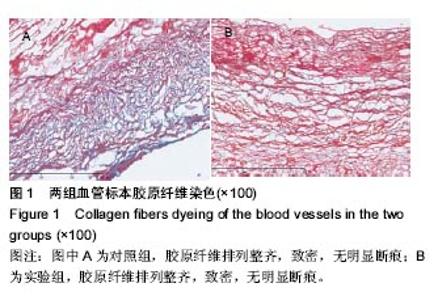
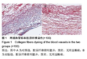
2.1 血管胶原纤维染色结果 对照组胶原纤维排列整齐,致密,无明显断痕;实验组胶原纤维排列整齐,致密,无明显断痕,见图1。"
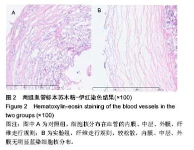
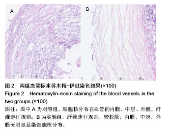
2.2 血管苏木精-伊红染色观察结果 对照组蓝色的细胞核分布在血管的内膜、中层、外膜,纤维走行规则;实验组纤维走行规则,较松散,内膜、中层、外膜无明显蓝染细胞核分布,内膜蓝染区为未洗涤干净的染剂,见图2。"
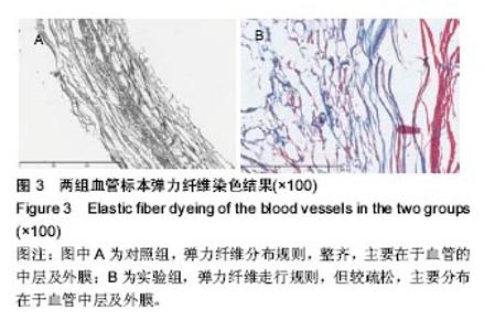
2.3 血管弹力纤维染色结果 对照组弹力纤维分布规则,整齐,主要在于血管的中层及外膜;实验组弹力纤维走行规则,但较疏松,主要分布在于血管中层及外膜,见图3。"


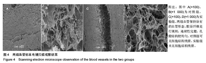
2.4 血管电镜扫描结果 经冷冻干燥后处理,电镜扫描下可见两组血管保持原有的血管形态,胶原纤维走行规则,连续性完整,孔隙结构较均匀;对照组可见细胞结构残留,实验组未见细胞结构残留,见图4。 2.5 血管厚度及拉力强度检测结果 厚度测试结构显示:对照组、实验组血管平均厚度分别为1.29,1.02 mm,组间比较差异有显著性意义 (P < 0.01),说明经过脱细胞处理血管较未经处理血管壁组成成分有一定程度的减少。 拉力强度测试结果:对照组、实验组血管平均拉力强度分别为2.96,3.04 N/mm2,组间比较差异无显著性意义(P > 0.05),说明经脱细胞处理血管与未经处理血管在抗张力强度上无明显差异。 2.6 血管压力承受值测试结果 压力测试发现:两组血管均可承受46.55 kPa压力值,实验组血管在该压力下大体观察可见轻度假瘤样扩张;在压力达到53.2 kPa时,对照组血管呈瘤样扩张,实验组血管破裂。 "
| [1]Sukovatykh BS,Belikov LN,Sukovatykh MB,et al.Results of using various methods of autovenous femoropopliteal bypass grafting below the knee-joint fissure.Angiol Sosud Khir. 2016; 22(4):137-143.[2]Ramanathan G,Singaravelu S,Muthukumar T,et al.Design and characterization of 3D hybrid collagen matrixes as a dermal substitute in skin tissue engineering.Mater Sci Eng C Mater Biol Appl. 2017;3(1);72:359-370.[3]Frueh FS,Menger MD,Lindenblatt N,et al.Current and emerging vascularization strategies in skin tissue engineering.Crit Rev Biotechnol.2017;37(5):613-625.[4]Busra MF,Chowdhury SR,bin Ismail F,et al. Tissue-Engineered skin substitute enhances wound healing after radiation therapy.Adv Skin Wound Care. 2016;29(3):120-129.[5]Waller CJ,Cogbill TH,Kallies KJ,et al.Contemporary management of subclavian and axillary artery injuries - A Western Trauma Association Multicenter Review.J Trauma Acute Care Surg.2017.doi: 10.1097/TA.0000000000001645.[Epub ahead of print][6]Valverde García S,Peña Cortés R,Torre Eiriz J et al.Disruption in "non-anastomotic section" of an axillofemoral bypass. A thorough critical review of the literature.[J] Ann Vasc Surg. 2017.7.13. pii: S0890-5096(17)30846-4. doi:10.1016/j.avsg.[7]Bakkali T,Zoulati M,Aghoutane N,Bounssir A,et al.False aneurysm of the carotid artery revealing Behçet disease.J Med Vasc.2017;42(3):185-188.[8]Heger T,Strauß S,Blessing E,et al.Short and long-term results after endovascular management of vascular complications during transfemoral aortic valve implantation.Acta Cardiol. 2017:1-9.[9]Li YH,Wang JS,Yao C,et al.Risk factors of rupture of internal carotid artery during surgical resection of carotid body tumor.Zhonghua Yi Xue Za Zhi.2017;97(22):1724-1728.[10]Hwang PT,Lim DJ,Fee T,et al.A bio-inspired hybrid nanosack for graft vascularization at the omentum.Acta Biomater. 2016;41:224-234.[11]Hulin-Curtis S,Williams H,Wadey KS,et al.Targeting Wnt/β-Catenin Activated Cells with Dominant-Negative N-cadherin to Reduce Neointima Formation.Mol Ther Methods Clin Dev.2017;5:191-199.[12]Yasuda N,Goto K,Ohchi Y,et al.The efficacy and safety of antithrombin and recombinant human thrombomodulin combination therapy in patients with severe sepsis and disseminated intravascular coagulation.J Crit Care. 2016;36: 29-34. [13]Renal Autotransplantation with Autologous Saphenous Vein Graft in a Patient with Takayasu Arteritis and Existing Renal Artery Stent in Her Solitary Kidney.Urol Int.2017.doi: 10.1159/000475509.[Epub ahead of print].[14]He Y,Xu X,Zhu T,et al.Resident Arterial Cells and Circulating Bone Marrow-Derived Cells both Contribute to IntimalHyperplasia in a Rat Allograft Carotid Transplantation Model.Cell Physiol Biochem.2017;42(4):1303-1312.[15]Good BC,Weiss WJ,Deutsch S,et al.Asynchronous Pumping of a Pulsatile Ventricular Assist Device in a Pediatric Anastomosis Model.World J Pediatr Congenit Heart Surg. 2017;8(4):511-519.[16]Tepe G,Gögebakan Ö,Redlich U,et al.Angiographic and Clinical Outcomes After Treatment of Femoro-Popliteal Lesions with a Novel Paclitaxel-Matrix-Coated Balloon Catheter.Cardiovasc Intervent Radiol.2017.10.1007/ s00270-017-1713-2.[Epub ahead of print][17]Baek SE,Jang MA,Lee SJ,et al.5-Lipoxygenase in monocytes emerges as a therapeutic target for intimal hyperplasia in a murine wire-injured femoral artery.Biochim Biophys Acta. 2017;1863(9):2210-2221.[18]Guo H,Zhang W,Wang Y,et al.A good water soluble Lillie-Mayer hematoxylin and eosin Y staining solution.Diag Path J.2015;22(10):223-226.[19]Song W,Wang J.The study on improved old tissue sections HE staining method by using GS staining.Diag Path J. 2015; 11:1167-1172.[20]Zhang YD,Zhao SC,Zhu ZS,et al.Cx43- and Smad-Mediated TGF/ BMP Signaling Pathway Promotes Cartilage Differentiation of Bone Marrow Mesenchymal Stem Cells and Inhibits Osteoblast Differentiation.Cell Physiol Biochem. 2017; 42(4):1277-1293.[21]Li Y,Zhang J,Xu Y,et al.Effects of 630 nm Red and 460 nm Blue Light Emitting Diode Irradiation on Healing of the Skin Wound in Japanese Big-ear White Rabbit.Chin Med Sci J. 2017;39(3):301-306.[22]Tingting D,Na L,Bin G,et al.Effects of Aging on the Proliferation and Differentiation Capacity of Human Periodontal Ligament Stem Cells.Chin Med Sci J. 2017;32(2): 83-91.[23]Chong RS,Lee YS,Chu SWL,et al.Inhibition of Monocyte Chemoattractant Protein 1 Prevents Conjunctival Fibrosis in an Experimental Model of Glaucoma Filtration Surgery.Invest Ophthalmol Vis Sci.2017;58(9):3432-3439. [24]Tang Z,Niu J,Huang H ,et al.Potential biodegradable Zn-Cu binary alloys developed for cardiovascular implant applications.J Mech Behav Biomed Mater.2017;72:182-191. [25]Davis LA,Stewart SE,Carsten CG 3rd,et al.Characterization of fracture behavior of human atherosclerotic fibrous caps using a miniature single edge notched tensile test.Acta Biomater. 2016;43:101.[26]Stephen S,Agnihotri M,Kaur S.A Randomized, Controlled Trial to Assess the Effect of Topical Insulin Versus Normal Saline in Pressure Ulcer Healing.Ostomy Wound Manage. 2016;62(6): 16-23.[27]Roth SP,Erbe I,Burk J.Decellularization of Large Tendon Specimens: Combination of Manually Performed Freeze-Thaw Cycles and Detergent Treatment.Methods Mol Biol.2017.doi: 10.1007/7651_2017_49. [Epub ahead of print][28]McFerrin MB,Chi X,Cutter G,et al.Dysregulation of 14-3-3 proteins in neurodegenerative diseases with Lewy body or Alzheimer pathology.Ann Clin Transl Neurol. 2017;4(7): 466-477.[29]Wang W,Shao A,Feng S,et al.Physicochemical characterization and gastrointestinal adhesion of S-layer proteins-coating liposomes.Int J Pharm. 2017;529(1-2): 227-237.[30]Wang H,Fan S,Li L,et al.Preparation of human umbilical vein acellular matrix.China Tissue Engineering research and clinical rehabilitation.2007;11:335-337.[31]Xie H,Campbell CE,Shaffer BS,et a1.Different outcomes in urethral reconstruction using elastin and collagen patches and conduits in rabbits.J Biomed Mater Res B Appl Biomater. 2007;11(8):116-120.[32]Im SH,Kim CY,Jung Y,et al.Biodegradable vascular stents with high tensile and compressive strength: a novel strategy for applying monofilaments via solid-state drawing and shaped-annealing processes.Biomater Sci. 2017;5(3): 422-431.[33]Masselter T,Haushahn T,Fink S,et al.Biomechanics of selected arborescent and shrubby monocotyledons.Beilstein J Nanotechnol.2016;7:1602-1619.[34]Chaves C,Gao C,Hunckler J,et al.Dual-acting biofunctionalised scaffolds for applications in regenerative medicine.J Mater Sci Mater Med.2017;28(2):32.[35]Khurshid S,Afzal M,Khalid R,et al.Potential of multi-component antigens for tuberculosis diagnosis. Biologicals.2017;48:109-113.[36]Feavers IM,Maiden MCJ.Recent Progress in the Prevention of Serogroup B Meningococcal Disease.Clin Vaccine Immunol.2017;24(5):pii: e00566-16.doi: 10.1128/CVI.00566-16.[37]Easterhoff D,Moody MA,Fera D,et al.Boosting of HIV envelope CD4 binding site antibodies with long variable heavy third complementarity determining region in the randomized double blind RV305 HIV-1 vaccine trial.PLoS Pathog.2017;13(2):e1006182.[38]Donald RG,Hawkins JC,Hao L,et al.Meningococcal serogroup B vaccines: Estimating breadth of coverage.Hum Vaccin Immunother.2017;13(2):255-265.[39]Qin W,Wang L,Zhai R,et al.Apa2H1, the first head domain of Apa2 trimeric autotransporter adhesin, activates mouse bone marrow-derived dendritic cells and immunization with Apa2H1 protects against Actinobacillus pleuropneumoniae infection. Mol Immunol.2017;81:108-117.[40]Navarrete-Perea J,Moguel B,Bobes RJ,et al.Protein profiles of Taenia solium cysts obtained from skeletal muscles and the central nervous system of pigs: Search for tissue-specific proteins.Exp Parasitol.2017;172:23-29.[41]Veljkovic V,Veljkovic N,Paessler S,et al.Predicted Enhanced Human Propensity of Current Avian-Like H1N1 Swine Influenza Virus from China.PLoS One.2016;11(11):e0165451.[42]Hayward CJ,Fradette J,Morissette Martin P,et al.Using human umbilical cord cells for tissue engineering: A comparison with skin cells.Differentiation 2014;87(3-4):172-181.[43]Bao RM,Yang HM,Yu CM,et al.An efficient protocol to enhance the extracellular production of recombinant protein from Escherichia coli by the synergistic effects of sucrose, glycine, and Triton X-100.Prot Expr Puri.2016;126(10):9-15. |
| [1] | Zhang Tongtong, Wang Zhonghua, Wen Jie, Song Yuxin, Liu Lin. Application of three-dimensional printing model in surgical resection and reconstruction of cervical tumor [J]. Chinese Journal of Tissue Engineering Research, 2021, 25(9): 1335-1339. |
| [2] | Zeng Yanhua, Hao Yanlei. In vitro culture and purification of Schwann cells: a systematic review [J]. Chinese Journal of Tissue Engineering Research, 2021, 25(7): 1135-1141. |
| [3] | Xu Dongzi, Zhang Ting, Ouyang Zhaolian. The global competitive situation of cardiac tissue engineering based on patent analysis [J]. Chinese Journal of Tissue Engineering Research, 2021, 25(5): 807-812. |
| [4] | Wu Zijian, Hu Zhaoduan, Xie Youqiong, Wang Feng, Li Jia, Li Bocun, Cai Guowei, Peng Rui. Three-dimensional printing technology and bone tissue engineering research: literature metrology and visual analysis of research hotspots [J]. Chinese Journal of Tissue Engineering Research, 2021, 25(4): 564-569. |
| [5] | Chang Wenliao, Zhao Jie, Sun Xiaoliang, Wang Kun, Wu Guofeng, Zhou Jian, Li Shuxiang, Sun Han. Material selection, theoretical design and biomimetic function of artificial periosteum [J]. Chinese Journal of Tissue Engineering Research, 2021, 25(4): 600-606. |
| [6] | Liu Fei, Cui Yutao, Liu He. Advantages and problems of local antibiotic delivery system in the treatment of osteomyelitis [J]. Chinese Journal of Tissue Engineering Research, 2021, 25(4): 614-620. |
| [7] | Li Xiaozhuang, Duan Hao, Wang Weizhou, Tang Zhihong, Wang Yanghao, He Fei. Application of bone tissue engineering materials in the treatment of bone defect diseases in vivo [J]. Chinese Journal of Tissue Engineering Research, 2021, 25(4): 626-631. |
| [8] | Zhang Zhenkun, Li Zhe, Li Ya, Wang Yingying, Wang Yaping, Zhou Xinkui, Ma Shanshan, Guan Fangxia. Application of alginate based hydrogels/dressings in wound healing: sustained, dynamic and sequential release [J]. Chinese Journal of Tissue Engineering Research, 2021, 25(4): 638-643. |
| [9] | Chen Jiana, Qiu Yanling, Nie Minhai, Liu Xuqian. Tissue engineering scaffolds in repairing oral and maxillofacial soft tissue defects [J]. Chinese Journal of Tissue Engineering Research, 2021, 25(4): 644-650. |
| [10] | Xing Hao, Zhang Yonghong, Wang Dong. Advantages and disadvantages of repairing large-segment bone defect [J]. Chinese Journal of Tissue Engineering Research, 2021, 25(3): 426-430. |
| [11] | Chen Siqi, Xian Debin, Xu Rongsheng, Qin Zhongjie, Zhang Lei, Xia Delin. Effects of bone marrow mesenchymal stem cells and human umbilical vein endothelial cells combined with hydroxyapatite-tricalcium phosphate scaffolds on early angiogenesis in skull defect repair in rats [J]. Chinese Journal of Tissue Engineering Research, 2021, 25(22): 3458-3465. |
| [12] | Wang Hao, Chen Mingxue, Li Junkang, Luo Xujiang, Peng Liqing, Li Huo, Huang Bo, Tian Guangzhao, Liu Shuyun, Sui Xiang, Huang Jingxiang, Guo Quanyi, Lu Xiaobo. Decellularized porcine skin matrix for tissue-engineered meniscus scaffold [J]. Chinese Journal of Tissue Engineering Research, 2021, 25(22): 3473-3478. |
| [13] | Mo Jianling, He Shaoru, Feng Bowen, Jian Minqiao, Zhang Xiaohui, Liu Caisheng, Liang Yijing, Liu Yumei, Chen Liang, Zhou Haiyu, Liu Yanhui. Forming prevascularized cell sheets and the expression of angiogenesis-related factors [J]. Chinese Journal of Tissue Engineering Research, 2021, 25(22): 3479-3486. |
| [14] | Liu Chang, Li Datong, Liu Yuan, Kong Lingbo, Guo Rui, Yang Lixue, Hao Dingjun, He Baorong. Poor efficacy after vertebral augmentation surgery of acute symptomatic thoracolumbar osteoporotic compression fracture: relationship with bone cement, bone mineral density, and adjacent fractures [J]. Chinese Journal of Tissue Engineering Research, 2021, 25(22): 3510-3516. |
| [15] | Liu Liyong, Zhou Lei. Research and development status and development trend of hydrogel in tissue engineering based on patent information [J]. Chinese Journal of Tissue Engineering Research, 2021, 25(22): 3527-3533. |
| Viewed | ||||||
|
Full text |
|
|||||
|
Abstract |
|
|||||