Chinese Journal of Tissue Engineering Research ›› 2018, Vol. 22 ›› Issue (6): 877-882.doi: 10.3969/j.issn.2095-4344.0062
Previous Articles Next Articles
Application of microsutures with vascular endothelial growth factor to improve vascular endothelial regeneration after small vessel anastomosis in the rats
- Department of Hands and Feet Microsurgery, Dalian Municipal Central Hospital, Dalian 116033, Liaoning Province, China
-
Received:2017-09-14Online:2018-02-28Published:2018-02-28 -
Contact:Zhong Sheng, Master, Attending physician, Department of Hands and Feet Microsurgery, Dalian Municipal Central Hospital, Dalian 116033, Liaoning Province, China -
About author:Zhang Tie-hui, Chief physician, Professor, Department of Hands and Feet Microsurgery, Dalian Municipal Central Hospital, Dalian 116033, Liaoning Province, China -
Supported by:the Project of Dalian Science and Technology Department, No. 2015E21SF008
CLC Number:
Cite this article
Zhang Tie-hui, Liang Wu, Ren Yuan-fei, Dong Yu-jin, Yang Wen-feng, Shang Yao-hua, Li Ju-tao, Zhong Sheng. Application of microsutures with vascular endothelial growth factor to improve vascular endothelial regeneration after small vessel anastomosis in the rats[J]. Chinese Journal of Tissue Engineering Research, 2018, 22(6): 877-882.
share this article
Add to citation manager EndNote|Reference Manager|ProCite|BibTeX|RefWorks

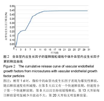
2.1 检测血管内皮因子缓释微粒大小 激光粒度分析仪测量测试15份微粒的粒径范围为83-121 nm,平均粒径为(104.25±17.03)nm。其中1份样本测量直径为53.27 μm,考虑原因为多个微粒聚合而成,予以排除该样本。 2.2 微粒中血管内皮因子的体外释放检测 孵育1 d时,微粒中的血管内皮生长因子释放量为15.54%,表现为爆发性释放,随后释放速度有所减慢,在第5天后又有一个快速释放期,但速度小于第一个快速释放期,第8天以后呈持续缓慢释放,第13天开始每日释放量明显减少并波动不大,第20天开始无明显释放量,后连续监测10 d 维持于43.71%,连续监测30 d最大累积值为43.71%(图2)。"
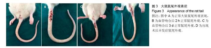
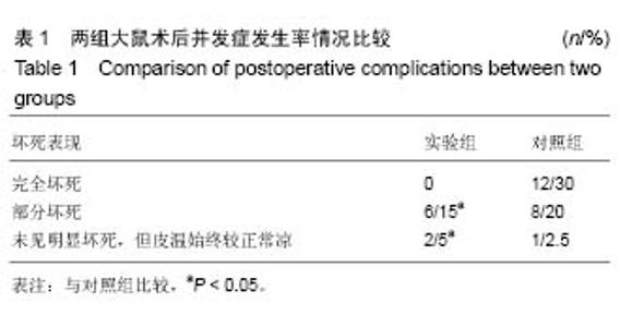
2.3 实验动物数量分析 90只大鼠均造模成功,其中4只在实验过程中死于建模后感染,予以剔除,并补充重新制作动物模型,最终90只大鼠进入结果分析。 2.4 两组大鼠术后尾部大体观察分析 正常大鼠鼠尾颜色红润,皮温暖,活动度良好。血管吻合后,若血管再通正常表现大鼠颜色、温度没有变化。实验组大鼠共有8只发生术后并发症:术后2 h,仅有5只鼠尾发生动脉缺血表现,鼠尾远端苍白,皮温凉;术后3 d,有3只鼠尾发生动脉缺血表现;至术后7 d时,上述8只鼠尾中,6只部分坏死,2只未见明显坏死,但皮温始终较正常凉。对照组大鼠共有21只发生术后并发症:术后2 h,16只动脉缺血表现;术后3 d时,新添5只发生动脉缺血表现;至术后7 d时,上述鼠尾完全坏死12只,部分坏死8只,1只未见明显坏死,但皮温始终较正常凉。实验组术后并发症发生率明显小于对照组(P < 0.05),病情程度也明显轻于对照组,见图3、表1。"

"

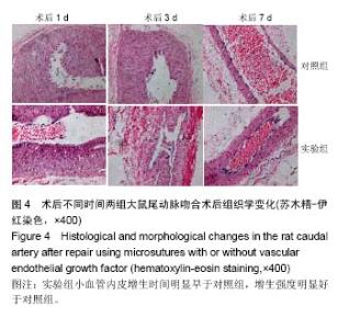
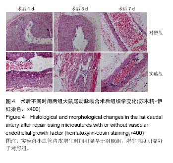
2.5 两组大鼠术后小动脉内皮细胞变化情况 ①对照组:血管吻合术后1 d,内皮细胞肿胀,部分脱落,内皮下层呈细胞浸润及创伤性增生反应,内弹性膜层消失,平滑肌细胞发生变性坏死,外膜层细胞浸润并呈创伤性增生反应;术后3 d,内皮细胞脱落区出现新生内皮细胞并出现生长移行,使吻合口开始有内皮细胞覆盖;术后5-7 d,新生的内皮细胞爬过吻合口裂隙并覆盖缝线;②实验组:血管吻合术后1 d,吻合口缝线附近可见增生的内皮细胞。术后3 d,小血管吻合口两端缝线附近可见大量增生的内皮细胞和内皮下组织并向管腔突出,形成突起,并完成覆盖缝线;术后7 d,内皮细胞及内弹性膜修复完全恢复正常,平滑肌细胞进一步增生,外膜恢复正常,实验组小血管内皮增生时间明显早于对照组,增生强度明显好于对照组(图4)。"
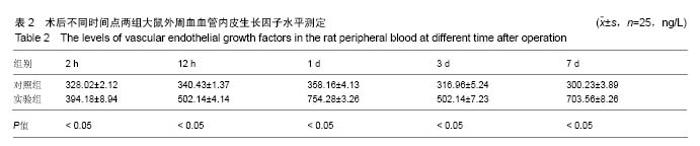
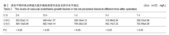
2.6 大鼠外周血血管内皮生长因子测定 实验组术后各时间点外周血血管内皮生长因子水平均高于对照组(P < 0.05);实验组术后1 d为爆发性释放,随后释放速度有所减慢,在第7天左右又有一个快速释放期,但速度小于第一个快速释放期并波动不大,与体外缓释微粒释放情况大致一致;对照组术后1 d外周血血管内皮生长因子水平也有所增加,但其增加量明显小于实验组,且随着时间点延长,血管内皮生长因子水平逐渐减少,见表2。"
| [1]Zhang L,Si T,Fischer AJ,et al.Coaxial Electrospray of Ranibizumab- Loaded Microparticles for Sustained Release of Anti-VEGF Therapies. PLoS One.2015;10(8):e0135608. [2]Park K.Delivery of definable numbers of PLGA microparticles within embryoid bodies.J Control Release,.2013;168(1):103. [3]Herrán E,Ruiz-Ortega JÁ,Aristieta A,et al.In vivo administration of VEGF- and GDNF-releasing biodegradable polymeric microspheres in a severe lesion model of Parkinson's disease.Eur J Pharm Biopharm. 2013;85(3 Pt B):1183-1190. [4]Simón-Yarza T,Tamayo E,Benavides C,et al.Functional benefits of PLGA particulates carrying VEGF and CoQ10 in an animal of myocardial ischemia.Int J Pharm.2013;454(2):784-790. [5]White LJ,Kirby GT,Cox HC,et al.Accelerating protein release from microparticles for regenerative medicine applications.Mater Sci Eng C Mater Biol Appl.2013;33(5):2578-2583. [6]Dhanda DS,Tyagi P,Mirvish SS,et al.Supercritical fluid technology based large porous celecoxib-PLGA microparticles do not induce pulmonary fibrosis and sustain drug delivery and efficacy for several weeks following a single dose.J Control Release.2013;168(3):239-250. [7]Qutachi O,Shakesheff KM,Buttery LD,et al.Delivery of definable number of drug or growth factor loaded poly(DL-lactic acid-co-glycolic acid microparticles within human embryonic stem cell derived aggregates.J Control Release.2013;168(1):18-27.[8]Bible E,Qutachi O,Chau DY,et al.Neo-vascularization of the stroke cavity by implantation of human neural stem cells on VEGF-releasing PLGA microparticles.Biomaterials.2012;33(30):7435-7446.[9]Simón-Yarza T,Formiga FR,Tamayo E,et al.PEGylated-PLGA microparticles containing VEGF for long term drug delivery.Int J Pharm.2013;440(1):13-18. [10]Xu H,Nguyen KT,Brilakis ES,et al.Enhanced endothelialization of a new stent polymer through surface enhancement and incorporation of growth factor-delivering microparticles.J Cardiovasc Transl Res. 2012;5(4):519-527. [11]Formiga FR,Pelacho B,Garbayo E,et al.Sustained release of VEGF through PLGA microparticles improves vasculogenesis and tissue remodeling in an acute myocardial ischemia-reperfusion model.J Control Release.2010;147(1):30-37. [12]Shahani K,Swaminathan SK,Freeman D,et al.Injectable sustained release microparticles of curcumin: a new concept for cancer chemoprevention.Cancer Res.2010;70(11):4443-4452. [13]Jay SM,Shepherd BR,Bertram JP,et al.Engineering of multifunctional gels integrating highly efficient growth factor delivery with endothelial cell transplantation.FASEB J.2008;22(8):2949-2956. [14]Amrite AC,Ayalasomayajula SP,Cheruvu NP,et al.Signgle periocular injection of celecoxib-PLGA microparticles inhibits diabetes-induced elevations in retinal PGE2, VEGF, and vascular leakage.Invest Ophthalmol Vis Sci.2006;47(3):1149-1160.[15]Bandi N,Ayalasomayajula SP,Dhanda DS,et al.Intratracheal budesonide-poly(lactide-co-glycolide) microparticles reduce oxidative stress,VEGFexpression, and vascular leakage in a benzo(a)pyrene-fed mouse model.J Pharm Pharmacol.2005;57(7):851-860.[16]Chen Y,Luo X,Xiao J,et al.Experimental study on polylactic glycolate acid microparticles with releasing-slowly vascular endothelial growth factor.Zhongguo Xiu Fu Chong Jian Wai Ke Za Zhi. 2005;19(4):262-263. [17]Ayalasomayajula SP,Kompella UB.Subconjunctivally administered celecoxib-PLGA microparticles sustain retinal drug levels and alleviate diabetes-induced oxidative stress in a rat model.Eur J Pharmacol. 2005;511(2-3):191-198.[18]陈扬,肖建德,李振宇,等.血管内皮生长因子缓释微粒的合成与制备[J].中国骨肿瘤骨病杂志,2006,3(3):150-151.[19]沈素祥,美小涛,张绍东,等.rhBMP-2/无定形磷酸钙纳米缓释微粒成骨活性的实验研究[J].生物医学工程与临床杂志, 2008,3(12):175-178.[20]潘兴纳,李亚雄,蒋立虹.组织工程血管支架材料的研究与进展[J].中国组织工程研究,2016,20(34):5149-5154.[21]Gaitskell K,Martinek I,Bryant A,et al.Angiogenesis inhibitors for the treatment of ovarian cancer. Cochrane Database Syst Rev.2011;(9): CD007930.[22]Kroep JR,Nortier JWR.The role of bevacizumab in advanced epithelial ovarian cancer.Curr Pharm Des.2012;18(25):3775-3783.[23]Yamazaki Y,Morita T.Molecular and functional diversity of vascular endothelial growth factors. Mol Divers. 2006;10(4):515-527.[24]Roy H,Bhardwaj S,Ylä-Herttuala S. Biology of vascular endothelial growth factors.FEBS Lett. 2006;580(12):2879-2887.[25]Jackson MR,Carney EW,Lye SJ,et al.Localization of two angiogenic growth factors (PDECGF and VEGF) in human placentae throughout gestation.Placenta.1994;15(4):341-353.[26]Wei MH,Popescu NC,Lerman MI,et al.Localization of the human vascular endothelial growth factor gene, VEGF, at chromosome 6p12.Hum Genet.1996;97(6):794–797.[27]Vincenti V,Cassano C,Rocchi M,et al.Assignment of the vascular endothelial growth factor gene to human chromosome 6p21.3. Circulation.1996;93(8):1493-1495.[28]Hsieh YY,Chang CC,Tsai FJ,et al.Tallele for VEGF-460 gene polymorphism at 5’-untranslated region is associated with higher susceptibility of leiomyoma.Biochem Genet.2008;46(5-6):356-361.[29]Feng Y,Lin X,Zhou S,et al.The associations between the polymorphisms of the ER-α gene and the risk of uterine leiomyoma(ULM).Tumour Biol.2013;34(5):3077-3082.[30]Hsieh YY,Chang CC,Tsai FJ,et al.Estrogen receptor thymine-adenine dinucleotide repeat polymorphism is associated with susceptibility to leiomyoma.Fertil Steril.2003;79(1):96-99.[31]Yaghmaei M,Salimi S,Namazi L,et al.Association of XRCC1 Arg399GIn and Tp53 Arg72Pro polymorphisms and increased risk of uterine leiomyoma-a case-control study.Genet Mol Biol. 2015;38(4): 444-449.[32]Wang F,Chen J,Wang L,et al.CYP1A1 genetic polymorphisms and uterine leiomyoma risk: a meta-analysis.Int J Clin Exp Med. 2015;8(3): 3590-3594.[33]Feng Y,Zhao X,Zhou C,et al.The associations between the Val158Met in the catechol-O-methyltransferase(COMT) gene and the risk of uterine leiomyoma(ULM). Gene.2013;529(2): 296-299.[34]Salimi S,Hajizadeh A,Khodamian M,et al.Age-dependent association of MDM2 promoter polymorphisms and uterine leiomyoma in South-East Iran: A preliminary report.J Obstet Gynaecol Res.2015; 41(5):729-734.[35]Stoner M,Wang F,Wormke M,et al.Inhibition of vascular endothelial growth factor expression in HEC1A endometrial cancer cells through interactions of estrogen receptor alpha and Sp3 proteins.J Biol Chem. 2000;275(30):22769-22779.[36]Adams J,Carder PJ,Downey S,et al.Vascular endothelial growth factor(VEGF) in breast cancer: comparison of plasma, serum, and tissue VEGF and microvessel density and effects of tamoxifen.Cancer Res.2000;60(11):2898-2905.[37]Chao Y,Li CP,Chau GY,et al.Prognostic significance of vascular endothelial growth factor, basic fibroblast growth factor, and angiogenin in patients with resectable hepatocellular carcinoma after surgery.Ann Surg Oncol.2003;10(4):355-362. |
| [1] | Zhang Tongtong, Wang Zhonghua, Wen Jie, Song Yuxin, Liu Lin. Application of three-dimensional printing model in surgical resection and reconstruction of cervical tumor [J]. Chinese Journal of Tissue Engineering Research, 2021, 25(9): 1335-1339. |
| [2] | Zeng Yanhua, Hao Yanlei. In vitro culture and purification of Schwann cells: a systematic review [J]. Chinese Journal of Tissue Engineering Research, 2021, 25(7): 1135-1141. |
| [3] | Xu Dongzi, Zhang Ting, Ouyang Zhaolian. The global competitive situation of cardiac tissue engineering based on patent analysis [J]. Chinese Journal of Tissue Engineering Research, 2021, 25(5): 807-812. |
| [4] | Wu Zijian, Hu Zhaoduan, Xie Youqiong, Wang Feng, Li Jia, Li Bocun, Cai Guowei, Peng Rui. Three-dimensional printing technology and bone tissue engineering research: literature metrology and visual analysis of research hotspots [J]. Chinese Journal of Tissue Engineering Research, 2021, 25(4): 564-569. |
| [5] | Chang Wenliao, Zhao Jie, Sun Xiaoliang, Wang Kun, Wu Guofeng, Zhou Jian, Li Shuxiang, Sun Han. Material selection, theoretical design and biomimetic function of artificial periosteum [J]. Chinese Journal of Tissue Engineering Research, 2021, 25(4): 600-606. |
| [6] | Liu Fei, Cui Yutao, Liu He. Advantages and problems of local antibiotic delivery system in the treatment of osteomyelitis [J]. Chinese Journal of Tissue Engineering Research, 2021, 25(4): 614-620. |
| [7] | Li Xiaozhuang, Duan Hao, Wang Weizhou, Tang Zhihong, Wang Yanghao, He Fei. Application of bone tissue engineering materials in the treatment of bone defect diseases in vivo [J]. Chinese Journal of Tissue Engineering Research, 2021, 25(4): 626-631. |
| [8] | Zhang Zhenkun, Li Zhe, Li Ya, Wang Yingying, Wang Yaping, Zhou Xinkui, Ma Shanshan, Guan Fangxia. Application of alginate based hydrogels/dressings in wound healing: sustained, dynamic and sequential release [J]. Chinese Journal of Tissue Engineering Research, 2021, 25(4): 638-643. |
| [9] | Chen Jiana, Qiu Yanling, Nie Minhai, Liu Xuqian. Tissue engineering scaffolds in repairing oral and maxillofacial soft tissue defects [J]. Chinese Journal of Tissue Engineering Research, 2021, 25(4): 644-650. |
| [10] | Xing Hao, Zhang Yonghong, Wang Dong. Advantages and disadvantages of repairing large-segment bone defect [J]. Chinese Journal of Tissue Engineering Research, 2021, 25(3): 426-430. |
| [11] | Chen Siqi, Xian Debin, Xu Rongsheng, Qin Zhongjie, Zhang Lei, Xia Delin. Effects of bone marrow mesenchymal stem cells and human umbilical vein endothelial cells combined with hydroxyapatite-tricalcium phosphate scaffolds on early angiogenesis in skull defect repair in rats [J]. Chinese Journal of Tissue Engineering Research, 2021, 25(22): 3458-3465. |
| [12] | Wang Hao, Chen Mingxue, Li Junkang, Luo Xujiang, Peng Liqing, Li Huo, Huang Bo, Tian Guangzhao, Liu Shuyun, Sui Xiang, Huang Jingxiang, Guo Quanyi, Lu Xiaobo. Decellularized porcine skin matrix for tissue-engineered meniscus scaffold [J]. Chinese Journal of Tissue Engineering Research, 2021, 25(22): 3473-3478. |
| [13] | Mo Jianling, He Shaoru, Feng Bowen, Jian Minqiao, Zhang Xiaohui, Liu Caisheng, Liang Yijing, Liu Yumei, Chen Liang, Zhou Haiyu, Liu Yanhui. Forming prevascularized cell sheets and the expression of angiogenesis-related factors [J]. Chinese Journal of Tissue Engineering Research, 2021, 25(22): 3479-3486. |
| [14] | Liu Chang, Li Datong, Liu Yuan, Kong Lingbo, Guo Rui, Yang Lixue, Hao Dingjun, He Baorong. Poor efficacy after vertebral augmentation surgery of acute symptomatic thoracolumbar osteoporotic compression fracture: relationship with bone cement, bone mineral density, and adjacent fractures [J]. Chinese Journal of Tissue Engineering Research, 2021, 25(22): 3510-3516. |
| [15] | Liu Liyong, Zhou Lei. Research and development status and development trend of hydrogel in tissue engineering based on patent information [J]. Chinese Journal of Tissue Engineering Research, 2021, 25(22): 3527-3533. |
| Viewed | ||||||
|
Full text |
|
|||||
|
Abstract |
|
|||||