Chinese Journal of Tissue Engineering Research ›› 2026, Vol. 30 ›› Issue (34): 8939-8945.doi: 10.12307/2026.888
Previous Articles Next Articles
Application of tissue clearing technology in a rat model of chronic spinal cord injury
Wang Zhizhuang1, Xu Bo1, Ma Guoliang1, Zhang Dan2, Qin Xiaokuan1, Feng Minshan1, Chen Xin1, Yang Kexin1, Yang Bowen1, Yin He1
- 1Wangjing Hospital, China Academy of Chinese Medical Sciences, Beijing 100102, China; 2Shared Instrument Platform of Biomedical Testing Center, Tsinghua University, Beijing 100804, China
-
Received:2025-10-14Revised:2026-02-08Online:2026-12-08Published:2026-04-13 -
Contact:Yin He, PhD, Associate chief physician, Wangjing Hospital, China Academy of Chinese Medical Sciences, Beijing 100102, China Co-corresponding author: Yang Bowen, PhD, Attending physician, Wangjing Hospital, China Academy of Chinese Medical Sciences, Beijing 100102, China -
About author:Wang Zhizhuang, MS, Wangjing Hospital, China Academy of Chinese Medical Sciences, Beijing 100102, China -
Supported by:The National Natural Science Foundation of China (Youth Science Fund Project), No. 82405442 (to YBW); The Excellent Young Scientific and Technological Talents Training Program of the Fundamental Research Funds for the China Academy of Chinese Medical Sciences, Nos. ZZ18-YQ-031 (to YBW) and ZZ14-YQ-022 (to YH)
CLC Number:
Cite this article
Wang Zhizhuang, Xu Bo, Ma Guoliang, Zhang Dan, Qin Xiaokuan, Feng Minshan, Chen Xin, Yang Kexin, Yang Bowen, Yin He. Application of tissue clearing technology in a rat model of chronic spinal cord injury[J]. Chinese Journal of Tissue Engineering Research, 2026, 30(34): 8939-8945.
share this article
Add to citation manager EndNote|Reference Manager|ProCite|BibTeX|RefWorks
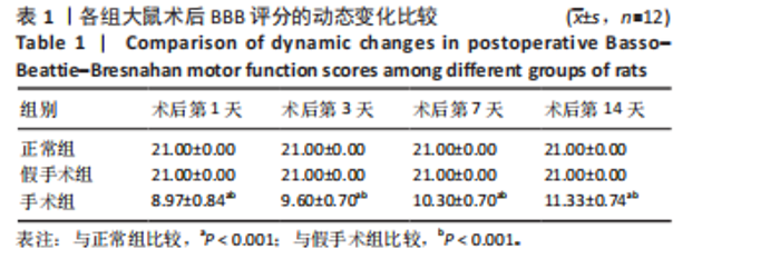
2.1 实验动物数量分析 36只SD大鼠全部进入结果分析。 2.2 各组大鼠运动功能BBB评分比较 造模后,手术组大鼠后肢运动无力、无法支撑体质量,运动不灵活;假手术组大鼠在术后较短时间内表现出运动功能的一过性轻度受损,短期内即恢复正常水平。手术组术后第1,3,7,14天的BBB评分低于正常组、假手术组(P < 0.001),见表1。"
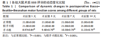
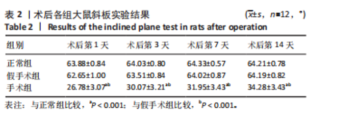
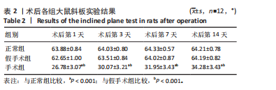
2.3 各组大鼠斜板实验结果比较 各组大鼠斜板实验角度测量值见表2。正常组大鼠的斜板实验角度值在实验期间保持相对稳定,术后第1天为(63.88±0.84)°,术后第14天为(64.21±0.78)°,表明正常组大鼠的平衡能力未受到显著影响。假手术组大鼠斜板实验角度值在实验期间也表现出一定的稳定性,术后第1天为(62.65±1.00)°,术后第14天为(64.19±0.82)°,与正常组相比无明显差异,说明假手术操作对大鼠的平衡能力影响较小。手术组大鼠实验期间的斜板实验角度值显著低于正常组和假手术组,术后第1天为(26.78±3.07)°,术后第14天为(34.28±3.43)°,结果表明手术操作对大鼠的平衡能力产生了显著的负面影响,并且这种影响在实验期间虽有一定程度恢复,但仍未恢复到正常水平。"
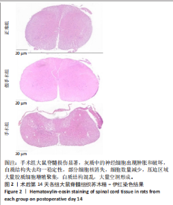
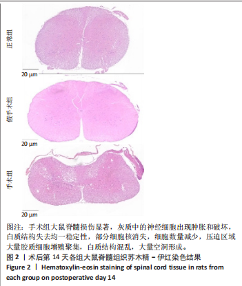
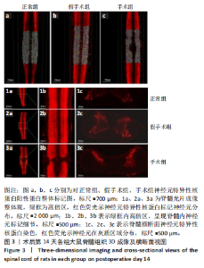
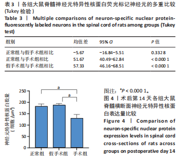
2.4 各组大鼠脊髓组织苏木精-伊红染色结果 见图2。 苏木精-伊红染色显示,正常组大鼠脊髓横断面显示出规整的形态和清晰的边界,神经细胞核染色深,结构完整,总体形态正常,胶质细胞分布较少、均匀,脊髓内偶尔可见空泡状改变;假手术组大鼠脊髓组织形态与正常组相似;手术组大鼠脊髓损伤显著,表现为神经细胞的肿胀、胞核固缩和坏死,部分神经元形态增大且结构模糊,灰质中的神经细胞出现肿胀和破坏,白质结构失去均一稳定性,部分细胞核消失,细胞数量减少,压迫区域大量胶质细胞增殖聚集,白质结构混乱,大量空洞形成。 2.5 各组大鼠脊髓组织组织透明化联合神经元特异性核蛋白免疫荧光标记结果 采用组织透明化技术联合神经元特异性核蛋白免疫荧光标记对各组脊髓组织进行三维重建分析和横断面视图分析(图3)。形态学观察显示,正常组与假手术组脊髓外观连续饱满,手术组脊髓外观凹陷甚至中断。横断面脊髓组织观察显示,正常组和假手术组神经元特异性核蛋白红色荧光信号均匀分布,灰质前角神经元呈层状密集排列,白质纤维束完整,未见荧光中断;手术组压迫节段神经元特异性核蛋白荧光强度明显减弱,灰质神经元层结构破坏,出现区域性荧光中断。神经元特异性核蛋白免疫荧光蛋白标记神经元定量计数显示,正常组与假手术组相比无明显差异(P=0.332 8),手术组损伤核心"
"
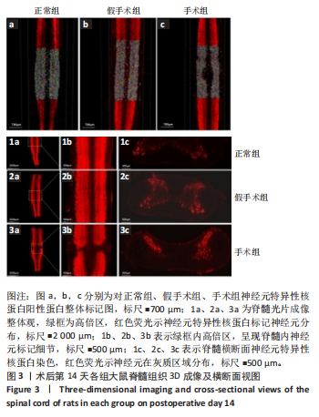

区神经元密度低于正常组、假手术组(P < 0.000 1),见表3,图4。三维结构塌陷与神经元定量丢失共同证实脊髓压迫导致不可逆性神经元损伤。"
| [1] HU X, XU W, REN Y, et al. Spinal cord injury: molecular mechanisms and therapeutic interventions. Signal Transduct Target Ther. 2023;8(1):245. [2] 王子恒,吴霜.脊髓损伤后氧化应激相关基因及分子机制:基于GEO数据库的数据分析及验证[J].中国组织工程研究,2025,29(32):6893-6904. [3] KARSY M, HAWRYLUK G. Modern Medical Management of Spinal Cord Injury. Curr Neurol Neurosci Rep. 2019;19(9):65. [4] 李宝莹,郑遵成.脊髓损伤后的病理改变与神经修复策略[J].医学信息,2025, 38(8):172-177. [5] QUADRI SA, FAROOQUI M, IKRAM A, et al. Recent update on basic mechanisms of spinal cord injury. Neurosurg Rev. 2020;43(2):425-441. [6] WYNDAELE M, WYNDAELE JJ. Incidence, prevalence and epidemiology of spinal cord injury: what learns a worldwide literature survey? Spinal Cord. 2006;44(9): 523-529. [7] NING GZ, YU TQ, FENG SQ, et al. Epidemiology of traumatic spinal cord injury in Tianjin, China. Spinal Cord. 2011;49(3):386-390. [8] ZHOU H, LOU Y, CHEN L, et al. Epidemiological and clinical features, treatment status, and economic burden of traumatic spinal cord injury in China: a hospital-based retrospective study. Neural Regen Res. 2024;19(5):1126-1133. [9] DE ALMEIDA FM, MARQUES SA, DOS SANTOS ACR, et al. Molecular approaches for spinal cord injury treatment. Neural Regen Res. 2023;18(1):23-30. [10] LI C, LUO Y, LI S. The roles of neural stem cells in myelin regeneration and repair therapy after spinal cord injury. Stem Cell Res Ther. 2024;15(1):204. [11] FAN S, WANG W, ZHENG X. Repetitive Transcranial Magnetic Stimulation for the Treatment of Spinal Cord Injury: Current Status and Perspective. Int J Mol Sci. 2025;26(2):825. [12] CSOBONYEIOVA M, POLAK S, ZAMBORSKY R, et al. Recent Progress in the Regeneration of Spinal Cord Injuries by Induced Pluripotent Stem Cells. Int J Mol Sci. 2019;20(15):3838. [13] TIAN T, LI X. Applications of tissue clearing in the spinal cord. Eur J Neurosci. 2020;52(9):4019-4036. [14] 高阳,高原,马炳江,等.骨科学领域组织透明化技术的应用与展望[J].中国组织工程研究,2025,29(15):3227-3234. [15] 张丹,连佳欢,冯倩倩.组织透明化方法对小鼠全脑可视化的影响[J].实验技术与管理,2025,42(3):9-17. [16] 田婷,杨朝阳,李晓光.组织透明化技术的研究与应用[J].中国组织工程研究, 2020;24(21):3363-3371. [17] 黄义源,杨亮,袁洁,等.三种组织透明化方法在神经科学应用中的比较[J].空军军医大学学报,2022,43(9):931-938. [18] 陈小玉,罗连响,潘韵琪,等.组织透明化技术在神经退行性疾病中的应用研究进展[J].解放军医学杂志,2022,47(3):305-313. [19] 唐倩,侯兰伟,刘宇宁,等.组织透明化技术在器官可视化应用中的初步探讨[J].中国临床解剖学杂志,2024,42(4):393-398. [20] TAINAKA K, KUNO A, KUBOTA SI, et al. Chemical Principles in Tissue Clearing and Staining Protocols for Whole-Body Cell Profiling. Annu Rev Cell Dev Biol. 2016;32:713-741. [21] JIN BH, WOO J, LEE M, et al. Optimization of the optical transparency of bones by PACT-based passive tissue clearing. Exp Mol Med. 2023;55(10):2190-2204. [22] ZHAN YJ, ZHANG SW, ZHU S, et al. Tissue Clearing and Its Application in the Musculoskeletal System. ACS Omega. 2023;8(2):1739-1758. [23] WANG J, JIANG P, DENG W, et al. Grafted human ESC-derived astroglia repair spinal cord injury via activation of host anti-inflammatory microglia in the lesion area. Theranostics. 2022;12(9):4288-4309. [24] WANG R, BAI J. Pharmacological interventions targeting the microcirculation following traumatic spinal cord injury. Neural Regen Res. 2024;19(1):35-42. [25] LIU J, YAN R, WANG B, et al. Decellularized extracellular matrix enriched with GDNF enhances neurogenesis and remyelination for improved motor recovery after spinal cord injury. Acta Biomater. 2024;180:308-322. [26] STEWARD O, ZHENG B, TESSIER-LAVIGNE M. False resurrections: distinguishing regenerated from spared axons in the injured central nervous system. J Comp Neurol. 2003;459(1):1-8. [27] POCRATSKY AM, BURKE DA, MOREHOUSE JR, et al. Reversible silencing of lumbar spinal interneurons unmasks a task-specific network for securing hindlimb alternation. Nat Commun. 2017;8(1):1963. [28] HILTON BJ, BLANQUIE O, TEDESCHI A, et al. High-resolution 3D imaging and analysis of axon regeneration in unsectioned spinal cord with or without tissue clearing. Nat Protoc. 2019;14(4):1235-1260. [29] WANG J, RONG W, HU X, et al. Hyaluronan tetrasaccharide in the cerebrospinal fluid is associated with self-repair of rats after chronic spinal cord compression. Neuroscience. 2012;210:467-480. [30] MCCREEDY DA, JALUFKA FL, PLATT ME, et al. Passive Clearing and 3D Lightsheet Imaging of the Intact and Injured Spinal Cord in Mice. Front Cell Neurosci. 2021;15:684792. [31] NI Y, NAWABI H, LIU X, et al. Characterization of long descending premotor propriospinal neurons in the spinal cord. J Neurosci. 2014;34(28):9404-9417. [32] ERTÜRK A, MAUCH CP, HELLAL F, et al. Three-dimensional imaging of the unsectioned adult spinal cord to assess axon regeneration and glial responses after injury. Nat Med. 2011;18(1):166-171. [33] ZHAI T, REN S, QIAN S, et al. Exercise training promotes nerve cell repair and regeneration after spinal cord injury. Neural Regen Res. 2026;21(6):2153-2168. [34] RICHARDSON DS, LICHTMAN JW. Clarifying Tissue Clearing. Cell. 2015;162(2): 246-257. [35] NAKANISHI K, SHINOZAKI M, NAGOSHI N, et al. biPACT: A method for three-dimensional visualization of mouse spinal cord circuits of long segments with high resolution. J Neurosci Methods. 2022;379:109672. [36] WEATHERSTONE JH, KOPP-SCHEINPFLUG C, PILATI N, et al. Maintenance of neuronal size gradient in MNTB requires sound-evoked activity. J Neurophysiol. 2017;117(2):756-766. [37] PINHO AG, CIBRÃO JR, LIMA R, et al. Immunomodulatory and regenerative effects of the full and fractioned adipose tissue derived stem cells secretome in spinal cord injury. Exp Neurol. 2022;351:113989. [38] ASSUNÇÃO-SILVA RC, PINHO A, CIBRÃO JR, et al. Adipose tissue derived stem cell secretome induces motor and histological gains after complete spinal cord injury in Xenopus laevis and mice. J Tissue Eng. 2024;15:20417314231203824. [39] SILVA D, SCHIRMER L, PINHO TS, et al. Sustained Release of Human Adipose Tissue Stem Cell Secretome from Star-Shaped Poly(ethylene glycol) Glycosaminoglycan Hydrogels Promotes Motor Improvements after Complete Transection in Spinal Cord Injury Rat Model. Adv Healthc Mater. 2023;12(17):e2202803. [40] SHAW DJ, CZEKÓOVÁ K, MAREČEK R, et al. The interacting brain: Dynamic functional connectivity among canonical brain networks dissociates cooperative from competitive social interactions. Neuroimage. 2023;269:119933. [41] TETREAULT LA, KWON BK, EVANIEW N, et al. A Clinical Practice Guideline on the Timing of Surgical Decompression and Hemodynamic Management of Acute Spinal Cord Injury and the Prevention, Diagnosis, and Management of Intraoperative Spinal Cord Injury: Introduction, Rationale, and Scope. Global Spine J. 2024;14(3_suppl):10s-24s. [42] WANG G, LI Q, LIU S, et al. An injectable decellularized extracellular matrix hydrogel with cortical neuron-derived exosomes enhances tissue repair following traumatic spinal cord injury. Mater Today Bio. 2024;28:101250. |
| [1] | Fu Lyupeng, Yu Peng, Liang Guoyan, Chang Yunbing. Electroactive materials applied in spinal surgery [J]. Chinese Journal of Tissue Engineering Research, 2026, 30(8): 2113-2123. |
| [2] | Cao Xinyan, Yu Zifu, Leng Xiaoxuan, Gao Shiai, Chen Jinhui, Liu Xihua. Effect of repetitive transcranial magnetic stimulation and transcranial direct current stimulation on motor function and gait in children with cerebral palsy: a network meta-analysis [J]. Chinese Journal of Tissue Engineering Research, 2026, 30(6): 1539-1548. |
| [3] | Guo Ying, Tian Feng, Wang Chunfang. Potential drug targets for the treatment of rheumatoid arthritis: large sample analysis from European databases [J]. Chinese Journal of Tissue Engineering Research, 2026, 30(6): 1549-1557. |
| [4] | Yang Zhijie, Zhao Rui, Yang Haolin, Li Xiaoyun, Li Yangbo, Huang Jiachun, Lin Yanping, Wan Lei, HuangHongxing. Postmenopausal osteoporosis: predictive values of muscle mass, grip strength, and appendicular skeletal muscle index [J]. Chinese Journal of Tissue Engineering Research, 2026, 30(5): 1073-1080. |
| [5] | Yin Yongcheng, Zhao Xiangrui, Yang Zhijie, Li Zheng, Li Fang, Ning Bin. Effect and mechanism of peroxiredoxin 1 in microglial inflammation after spinal cord injury [J]. Chinese Journal of Tissue Engineering Research, 2026, 30(5): 1106-1113. |
| [6] | Yu Huifen, Mo Licun, Cheng Leping. The position and role of 5-hydroxytryptamine in the repair of tissue injury [J]. Chinese Journal of Tissue Engineering Research, 2026, 30(5): 1196-1206. |
| [7] | Jiang Li, Peng Guoqiang, Li Sen. Interleukin-10 alleviates inflammatory responses after acute tendon injury [J]. Chinese Journal of Tissue Engineering Research, 2026, 30(34): 8906-8913. |
| [8] | Yin Haoran, Wang Fangyong. Mechanism of epothilone B improving spinal cord microcirculation after spinal cord injury in rats [J]. Chinese Journal of Tissue Engineering Research, 2026, 30(34): 8930-8938. |
| [9] | Wang Lei, Hu Baoyang, Fang Fang. Bibliometric analysis of research hotspots on mitochondria and spinal cord injury treatment [J]. Chinese Journal of Tissue Engineering Research, 2026, 30(29): 7764-7772. |
| [10] | He Long, Gao Shuang, Chen Chao, Qin Guozhong, Ran Qingsen, Wang Zhengchun, Yang Yafeng, Ren Hang, Qiu Yunkai, Yang Yang, Li Wei. A new strategy for preventing and treating orthopedic diseases by regulating ferroptosis through signaling pathways [J]. Chinese Journal of Tissue Engineering Research, 2026, 30(29): 7619-7631. |
| [11] | Wang Ziqi, Bu Xianzhong, Guo Xiaohui, Li Hanxi, Qian Yuhao, Wang Yixin, Bu Baoxian. Integrated proteomics and transcriptomics analysis of the mechanism of Buyang Huanwu Tang in protecting the acute spinal cord injury rat model [J]. Chinese Journal of Tissue Engineering Research, 2026, 30(25): 6472-6488. |
| [12] | Wei Xinyi, Zheng Yan, Chen Qian, Ren Jiajia, Li Jian. Molecular dynamic characteristics of rat gastrocnemius muscle under acute and short-term exercise intervention during the subacute phase of spinal cord injury [J]. Chinese Journal of Tissue Engineering Research, 2026, 30(24): 6196-6206. |
| [13] | Zhang Xiaoxu, Tian Zhenli, Xie Tingting. Roles of pregnane X receptor in sodium arsenite-induced oxidative stress and inflammatory injury in human normal hepatocytes [J]. Chinese Journal of Tissue Engineering Research, 2026, 30(24): 6259-6266. |
| [14] | Zheng Peng, Jia Xiaoning, Tao Jingwei, Fan Xiao. Tetramethylpyrazine improves iron metabolism disorders in a rat model of spinal cord injury via the Keap-1/Nrf2 signaling pathway [J]. Chinese Journal of Tissue Engineering Research, 2026, 30(23): 6134-6141. |
| [15] | Gao Jiabin, Li Tianqi, Xu Kun, Zhu Hanmin, Zhou Xi, Li Wei. Mitophagy regulates osteoclasts: a new perspective for osteoporosis treatment [J]. Chinese Journal of Tissue Engineering Research, 2026, 30(23): 5982-5991. |
| Viewed | ||||||
|
Full text |
|
|||||
|
Abstract |
|
|||||