Chinese Journal of Tissue Engineering Research ›› 2026, Vol. 30 ›› Issue (34): 8930-8938.doi: 10.12307/2026.880
Previous Articles Next Articles
Mechanism of epothilone B improving spinal cord microcirculation after spinal cord injury in rats
Yin Haoran1, 2, Wang Fangyong1, 2
- 1School of Rehabilitation Medicine, Capital Medical University, Beijing 100071, China; 2Department of Spinal and Spinal Cord Surgery, Beijing Boai Hospital, China Rehabilitation Research Center, Beijing 100068, China
-
Received:2025-09-17Revised:2026-02-28Online:2026-12-08Published:2026-04-13 -
Contact:Wang Fangyong, Professor, Chief physician, Doctoral supervisor, School of Rehabilitation Medicine, Capital Medical University, Beijing 100071, China; Department of Spinal and Spinal Cord Surgery, Beijing Boai Hospital, China Rehabilitation Research Center, Beijing 100068, China -
About author:Yin Haoran, MS candidate, School of Rehabilitation Medicine, Capital Medical University, Beijing 100071, China; Department of Spinal and Spinal Cord Surgery, Beijing Boai Hospital, China Rehabilitation Research Center, Beijing 100068, China -
Supported by:National Key R&D Program of China, No. 2021YFF0501604 (to WFY)
CLC Number:
Cite this article
Yin Haoran, Wang Fangyong. Mechanism of epothilone B improving spinal cord microcirculation after spinal cord injury in rats[J]. Chinese Journal of Tissue Engineering Research, 2026, 30(34): 8930-8938.
share this article
Add to citation manager EndNote|Reference Manager|ProCite|BibTeX|RefWorks
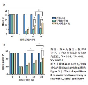
2.1 实验动物数量分析 造模后饲养过程中,脊髓损伤组死亡1只、埃博霉素B组死亡2只,考虑是疼痛导致体质量急剧下降/泌尿系感染所致,后续实验予以补全。 2.2 埃博霉素B改善了脊髓损伤大鼠的运动功能 2.2.1 BBB评分与斜板实验结果 造模后第1天,脊髓损伤组与埃博霉素B组BBB评分均为0分,表示造模成功;造模后第14,28天,埃博霉素B组大鼠BBB评分高于脊髓损伤组(P < 0.05,P < 0.01);造模后第28天,埃博霉素B组大鼠斜板实验角度大于脊髓损伤组(P < 0.05);脊髓损伤组造模后14 d的斜板实验角度大于造模后7 d(P < 0.000 1),埃博霉素B组造模后28 d的BBB评分、斜板实验角度均大于造模后14 d (P < 0.000 1),见图5。"

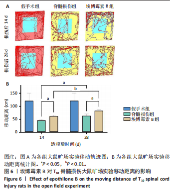
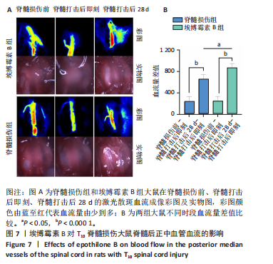
2.2.2 旷场轨迹图及移动距离比较 造模后第14,28天,脊髓损伤组大鼠旷场实验移动距离均小于埃博霉素B组[14 d:(44.55±5.174),(60.93±4.841) cm;28 d:(62.12±8.407),(81.91± 5.733) cm,P < 0.05];埃博霉素B组、脊髓损伤组大鼠造模后28 d的旷场实验移动距离均大于造模后14 d(P < 0.05,P < 0.01),见图6。 2.3 埃博霉素B增加了脊髓损伤大鼠的血流恢复 造模后第28天进行激光散斑血流成像检测时,脊髓损伤组和埃博霉素B组均有4只因伤口粘连过于严重打开椎板时血管破裂无法继续观察,其余大鼠均进行激光散斑血流成像检测。造模前,两组大鼠脊髓实物图中可以看到完整有脊膜包被的脊髓,下方正中存在一条粗大的血管,血流量越高激光散斑血流成像彩图中彩图颜色就越偏向红色,血流量越低彩图越偏向蓝色;造模后即刻可见脊膜仍然完整,但脊髓结构不清,出现血肿,正中血管血流量也有所下降;造模后第28天再次打开原造模切口时发现脊髓表面与周围存在不同程度粘连,脊髓结构部分恢复,正中血管血流量较造模或即刻有明显增加,见图7A。 由于大鼠血流基线数据个体之间差异较大,直接比较容易出现第一类错误,故对数据进行二次变换,将造模前-造模后即刻和造模后第28天-造模后即刻的脊髓血流量差值作为新的数据进行分析,避免了因方差过大造成的假阳性错误。"

埃博霉素B组与脊髓损伤组大鼠造模前-造模后即刻的脊髓血流量差值比较差异无显著性意义(P > 0.05),证明将造模前后血流变化量作为新的基线可行。两组造模后第28天-造模后即刻的脊髓血流量差值均大于造模前-造模后即刻的脊髓血流量差值(P < 0.000 1),说明随着时间的延长,局部血管血流量在逐渐恢复,这种恢复趋势与运动学评分的增加呈正相关,证明局部血流循环的重建能够促进大鼠神经功能障碍的恢复;埃博霉素B组造模后第28天-造模后即刻的脊髓血流量差值大于脊髓损伤组(P < 0.05),说明埃博霉素B从宏观上促进了血流的恢复,见图7B。"
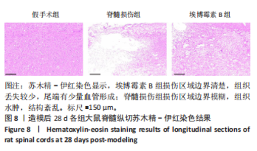
2.4 埃博霉素B减少损伤脊髓组织的丢失 大鼠脊髓损伤后第28天,深度麻醉处死脊髓损伤组与埃博霉素B组各3只大鼠,假手术组3只大鼠,取脊髓组织进行切片苏木精经-伊红染色,结果见图8。在低倍镜下观察3组损伤相应区域脊髓,可见脊髓损伤组打击处组织丢失严重,埃博霉素B组脊髓空洞面积占比[(17.70±3.40)%]较脊髓损伤组小[(52.27±1.07)%]。埃博霉素B组打击区域有炎症细胞浸润,但组织丢失较少,有少量血管形成,脊髓损伤组损伤区域边界模糊,大量组织丢失,结构紊乱。"

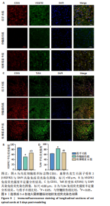
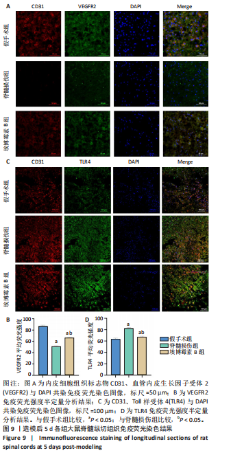
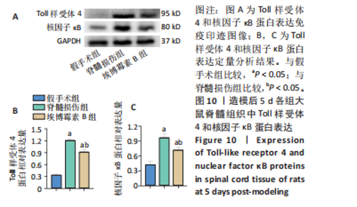
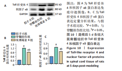
2.5 埃博霉素B减轻炎症因子分泌、促进血管再生 2.5.1 免疫荧光染色结果 图9显示3组大鼠损伤脊髓横切面免疫荧光染色及定量分析结果。免疫荧光染色显示,与假手术组相比,脊髓损伤组血管数(CD31表达)和血管内皮生长因子受体2表达明显减少;与脊髓损伤组相比,埃博霉素B组血管数量和血管内皮生长因子受体2表达增加,提示埃博霉素B促进血管再生;脊髓损伤组Toll样受体4表达多于假手术组、埃博霉素B组。 2.5.2 Western Blot检测结果 脊髓损伤组损伤脊髓组织中Toll样受体4和核因子?B蛋白表达高于假手术组、埃博霉素B组(P < 0.05),见图10。"
"
| [1] GBD 2016 Traumatic Brain Injury and Spinal Cord Injury Collaborators. Global, regional, and national burden of traumatic brain injury and spinal cord injury, 1990-2016: a systematic analysis for the Global Burden of Disease Study 2016. Lancet Neurol. 2019;18(1):56-87. [2] CHEN C, QIAO X, LIU W, et al. Epidemiology of spinal cord injury in China: A systematic review of the Chinese and English literature. Spinal Cord. 2022; 60(12):1050-1061. [3] 张浩,刘宇,肖世宁,等.中国创伤性脊髓损伤患者流行病学特征的Meta分析[J].中国脊柱脊髓杂志,2023,33(5):397-407. [4] JÖRGENSEN S, IWARSSON S, LEXELL J. Secondary health conditions, activity limitations, and life satisfaction in older adults with long-term spinal cord injury. PM R. 2017;9(4):356-366. [5] HOLTZ KA, LIPSON R, NOONAN VK, et al. Prevalence and effect of problematic spasticity after traumatic spinal cord injury. Arch Phys Med Rehabil. 2017;98(6): 1132-1138. [6] 周庆,郝璐,周泽强.固有免疫系统Toll样受体的研究进展[J].生物学杂志, 2016,33(3):83-87. [7] ZHOU JD, LIN H, LV TT, et al. Inappropriate Activation of TLR4/NF-κB is a Cause of Heart Failure. Cardiovasc Innov Appl. 2022;7(1). doi:10.15212/CVIA.2022.0020 [8] LUND MC, CLAUSEN BH, BRAMBILLA R, et al. The Role of Tumor Necrosis Factor Following Spinal Cord Injury: A Systematic Review. Cell Mol Neurobiol. 2023;43(3):925-950. [9] 李云鹏,吴明莉,冯晓东.脊髓损伤后线粒体融合介导的神经修复机制及干预措施研究进展[J].解放军医学杂志,2025,50(8):1015-102. [10] 张孝炜,闫炳翰,仇道迪,等.天然产物调控氧化应激治疗脊髓损伤[J].中国组织工程研究,2025,29(12):2560-2568. [11] CLARK JM, MARSHALL R. Nature of the Non-traumatic Spinal Cord Injury Literature: A Systematic Review. Top Spinal Cord Inj Rehabil. 2017;23(4):353-367. [12] GWAK YS, HULSEBOSCH CE. GABA and central neuropathic pain following spinal cord injury. Neuropharmacology. 2011;60(5):799-808. [13] 郭昱.缓释VEGF脊髓脱细胞支架移植治疗大鼠脊髓半切损伤的血管重建作用[D].福州:福建医科大学,2017. [14] 张羽,窦荣声,闫浩,等.基于微环境调控的脊髓损伤修复策略及相关研究进展[J].河北医药,2024,46(3):444-448. [15] HUANG JH, HE H, CHEN YN, et al. Exosomes derived from M2 Macrophages Improve Angiogenesis and Functional Recovery after Spinal Cord Injury through HIF-1α/VEGF Axis. Brain Sci. 2022;12(10):1322. [16] 周源.川芎嗪对大鼠急性脊髓损伤后血管新生及微血管三维形态的影响[D].长沙:中南大学,2014. [17] DING H, YU J, CHANG W, et al. Searching for differentially expressed proteins in spinal cord injury based on the proteomics analysis. Life Sci. 2020;242:117235. [18] 段文帅,鄢卫平,张会,等.血管内皮生长因子信号通路在脊髓损伤中的作用及其中医药干预研究进展[J].上海中医药杂志,2025,59(1):95-100. [19] MU J, LI L, WU J, et al. Hypoxia-stimulated mesenchymal stem cell-derived exosomes loaded by adhesive hydrogel for effective angiogenic treatment of spinal cord injury. Biomater Sci. 2022;10(7):1803-1811. [20] ZHONG D, CAO Y, LI CJ, et al. Neural stem cell-derived exosomes facilitate spinal cord functional recovery after injury by promoting angiogenesis. Exp Biol Med (Maywood). 2020;245(1):54-65.. [21] 邓博文.ASCI 后患者早期血液学指标改变情况及川芎嗪联合导电水凝胶修复脊髓损伤的机制研究[D].北京:北京中医药大学,2022. [22] TSIVELEKAS K, EVANGELOPOULOS DS, PALLIS D, et al. Angiogenesis in Spinal Cord Injury: Progress and Treatment. Cureus. 2022;14(5):e25475. [23] 吴典.腺病毒介导的VEGF-165基因过表达对缺氧性肺动脉高压新生大鼠肺血管重塑的影响研究[D].乌鲁木齐:新疆医科大学,2020. [24] RUSCHEL J, BRADKE F. Systemic administration of epothilone D improves functional recovery of walking after rat spinal cord contusion injury. Exp Neurol. 2018;306:243-249. [25] DUAN YY, CHAI Y, ZHANG NL, et al. Microtubule Stabilization Promotes Microcirculation Reconstruction After Spinal Cord Injury. J Mol Neurosci. 2021; 71(3):583-595. [26] DAVIS GE, STRATMAN AN, SACHARIDOY A, et al. Molecular basis for endothelial lumen formation and tubulogenesis during vasculogenesis and angiogenic sproutingInt. RevCell Mol Biol. 2011;288:101-165. [27] BOWERS SL, NORDEN PR, DAVIS GE. Molecular signaling pathways controlling vascular tube morphogenesis and pericyte-induced tube maturation in 3D extracellular matrices. Adv Pharmacol. 2016;77:241-280. [28] 王思凯,王玉玞,闫景龙.周细胞在脊髓损伤中的作用[J].脊柱外科杂志, 2021,19(6):416-419. [29] SANDNER B, PUTTAGUNTA R, MOTSCH M, et al. Systemic epothilone D improves hindlimb function after spinal cord contusion injury in rats. Exp Neurol. 2018; 306:250-259. [30] ZHAO W, CHAI Y, HOU Y, et al. Mechanisms responsible for the inhibitory effects of epothilone B on scar formation after spinal cord injury. Neural Regen Res. 2017;12(3):478-485. [31] RUSCHEL J, HELLAL F, FLYNN KC, et al. Axonal regeneration. Systemic administration of epothilone B promotes axon regeneration after spinal cord injury. Science. 2015;348(6232):347-352. [32] ZHU Y, UEZONO N, YASUI T, et al. Combinatrial treatment of anti-High Mobility Group Box-1 monoclonal antibody and epothilone B improves functional recovery after spinal cord contusion injury. Neurosci Res. 2021;172:13-25. [33] 曾琼祯.紫杉醇通过促进巨噬细胞NLPR3炎症小体活化发挥抗感染作用的机制研究[D].广州:暨南大学,2019. [34] 王咏红,申晨.TLR4在Hela细胞有丝分裂中的定位特点及其与微管的相互作用研究[J].标记免疫分析与临床,2018,25(10):1517-1521. [35] ZHANG D, LI Y, LIU Y, et al. Paclitaxel ameliorates lipopolysaccharide-induced kidney injury by binding myeloid differentiation protein-2 to block Toll-like receptor 4-mediated nuclear factor-κB activation and cytokine production. J Pharmacol Exp Ther. 2013;345(1):69-75. [36] 徐丽丽,高明,王利存,等.研究银杏二萜内酯调控Toll样受体4/核因子-κB通路对糖尿病大鼠肾脏病变的影响及机制[J].临床肾脏病杂志,2023, 23(9):750-759. [37] GRIFFIN JM, HINGORANI JAI PRAKASH S, BOCKEMÜHL T, et al. Rehabilitation enhances epothilone-induced locomotor recovery after spinal cord injury. Brain Commun. 2023;5(1):fcad005. [38] ZHANG L, WEI X, WANG Z, et al. NF-κB activation enhances STING signaling by altering microtubule-mediated STING trafficking. Cell Rep. 2023;42(3):112185. [39] LAMPSON BL, RAMΊREZ AS, BARO M, et al. Positive selection CRISPR screens reveal a druggable pocket in an oligosaccharyltransferase required for inflammatory signaling to NF-κB. Cell. 2024;187(9):2209-2223.e16. |
| [1] | Jiang Xianglong, Li Zhongshan, Che Tongtong. Application effects and mechanisms of low-frequency pulsed electromagnetic fields in muscle repair and growth [J]. Chinese Journal of Tissue Engineering Research, 2026, 30(9): 2350-2360. |
| [2] | Zeng Xuan, Weng Rui, Ye Shicheng, Tang Jiadong, Mo Ling, Li Wenchao. Two lumbar rotary manipulation techniques in treating lumbar disc herniation: a finite element analysis of biomechanical differences [J]. Chinese Journal of Tissue Engineering Research, 2026, 30(9): 2153-2161. |
| [3] | Jiang Xinghai, Song Yulin, Li Dejin, Shao Jianmin, Xu Junzhi, Liu Huakai, Wu Yingguo, Shen Yuehui, Feng Sicheng. Vascular endothelial growth factor 165 genes transfected into bone marrow mesenchymal stem cells to construct a vascularized amphiphilic peptide gel module [J]. Chinese Journal of Tissue Engineering Research, 2026, 30(8): 1903-1911. |
| [4] | Cai Ziming, Yu Qinghe, Ma Pengfei, Zhang Xin, Zhou Longqian, Zhang Chongyang, Lin Wenping. Heme oxygenase-1 alleviates lipopolysaccharide-induced inflammatory response in nucleus pulposus mesenchymal stem cells [J]. Chinese Journal of Tissue Engineering Research, 2026, 30(7): 1624-1631. |
| [5] | Zhong Caihong, Xiao Xiaoge, Li Ming, Lin Jianhong, Hong Jing. Biomechanical mechanism of sports-related patellar tendinitis [J]. Chinese Journal of Tissue Engineering Research, 2026, 30(6): 1417-1423. |
| [6] | Gao Zengjie, , Pu Xiang, Li Lailai, Chai Yihui, Huang Hua, Qin Yu. Increased risk of osteoporotic pathological fractures associated with sterol esters: evidence from IEU-GWAS and FinnGen databases [J]. Chinese Journal of Tissue Engineering Research, 2026, 30(5): 1302-1310. |
| [7] | Yin Yongcheng, Zhao Xiangrui, Yang Zhijie, Li Zheng, Li Fang, Ning Bin. Effect and mechanism of peroxiredoxin 1 in microglial inflammation after spinal cord injury [J]. Chinese Journal of Tissue Engineering Research, 2026, 30(5): 1106-1113. |
| [8] | Guo Jiachen, Gao Jun, Dai Wenhao, Liao Huayuan, Jiang You, Zhang Xi . Effect of compressive stress microenvironment on cytokines during fracture healing [J]. Chinese Journal of Tissue Engineering Research, 2026, 30(4): 908-916. |
| [9] | Wen Fan, Zhu Huan, Hu Qinghua, Tuo Yanfang, Zhou Shufeng, Hu Jiangping, Wang Kangfeng. Eight-week aerobic exercise and aerobic exercise combined with full-body vibration training improve the microcirculation function of obese college students [J]. Chinese Journal of Tissue Engineering Research, 2026, 30(34): 8994-9001. |
| [10] | Liu Yuxiao, Huang Sijing, Geng Longyu, Gao Beiyao, Yang Guang, Ge Ruidong, Gao Qi. The functions and underlying molecular mechanisms of PIEZO channels in nervous system diseases [J]. Chinese Journal of Tissue Engineering Research, 2026, 30(34): 9017-9023. |
| [11] | Luan Chuankai, Zhu Lei. Role and mechanism of exercise-regulated miRNAs in cardiac remodeling after acute myocardial infarction [J]. Chinese Journal of Tissue Engineering Research, 2026, 30(34): 9024-9031. |
| [12] | Jiang Li, Peng Guoqiang, Li Sen. Interleukin-10 alleviates inflammatory responses after acute tendon injury [J]. Chinese Journal of Tissue Engineering Research, 2026, 30(34): 8906-8913. |
| [13] | Wang Zhengye, Liu Wanlin, Zhao Zhenqun. Mechanism by which vascular endothelial growth factor A targets regulation of angiogenesis in the treatment of steroid-induced osteonecrosis of the femoral head [J]. Chinese Journal of Tissue Engineering Research, 2026, 30(3): 671-679. |
| [14] | Yang Yunhong, Guo Lihua, Tang Han, Lin Lvping, Kuang Hongjun, Zhao Hong. Proteomic analysis of the mechanism of moxibustion intervention in a rat model of atopic dermatitis [J]. Chinese Journal of Tissue Engineering Research, 2026, 30(29): 7581-7591. |
| [15] | Du Yaxin, Zhao Xiaojuan, Wu Ruixia, Dong Yizhi, Song Xinyue, Fu Hongyang, She Yitong, Zhang Jialin, Zhu Yong. Interaction between vascular-lymphatic system imbalance and immune microenvironment in intervertebral disc degeneration [J]. Chinese Journal of Tissue Engineering Research, 2026, 30(29): 7612-7618. |
| Viewed | ||||||
|
Full text |
|
|||||
|
Abstract |
|
|||||