Chinese Journal of Tissue Engineering Research ›› 2026, Vol. 30 ›› Issue (31): 8302-8308.doi: 10.12307/2026.794
Bibliometric analysis of studies on organoid air-liquid interface culture
Tong Man, Wang Yue, Wang Ruoyu, Wang Zhe, Fang Yanhua, Liang Shanshan
- Key Laboratory of Biomarker High Throughput Screening and Target Translation of Breast and Gastrointestinal Tumor, Affiliated Zhongshan Hospital of Dalian University, Dalian 116001, Liaoning Province, China
-
Received:2025-08-26Accepted:2025-12-22Online:2026-11-08Published:2026-05-26 -
Contact:Fang Yanhua, MD, Key Laboratory of Biomarker High Throughput Screening and Target Translation of Breast and Gastrointestinal Tumor, Affiliated Zhongshan Hospital of Dalian University, Dalian 116001, Liaoning Province, China. Co-corresponding author: Liang Shanshan, MD, Associate researcher, Key Laboratory of Biomarker High Throughput Screening and Target Translation of Breast and Gastrointestinal Tumor, Affiliated Zhongshan Hospital of Dalian University, Dalian 116001, Liaoning Province, China -
About author:Tong Man, MS, Key Laboratory of Biomarker High Throughput Screening and Target Translation of Breast and Gastrointestinal Tumor, Affiliated Zhongshan Hospital of Dalian University, Dalian 116001, Liaoning Province, China. Wang Yue, MS, Key Laboratory of Biomarker High Throughput Screening and Target Translation of Breast and Gastrointestinal Tumor, Affiliated Zhongshan Hospital of Dalian University, Dalian 116001, Liaoning Province, China. Tong Man and Wang Yue contributed equally to this work. -
Supported by:Liaoning Provincial International Science and Technology Cooperation Project, No. 2024JH2/101900006 (to LSS); Xingliao Yingcai Program-Medical Eminent Expert Project, No. YXMJ-JC-10 (to WRY); Liaoning Provincial Science and Technology Program Joint Project-Key Technology Research Project, No. 2024JH2/102600067 (to WZ)

CLC Number:
Cite this article
Tong Man, Wang Yue, Wang Ruoyu, Wang Zhe, Fang Yanhua, Liang Shanshan . Bibliometric analysis of studies on organoid air-liquid interface culture[J]. Chinese Journal of Tissue Engineering Research, 2026, 30(31): 8302-8308.
share this article
Add to citation manager EndNote|Reference Manager|ProCite|BibTeX|RefWorks
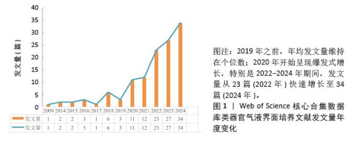
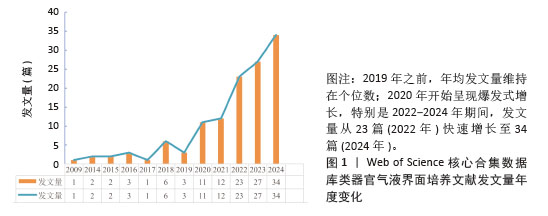
2.1 类器官气液界面培养文献发文量年度变化 根据检索条件,筛选出近20年来发表的关于类器官气液界面培养的文献共计127篇。其中96篇为“研究性论文”,31篇为“综述论文”。通过分析文献的年增长率,可将时间线划分为3个特征阶段:第一阶段(2005-2008年)、第二阶段(2009-2019年)和第三阶段(2020-2024年)。如图1所示,第一阶段未见相关文献发表。虽然气液界面培养研究始于1983年,但类器官的研究于2009年起步,该时期尚未建立连接这两个领域的理论基础。第二阶段年均发文量约2.6篇,表明类器官与气液界面培养的融合研究处于萌芽状态。值得注意的是,2009-2014年间发文量为0,这是由于类器官研究尚处早期发展阶段,两个领域的交叉研究极为有限。2013年《Science》杂志将类器官技术评为“十大突破性技术”之一,由此推动研究者尝试结合两种技术以优化类器官培养体系。第三阶段呈现爆发式增长态势,年均发文量达21.4篇,其中2024年发表34篇相关论文,较2018年增长4倍。近3年类器官气液界面培养文献呈指数级增长,这一现象与2019年末全球暴发的SARS-CoV-2疫情密切相关。传统肺泡细胞体外培养存在技术瓶颈,而细胞系或原代细胞培养存在细胞组成单一、无法模拟组织微环境的局限性,动物模型虽能提供研究数据但成本高昂且存在种属差异。类器官是一种能高度模拟宿主组织的体外细胞结构,结合气液界面培养技术可以促进类器官中细胞的分化,已成为研究冠状病毒的可靠生物学模型[24-25]。因此,2019年,COVID-19疫情的暴发推动了相关研究的持续增长。 "
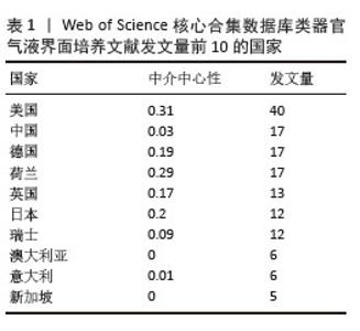
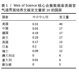
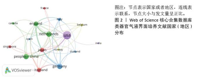
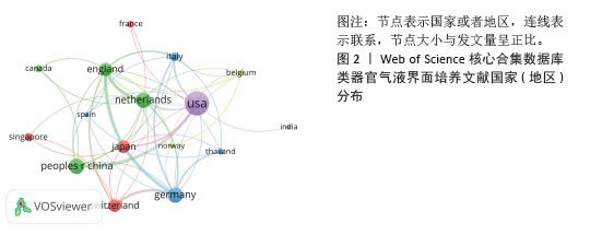
2.2 类器官气液界面培养文献国家(地区)分布 文献来源覆盖24个国家。发文量前10的国家主要分布在欧洲和亚洲。美国以40篇文献(31.50%)位居首位,中国(17篇,13.39%)、荷兰(17篇,13.39%)、德国(17篇,13.39%)紧随其后(表1)。中介中心性衡量节点在网络中的桥梁作用,数值越高,表明该国家在合作网络中占据更核心的位置。美国表现出最高的中介中心性(0.31),显示其在合作网络中的核心影响力。荷兰的中介中心性为0.29,紧随美国之后,表明其在国际合作中的重要性。日本和德国分别以0.2和0.19的中介中心性,展现了较高的影响力。进一步筛选出发文量≥2篇的18个国家,基于各国发文量及合作关系构建了国际合作网络图谱(图2)。结果显示,国家间存在显著正向合作关联:例如美国与中国、德国、荷兰等国家均保持密切合作。 "
"
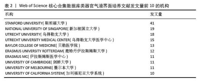
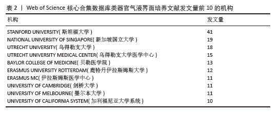
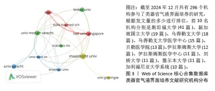
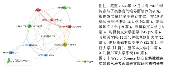
2.3 类器官气液界面培养文献研究机构分布 在排名前10的研究机构中(表2),约40%位于荷兰,美国占30%。斯坦福大学(41篇,32.28%)、新加坡国立大学(19篇,14.96%)与乌得勒支大学(18篇,14.17%)位列机构发文量前3名。基于最低发文量≥3篇的标准,选取22所机构进行可视化分析,结合各机构发文量及合作关系构建协作网络。如图3所示,斯坦福大学虽然发文量最高,但由于其研究独立性较强,并未与其他机构形成深度合作关系。相比之下,荷兰马斯里赫特大学、荷兰乌得勒支大学、新加坡科技研究局以及荷兰莱顿大学等通过紧密合作形成了多节点网络,合作强度可能与机构的地理邻近性或学科互补性相关。中国机构未出现在合作网络中,可能以本土研究为主,或国际合著较少。 "
"
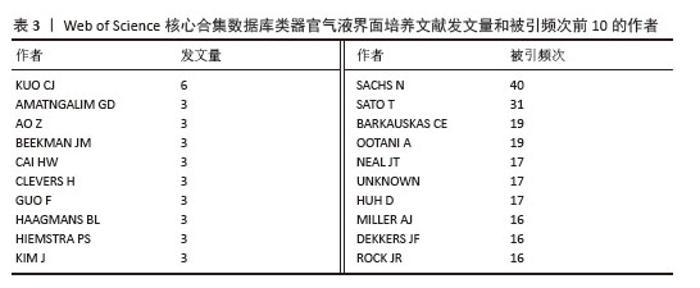
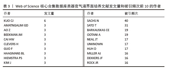
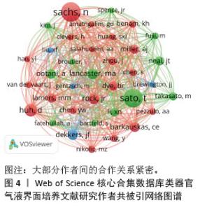
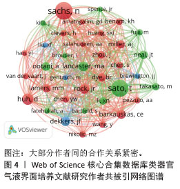
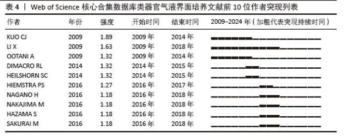
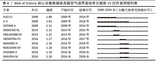
2.4 类器官气液界面培养研究作者分布 共有1 037位学者参与了类器官气液界面培养的学术探索。其中,KUO CJ以6篇发文量成为最高产作者(表3)。在7 333位被引作者中,有10位被引频次超过15次(表3),SACHS N以40次被引居首,SATO T(31次)列第二位。进一步筛选被引频次≥11次的作者构建共被引网络(图4),结果显示SACHS N、SATO T和DEKKERS JF等核心学者间存在紧密的学术关联。而KUO CJ虽然是类器官气液界面培养领域发文量第一的作者(6篇),但被引频次并不显著,结合前10名作者突现表(表4)发现,KUO CJ持续突现的时间处于类器官气液界面培养发展的早期阶段(2009-2014年),表明其研究虽具有技术开创性,但可能因方法被新技术迭代替代而影响力衰减。 "
"
"
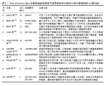
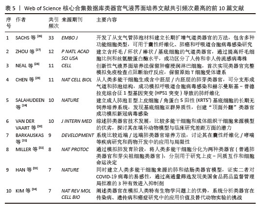
2.5 类器官气液界面培养文献共被引网络 学术文献中的引用行为可视为特定研究领域的基础支撑,被多篇文献共同引用的论文,往往标志着它在该领域的奠基性地位。其中,最高被引文献超过33次。数据分析显示(表5),共被引频次前10的文献主要涉及3大方向:气液界面培养参与类器官技术开发、疾病模型与应用、临床应用与转化——这些领域恰恰凸显了气液界面培养相较于传统技术的独特优势[24,26-34]。通过筛选共被引频次≥8次的文献构建网络图谱(图5),可清晰观察到3组活跃的共被引关系群(以红、绿、蓝标示);“Long-term expanding human airway organoids for disease modeling[26]”“Single Lgr5 stem cells build crypt-villus structures in vitro without a mesenchymal niche[35]”“In vitro generation of human pluripotent stem cell derived lung organoids”等核心文献形成显著的知识节点[36]。 "
"
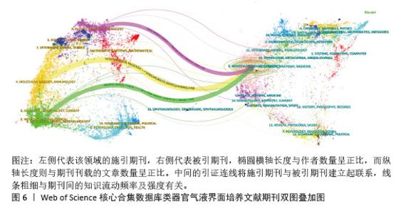
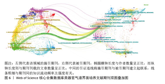
2.6 类器官气液界面培养文献期刊双图叠加分析 基于文献计量学的双图叠加分析方法,构建了期刊引文网络的可视化图谱(图6)。左侧空间分布表示施引期刊集群,右侧空间呈现被引期刊集合。在几何表征方面,椭圆的横向维度与活跃作者群体规模呈正相关,纵向维度则与期刊载文量保持比例关系。施引期刊与被引期刊之间的引证关系通过动态加权连线呈现,连线的视觉粗细及连线的f值严格对应于期刊间引证关系的强度,即引用频次越高,连线越粗,f值越大,直观反映知识流动的活跃程度;而连线的箭头指向明确标识知识流动方向,即从施引期刊指向被引期刊。如图6所示,引证连线由上至下依次表明:发表在“Physics(物理学)/Materials(材料学)/Chemistry(化学)”类期刊的研究、发表在“Molecular(分子学)/Biology(生物学)/Immunology(免疫学)”类期刊的研究以及发表在“Medicine(医学)/Medical(内科)/Clinical(临床)”类期刊的研究,常引用“Molecular(分子学)/Biology(生物学)/Genetics(遗传学)”类期刊的文献,这揭示分子生物学和遗传学在类器官气液界面培养研究中的核心地位,研究成果为多个应用学科提供了关键的理论基础和技术支撑。 "
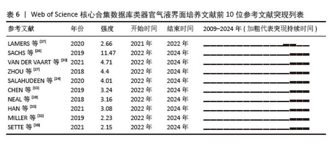
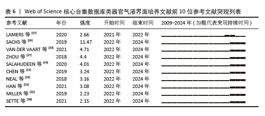
2.7 高突现性文献分析 “引文突现”指特定文献在某个时间段内被集中高频引用的现象。通过CiteSpace软件识别出10篇具有显著引文突现特征的文献(表6)[13,24,26-28,30,32-33,37-38]。图中每根横条代表一个年份,黑色条段表示该文献被密集引用的活跃期,这些突现文献最早出现于2018年,最近可追溯至2021年。 SACHS N团队发表的“Long-term expanding human airway organoids for disease modeling”展现出最强的引文突现强度(11.47) [26],突现期为2022-2024年;紧随其后的是VAN DER VAART J 团队“Airway organoids as models of human disease”[30],突现强度达4.71(2022-2024年)。表6中10篇标志性文献的突现强度介于11.47-2.66之间,突现持续时间为两三年。 "
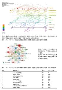
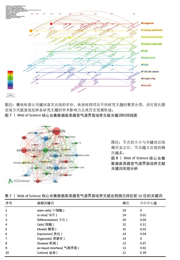
2.8 类器官气液界面培养研究热点分析 通过关键词时间线图分析可揭示研究领域的动态演进规律。关键词出现的时间跨度反映了该研究主题的起源时间和持续影响力——时间跨度越长,表明该研究方向起步越早且具有更持久的学术生命力。如图7所示,airway epithelium(气道上皮,#1)和skin(皮肤,#6)出现较早且持续至今。 关键词共现分析是快速识别领域研究焦点的重要工具。表7列出了类器官气液界面培养研究中最常出现的10个关键词。其中“stem cells (干细胞)”“in vitro(体外)”与“differentiation(分化)”出现频次均不低于20次。通过VOSviewer对出现频次≥6次的关键词进行共现分析(图7),节点间连线越密集表明关键词关联性越强,图7根据不同颜色展现了4个分类群。基于表5、表7、图7和图8,可将类器官气液界面培养的研究方向划分为4个主要领域。 类器官基础构建与癌症模型研究:该领域主要聚焦于利用干细胞(尤其是小鼠模型)构建类器官体系,深入研究生长特性、分化潜能及基因表达调控机制。同时,通过建立高度仿生的类器官模型,可有效模拟肿瘤微环境,为癌症发生发展机制研究、抗肿瘤药物筛选及个性化治疗方案制定提供重要平台。 病毒感染性模型的建立:该方向重点构建呼吸道感染模型,通过优化类器官气液界面培养条件建立的气道类器官系统,为COVID-19、呼吸道合胞病毒等病毒感染机制研究提供理想模型。值得注意的是,保留肿瘤浸润淋巴细胞等免疫组分的类器官模型,为深入解析感染免疫反应机制和开发新型抗病毒药物提供了独特的研究工具。 诱导多能干细胞分化进行再生医学研究:该领域创新性地将囊性纤维化等遗传性疾病类器官模型与诱导多能干细胞技术相结合,为阐明遗传病发病机制开辟了新途径。同时,通过精确调控人支气管干细胞的分化过程生成功能性肺类器官,这些研究成果为肺纤维化等疾病的修复治疗和器官再生提供了重要的实验基础。 气道基底细胞与稳态调控研究:该方向着重探究类器官气液界面培养条件下基底细胞向纤毛细胞/杯状细胞分化的分子调控机制,如Notch等关键信号通路的调控作用。此外,通过对基底细胞异常增殖现象的深入研究,为阐明哮喘、慢性阻塞性肺病等慢性呼吸道疾病的发病机制提供了新的理论依据。"
| [1] LANCASTER MA, KNOBLICH JA. Organogenesis in a dish: modeling development and disease using organoid technologies. Science. 2014; 345(6194):1247125. [2] FATEHULLAH A, TAN SH, BARKER N. Organoids as an in vitro model of human development and disease. Nat Cell Biol. 2016;18(3):246-254. [3] KRUCZEK K, SWAROOP A. Pluripotent stem cell-derived retinal organoids for disease modeling and development of therapies. Stem Cells. 2020; 38(10):1206-1215. [4] SHIMOKAWA M, OHTA Y, NISHIKORI S, et al. Visualization and targeting of LGR5+ human colon cancer stem cells. Nature. 2017;545(7653):187-192. [5] LI X, LARSSON P, LJUSLINDER I, et al. Ex Vivo Organoid Cultures Reveal the Importance of the Tumor Microenvironment for Maintenance of Colorectal Cancer Stem Cells. Cancers (Basel). 2020;12(4):923. [6] MAGLIARO C, RINALDO A, AHLUWALIA A. Allometric Scaling of physiologically-relevant organoids. Sci Rep. 2019;9(1):11890. [7] LEE HN, CHOI YY, KIM JW, et al. Effect of biochemical and biomechanical factors on vascularization of kidney organoid-on-a-chip. Nano Converg. 2021;8(1):35. [8] POUMAY Y, COQUETTE A. Modelling the human epidermis in vitro: tools for basic and applied research. Arch Dermatol Res. 2007;298(8):361-369. [9] CAO X, COYLE JP, XIONG R, et al. Invited review: human air-liquid-interface organotypic airway tissue models derived from primary tracheobronchial epithelial cells-overview and perspectives. In Vitro Cell Dev Biol Anim. 2021; 57(2):104-132. [10] HOPCRAFT SE, MOODY CA, DAMANIA B. Air-Liquid Interface System To Understand Epstein-Barr Virus-Associated Nasopharyngeal Carcinoma. mSphere. 2018;3(4):e00350-18. [11] KOOTER IM, ALBLAS MJ, JEDYNSKA AD, et al. Alveolar epithelial cells (A549) exposed at the air-liquid interface to diesel exhaust: First study in TNO’s powertrain test center. Toxicol In Vitro. 2013;27(8):2342-2349. [12] CARSON JL, BRIGHTON LE, JASPERS I. Phenotypic modification of human airway epithelial cells in air-liquid interface culture induced by exposure to the tobacco-specific nitrosamine 4-(methylnitrosamino)-1-(3-pyridyl)-1-butanone (NNK). Ultrastruct Pathol. 2015;39(2):104-109. [13] CHEN S, SCHOEN J. Air-liquid interface cell culture: From airway epithelium to the female reproductive tract. Reprod Domest Anim. 2019; 54 Suppl 3:38-45. [14] CANTIN-WARREN L, GUIGNARD R, CORTEZ GHIO S, et al. Specialized Living Wound Dressing Based on the Self-Assembly Approach of Tissue Engineering. J Funct Biomater. 2018;9(3):53. [15] WENGST A, REICHL S. RPMI 2650 epithelial model and three-dimensional reconstructed human nasal mucosa as in vitro models for nasal permeation studies. Eur J Pharm Biopharm. 2010;74(2):290-297. [16] AUFDERHEIDE M, ITO S, ISHIKAWA S, et al. Metaplastic phenotype in human primary bronchiolar epithelial cells after repeated exposure to native mainstream smoke at the air-liquid interface. Exp Toxicol Pathol. 2017;69(5):307-315. [17] OOTANI A, LI X, SANGIORGI E, et al. Sustained in vitro intestinal epithelial culture within a Wnt-dependent stem cell niche. Nat Med. 2009;15(6): 701-706. [18] YUKI K, CHENG N, NAKANO M, et al. Organoid Models of Tumor Immunology. Trends Immunol. 2020;41(8):652-664. [19] KE L, LU C, SHEN R, et al. Knowledge Mapping of Drug-Induced Liver Injury: A Scientometric Investigation (2010-2019). Front Pharmacol. 2020;11:842. [20] VAN ECK NJ, WALTMAN L. Software survey: VOSviewer, a computer program for bibliometric mapping. Scientometrics. 2010;84(2):523-538. [21] YEUNG AWK, TZVETKOV NT, BALACHEVA AA, et al. Lignans: Quantitative Analysis of the Research Literature. Front Pharmacol. 2020;11:37. [22] SYNNESTVEDT MB, CHEN C, HOLMES JH. CiteSpace II: visualization and knowledge discovery in bibliographic databases. AMIA Annu Symp Proc. 2005;2005:724-728. [23] ZHANG XL, ZHENG Y, XIA ML, et al. Knowledge Domain and Emerging Trends in Vinegar Research: A Bibliometric Review of the Literature from WoSCC. Foods. 2020;9(2):166. [24] SALAHUDEEN AA, CHOI SS, RUSTAGI A, et al. Progenitor identification and SARS-CoV-2 infection in long-term human distal lung organoid cultures. bioRxiv [Preprint]. 2020:2020.07.27.212076. [25] LAMERS MM, VAN DER VAART J, KNOOPS K, et al. An organoid-derived bronchioalveolar model for SARS-CoV-2 infection of human alveolar type II-like cells. EMBO J. 2021;40(5):e105912. [26] SACHS N, PAPASPYROPOULOS A, ZOMER-VAN OMMEN DD, et al. Long-term expanding human airway organoids for disease modeling. EMBO J. 2019;38(4):e100300. [27] ZHOU J, LI C, SACHS N, et al. Differentiated human airway organoids to assess infectivity of emerging influenza virus. Proc Natl Acad Sci U S A. 2018;115(26):6822-6827. [28] NEAL JT, LI X, ZHU J, et al. Organoid Modeling of the Tumor Immune Microenvironment. Cell. 2018;175(7):1972-1988.e16. [29] CHEN YW, HUANG SX, DE CARVALHO ALRT, et al. A three-dimensional model of human lung development and disease from pluripotent stem cells. Nat Cell Biol. 2017;19(5):542-549. [30] VAN DER VAART J, CLEVERS H. Airway organoids as models of human disease. J Intern Med. 2021; 289(5):604-613. [31] BARKAUSKAS CE, CHUNG MI, FIORET B, et al. Lung organoids: current uses and future promise. Development. 2017;144(6):986-997. [32] MILLER AJ, DYE BR, FERRER-TORRES D, et al. Generation of lung organoids from human pluripotent stem cells in vitro. Nat Protoc. 2019; 14(2):518-540. [33] HAN Y, DUAN X, YANG L, et al. Identification of SARS-CoV-2 inhibitors using lung and colonic organoids. Nature. 2021;589(7841):270-275. [34] KIM J, KOO BK, KNOBLICH JA. Human organoids: model systems for human biology and medicine. Nat Rev Mol Cell Biol. 2020;21(10):571-584. [35] SATO T, VRIES RG, SNIPPERT HJ, et al. Single Lgr5 stem cells build crypt-villus structures in vitro without a mesenchymal niche. Nature. 2009; 459(7244):262-265. [36] DYE BR, HILL DR, FERGUSON MA, et al. In vitro generation of human pluripotent stem cell derived lung organoids. Elife. 2015;4:e05098. [37] LAMERS MM, BEUMER J, VAN DER VAART J, et al. SARS-CoV-2 productively infects human gut enterocytes. Science. 2020;369(6499):50-54. [38] SETTE G, LO CICERO S, BLACONÀ G, et al. Theratyping cystic fibrosis in vitro in ALI culture and organoid models generated from patient-derived nasal epithelial conditionally reprogrammed stem cells. Eur Respir J. 2021; 58(6):2100908. [39] PRUNIÉRAS M, RÉGNIER M, WOODLEY D. Methods for cultivation of keratinocytes with an air-liquid interface. J Invest Dermatol. 1983;81(1 Suppl):28s-33s. [40] WHITCUTT MJ, ADLER KB, WU R. A biphasic chamber system for maintaining polarity of differentiation of cultured respiratory tract epithelial cells. In Vitro Cell Dev Biol. 1988;24(5):420-428. [41] SUN T, NORTON D, HAYCOCK JW, et al. Development of a closed bioreactor system for culture of tissue-engineered skin at an air-liquid interface. Tissue Eng. 2005;11(11-12):1824-1831. [42] HARMAN RM, BUSSCHE L, LEDBETTER EC, et al. Establishment and characterization of an air-liquid canine corneal organ culture model to study acute herpes keratitis. J Virol. 2014;88(23):13669-13677. [43] CAPUANA E, FUCARINO A, BURGIO S, et al. A dynamic air-liquid interface system for in vitro mimicking of the nasal mucosa. Biotechnol Bioeng. 2022;119(7):2004-2009. [44] PORTIER F, KANIA R, PLANÈS C, et al. Enhanced sodium absorption in middle ear epithelial cells cultured at air-liquid interface. Acta Otolaryngol. 2005;125(1):16-22. [45] YOKOYAMA F, SAKATA Y, OOTANI A, et al. Differentiation of gastric surface mucous cells (GSM06) induced by air-liquid interface is regulated partly through mitogen-activated protein kinase pathway. J Gastroenterol Hepatol. 2007;22(12):2310-2315. [46] BOCCELLATO F, WOELFFLING S, IMAI-MATSUSHIMA A, et al. Polarised epithelial monolayers of the gastric mucosa reveal insights into mucosal homeostasis and defence against infection. Gut. 2019;68(3):400-413. [47] HAWKINS FJ, SUZUKI S, BEERMANN ML, et al. Derivation of Airway Basal Stem Cells from Human Pluripotent Stem Cells. Cell Stem Cell. 2021;28(1): 79-95.e8. [48] TAKASATO M, ER PX, CHIU HS, et al. Generation of kidney organoids from human pluripotent stem cells. Nat Protoc. 2016;11(9):1681-1692. [49] USUI T, SASAKI K. Study on colorectal cancer using air-liquid interface organoid culture method. Nihon Yakurigaku Zasshi. 2019;154(2):50-55. [50] WAKAMATSU T, OGAWA H, YOSHIDA K, et al. Establishment of Organoids From Human Epithelioid Sarcoma With the Air-Liquid Interface Organoid Cultures. Front Oncol. 2022;12:893592. [51] AO Z, CAI H, HAVERT DJ, et al. One-Stop Microfluidic Assembly of Human Brain Organoids To Model Prenatal Cannabis Exposure. Anal Chem. 2020;92(6):4630-4638. [52] FATIMAH SS, CHUA K, TAN GC, et al. Organotypic culture of human amnion cells in air-liquid interface as a potential substitute for skin regeneration. Cytotherapy. 2013;15(8):1030-1041. [53] KIM Y, PARK N, RIM YA, et al. Establishment of a complex skin structure via layered co-culture of keratinocytes and fibroblasts derived from induced pluripotent stem cells. Stem Cell Res Ther. 2018;9(1):217. [54] XU F, WEI Y, TANG Z, et al. Tumorassociated macrophages in lung cancer: Friend or foe? (Review). Mol Med Rep. 2020;22(5):4107-4115. [55] DART A. Organoid 2.0. Nat Rev Cancer. 2019; 19(3):126-127. [56] ESSER LK, BRANCHI V, LEONARDELLI S, et al. Cultivation of Clear Cell Renal Cell Carcinoma Patient-Derived Organoids in an Air-Liquid Interface System as a Tool for Studying Individualized Therapy. Front Oncol. 2020;10:1775. [57] MORTAZ E, ADCOCK IA. Limitation of COPD Studies in Animal Modeling. Tanaffos. 2012;11(3):7-8. [58] PASPARAKIS M, HAASE I, NESTLE FO. Mechanisms regulating skin immunity and inflammation. Nat Rev Immunol. 2014;14(5):289-301. [59] LACROIX G, KOCH W, RITTER D, et al. Air-Liquid Interface In Vitro Models for Respiratory Toxicology Research: Consensus Workshop and Recommendations. Appl In Vitro Toxicol. 2018;4(2):91-106. [60] UPADHYAY S, PALMBERG L. Air-Liquid Interface: Relevant In Vitro Models for Investigating Air Pollutant-Induced Pulmonary Toxicity. Toxicol Sci. 2018;164(1):21-30. [61] PERRIN S. Preclinical research: Make mouse studies work. Nature. 2014;507(7493):423-425. [62] MATSUI T, SHINOZAWA T. Human Organoids for Predictive Toxicology Research and Drug Development. Front Genet. 2021;12:767621. [63] AMATNGALIM GD, RODENBURG LW, AALBERS BL, et al. Measuring cystic fibrosis drug responses in organoids derived from 2D differentiated nasal epithelia. Life Sci Alliance. 2022;5(12):e202101320. [64] SILVA S, BICKER J, FALCÃO A, et al. Air-liquid interface (ALI) impact on different respiratory cell cultures. Eur J Pharm Biopharm. 2023;184:62-82. [65] USUI T, SAKURAI M, ENJOJI S, et al. Establishment of a Novel Model for Anticancer Drug Resistance in Three-Dimensional Primary Culture of Tumor Microenvironment. Stem Cells Int. 2016;2016:7053872. [66] AUFDERHEIDE M, FÖRSTER C, BESCHAY M, et al. A new computer-controlled air-liquid interface cultivation system for the generation of differentiated cell cultures of the airway epithelium. Exp Toxicol Pathol. 2016;68(1):77-87. [67] ZSCHEPPANG K, BERG J, HEDTRICH S, et al. Human Pulmonary 3D Models For Translational Research. Biotechnol J. 2018;13(1):1700341. [68] ESCAFFRE O, BORISEVICH V, VERGARA LA, et al. Characterization of Nipah virus infection in a model of human airway epithelial cells cultured at an air-liquid interface. J Gen Virol. 2016;97(5):1077-1086. [69] LEE Y, DIZZELL SE, LEUNG V, et al. Effects of Female Sex Hormones on Susceptibility to HSV-2 in Vaginal Cells Grown in Air-Liquid Interface. Viruses. 2016; 8(9):241. [70] ESCAFFRE O, POPOV V, HAGER E, et al. Characterization of an air-liquid interface primary human vaginal epithelium to study Ebola virus infection and testing of antivirals. Antiviral Res. 2023;211:105551. [71] STROULIOS G, BROWN T, MORENI G, et al. Apical-out airway organoids as a platform for studying viral infections and screening for antiviral drugs. Sci Rep. 2022;12(1):7673. [72] RAJAN A, WEAVER AM, ALOISIO GM, et al. The Human Nose Organoid Respiratory Virus Model: an Ex Vivo Human Challenge Model To Study Respiratory Syncytial Virus (RSV) and Severe Acute Respiratory Syndrome Coronavirus 2 (SARS-CoV-2) Pathogenesis and Evaluate Therapeutics. mBio. 2021;13(1):e0351121. [73] SANO E, SUZUKI T, HASHIMOTO R, et al. Cell response analysis in SARS-CoV-2 infected bronchial organoids. Commun Biol. 2022;5(1):516. [74] ZIEGLER P, TIAN Y, BAI Y, et al. A primary nasopharyngeal three-dimensional air-liquid interface cell culture model of the pseudostratified epithelium reveals differential donor- and cell type-specific susceptibility to Epstein-Barr virus infection. PLoS Pathog. 2021;17(4):e1009041. |
| [1] | Xu Canli, He Wenxing, Wang Yuping, Ba Yinying, Chi Li, Wang Wenjuan, Wang Jiajia. Research context and trend of TBK1 in autoimmunity, signaling pathways, gene expression, tumor prevention and treatment [J]. Chinese Journal of Tissue Engineering Research, 2026, 30(在线): 1-11. |
| [2] | Zhu Xiaolong, Zhang Wei, Yang Yang. Visualization analysis of research hotspots and cutting-edge information in the field of intervertebral disc regeneration and repair [J]. Chinese Journal of Tissue Engineering Research, 2026, 30(9): 2391-2402. |
| [3] | Wen Fayan, Li Yan, Qiang Tianming, Yang Chen, Shen Linming, Li Yadong, Liu Yongming. Unilateral biportal endoscopic technology for treatment of lumbar degenerative diseases: global research status and changing trends [J]. Chinese Journal of Tissue Engineering Research, 2026, 30(9): 2380-2390. |
| [4] | Lai Yu, Chen Yueping, Zhang Xiaoyun. Research hotspots and frontier trends of bioactive materials in treating bone infections [J]. Chinese Journal of Tissue Engineering Research, 2026, 30(8): 2132-2144. |
| [5] | Ye Qianqian, Pan Hang, Tian Chuan, Zhu Xiangqing, Ye Li, Zhao Xiaojuan, Shu Liping, Pan Xinghua. Effects of highly active umbilical cord mesenchymal stem cells on structure and function of thymus in elderly tree shrews [J]. Chinese Journal of Tissue Engineering Research, 2026, 30(7): 1720-1729. |
| [6] | Liu Xingyu, Li Lijie. Secretome of stem cells from human exfoliated deciduous teeth: a new hotspot in tissue engineering and stem cell therapy [J]. Chinese Journal of Tissue Engineering Research, 2026, 30(7): 1858-1868. |
| [7] | Zhang Haiwen, Zhang Xian, Xu Taichuan, Li Chao. Bibliometric and visual analysis of the research status and trends of senescence in osteoporosis [J]. Chinese Journal of Tissue Engineering Research, 2026, 30(6): 1580-1591. |
| [8] | Huang Jie, Zeng Hao, Wang Wenchi, Lyu Zhucheng, Cui Wei. Visualization analysis of literature on the effect of lipid metabolism on osteoporosis [J]. Chinese Journal of Tissue Engineering Research, 2026, 30(6): 1558-1568. |
| [9] | Liu Xinyue, Li Chunnian, Li Yizhuo, Xu Shifang. Regeneration and repair of oral alveolar bone defects [J]. Chinese Journal of Tissue Engineering Research, 2026, 30(5): 1247-1259. |
| [10] | Yang Zeyu, Zhi Liang, Wang Jia, Zhang Jingyi, Zhang Qingfang, Wang Yulong, Long Jianjun. A visualized analysis of research hotspots in high-frequency repetitive transcranial magnetic stimulation from the macroscopic perspective [J]. Chinese Journal of Tissue Engineering Research, 2026, 30(5): 1320-1330. |
| [11] | Yu Shiyu, Yu Sutong, Xu Yang, Zhen Xiangyan, Han Fengxuan. Advances in research and application of tissue engineering therapeutic strategies in oral submucous fibrosis [J]. Chinese Journal of Tissue Engineering Research, 2026, 30(4): 936-948. |
| [12] | Bei Chaoyong, Zhu Jialin, Yu Zhengze, Shi Niujin, Li Huarui, Peng Fenglin. Visualization analysis of research hotspots and trends in the treatment of knee osteoarthritis [J]. Chinese Journal of Tissue Engineering Research, 2026, 30(36): 9632-9639. |
| [13] | Wang Xiaojing, Wei Jingyi, Liu Xihua. A visualized analysis of vagus nerve stimulation in the field of stroke rehabilitation [J]. Chinese Journal of Tissue Engineering Research, 2026, 30(35): 9316-9326. |
| [14] | Wang Quan, Tang Zhenglong. Visualization analysis of literature on medication-related osteonecrosis of the jaw [J]. Chinese Journal of Tissue Engineering Research, 2026, 30(35): 9327-9335. |
| [15] | Chen Xiaoxia, Zhao Lihua, Li Taowen, Qin Yimei, Liang Jinyu. Postmenopausal cognitive impairment: a bibliometric analysis of developmental context and hot trends [J]. Chinese Journal of Tissue Engineering Research, 2026, 30(35): 9336-9344. |
| Viewed | ||||||
|
Full text |
|
|||||
|
Abstract |
|
|||||