Chinese Journal of Tissue Engineering Research ›› 2026, Vol. 30 ›› Issue (31): 8293-8301.doi: 10.12307/2026.926
Previous Articles Next Articles
Bone tissue repair with animal/human-derived exosomes: visualization analysis of development trends
Jiao Ziyi, Enhejirigala, Shi Guopeng, Yan Dayang, Wang Wei, Wang Xing, Liu Fuling, Wang Haiyan, Li Xiaohe
- School of Basic Medical Sciences, Inner Mongolia Medical University, Hohhot 010031, Inner Mongolia Autonomous Region, China
-
Received:2026-04-08Accepted:2026-04-17Online:2026-11-08Published:2026-05-26 -
Contact:Wang Haiyan, MS, Professor, Master’s supervisor, School of Basic Medical Sciences, Inner Mongolia Medical University, Hohhot 010031, Inner Mongolia Autonomous Region, China. Co-corresponding author: Li Xiaohe, PhD, Professor, Doctoral supervisor, School of Basic Medical Sciences, Inner Mongolia Medical University, Hohhot 010031, Inner Mongolia Autonomous Region, China -
About author:Jiao Ziyi, MS candidate, School of Basic Medical Sciences, Inner Mongolia Medical University, Hohhot 010031, Inner Mongolia Autonomous Region, China. Enhejirigala, PhD, Associate professor, Master’s supervisor, School of Basic Medical Sciences, Inner Mongolia Medical University, Hohhot 010031, Inner Mongolia Autonomous Region, China. Jiao Ziyi and Enhejirigala contributed equally to this work. -
Supported by:Innovative Research Team in Universities of Inner Mongolia Autonomous Region, No. NMGIRT2419 (to WHY); Natural Science Foundation of Inner Mongolia Autonomous Region, No. 2025LHMS08028 (to WHY); Key Research and Development and Achievement Transformation Program of Inner Mongolia Autonomous Region (Science and Technology Support for Ecological Protection and High-Quality Development of the Yellow River Basin), No. 2025YFHH0237 (to LXH); Program for Innovative Research Team in Universities of Inner Mongolia Department of Education, No. NMGIRT2227 (to LXH); Project Supported by the Central Government Guidance Fund for Local Science and Technology, No. 2024ZY0127 (to Enhejirigala); Project of the “Young Scientific and Technological Talents Support Program” for Colleges and Universities in Inner Mongolia Autonomous Region, No. NJYT24031 (to Enhejirigala); “Talent Cultivation” Program for Scientific and Technological Innovation of College Students, No. YCPY2025023 (to LFL)

CLC Number:
Cite this article
Jiao Ziyi, Enhejirigala, Shi Guopeng, Yan Dayang, Wang Wei, Wang Xing, Liu Fuling, Wang Haiyan, Li Xiaohe. Bone tissue repair with animal/human-derived exosomes: visualization analysis of development trends[J]. Chinese Journal of Tissue Engineering Research, 2026, 30(31): 8293-8301.
share this article
Add to citation manager EndNote|Reference Manager|ProCite|BibTeX|RefWorks
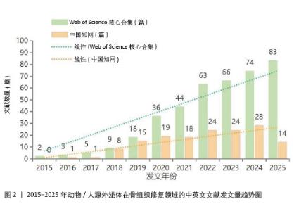
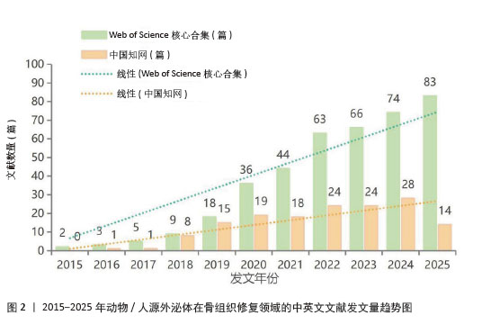
2.1 年发文量分析 运用CiteSpace软件分析中英文文献年发文量,将数据导入Microsoft Excel中绘制带趋势线的簇状柱形图,得到2015-2025年期间动物/人源外泌体在骨组织修复领域的中英文文献年度发文量分布和增长趋势。Web of Science核心合集英文发文量自2019年起增速加快,并于2025年达到峰值(83篇),显示国际学术界对该领域的探索仍处于高速发展期。中国知网中文文献增长相对平缓,2024年达产出高点(28篇)后趋于平稳,显示国内研究已进入稳定发展阶段。此外,中文文献中综述占比(85/152)高于论著,表明国内研究目前侧重于理论总结,高质量原创性论著产出仍有上升空间(图2)。 "
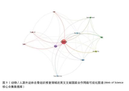
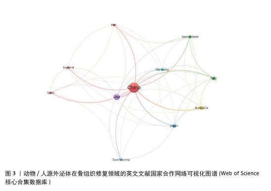
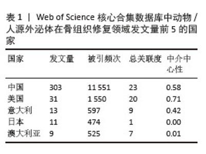
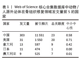
2.2 国家发文分析 基于Web of Science数据库,综合运用VOSviewer、CiteSpace和R包bibliometrix对动物/人源外泌体在骨组织修复领域发表的英文文献的国家分布和合作情况进行统计分析。国家合作网络可视化图谱中的节点大小代表发文量的多少,节点间的连线代表国家间的合作关系,连线的粗细代表合作的强度。从图3和表1中可以看出中国发文量(303篇)位居榜首,远超其他国家,显示了极高的产出活跃度。美国(31篇)与意大利(13篇)分列二、三位。然而,发文量并非衡量影响力的唯一指标。美国的中介中心性(0.71)位居全球首位,高于中国(0.58),说明美国在国际科研合作网络中发挥着关键的枢纽作用。相比之下,日本发文量虽位列前五,但中介中心性为0,提示其研究模式倾向于独立探索,在国际合作中相对边缘化。 "
"
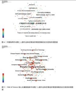
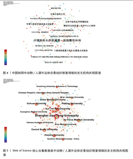
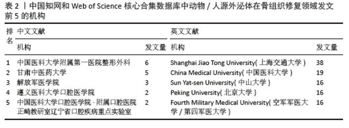
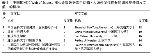
2.3 机构发文分析 基于Web of Science数据库和中国知网,节点类型选择为“机构”,运用CiteSpace软件对动物/人源外泌体在骨组织修复领域发表的中英文文献的机构进行统计分析,其中中文文献共纳入170个节点,95条连线,网络密度为0.006 6,英文文献共纳入185个节点,329条连线,网络密度为0.019 3。由此可见,该领域国内外机构间合作尚不紧密。中文文献中,中国医科大学附属第一医院整形外科发文量居首,甘肃中医药大学与解放军医学院次之。英文文献中,上海交通大学以38篇发文量位居第一,且中介中心性(0.11)最高,在合作网络中占据关键地位。由于多数机构中介中心性均未达到0.1,说明该领域研究虽在多家机构产出,但深层合作网络尚未完全成熟,跨机构协作有待加强(图4,5,表2)。 "
"
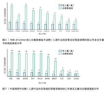
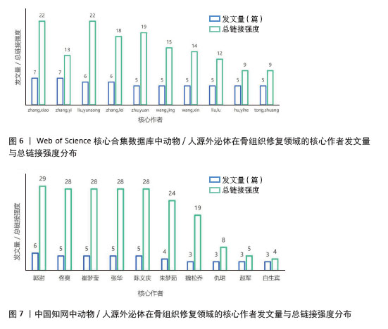
2.4 作者发文分析 基于Web of Science数据库和中国知网,运用Microsoft Office Excel2019对动物/人源外泌体在骨组织修复领域发表的中英文文献的作者进行统计分析。图6,7直观显示了该领域的科研力量分布与领军人物。在英文文献中,Zhang Xiao与Zhang Yi均以7篇的发文量位居榜首,Liu Yunsong、Zhang Lei以6篇紧随其后,其中Zhang Xiao与Liu Yunsong的总链接强度均为22,更加凸显其研究成果在国际学术界的影响力和认可度。在中文文献中,呈现以郭澍为核心的态势,发文量达到6篇,总链接强度为29,而佟爽、崔梦莹、张华及陈义庆学者均以5篇的产出并列第二,总链接强度均为28。 "
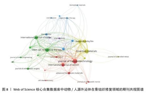
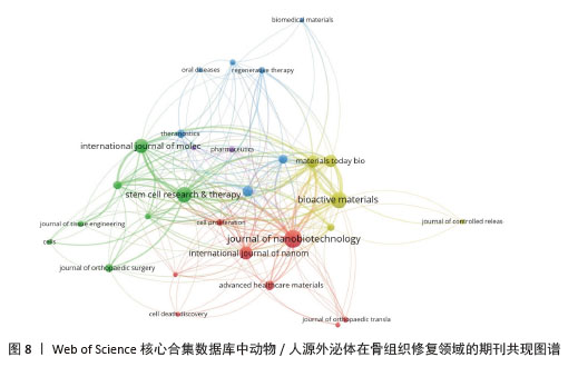
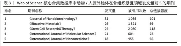
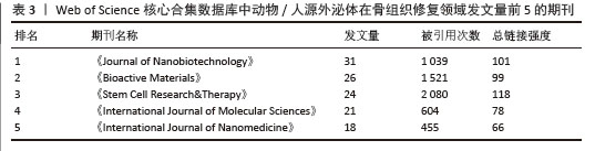
2.5 期刊可视化分析 运用VOSviewer软件对动物/人源外泌体在骨组织修复领域发表的英文文献的期刊来源进行可视化分析。在构建图谱时,把期刊发文量阈值(Minimum number of documents of a source)设置为4,最终从108种来源期刊中筛选出29种满足阈值要求的核心期刊构建可视化图谱(图8)。结合表3的统计数据,可以更深入地解析该领域的期刊分布特征。发文量分析显示, 《Journal of Nanobiotechnology》《Bioactive Materials》和《Stem Cell Research&Therapy》是该领域的主要成果发布平台。其中《Stem Cell Research&Therapy》被引用频次(2 080次)及总链接强度(118)最高,处于学术网络的核心地位。聚类分析显示,以纳米载体技术(红色聚类)与生物学机制(绿色聚类)为代表的研究方向紧密交织,表明该领域正由单一机制解析向生物材料与生物医学协同应用方向发展。"
"
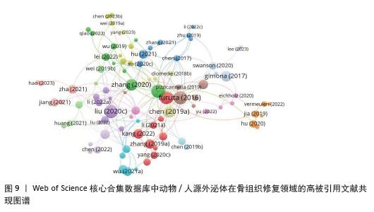
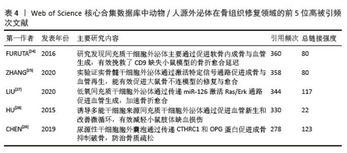
2.6 高被引文献结果分析 运用VOSviewer软件中“ bibliographic coupling-Documents”模块对动物/人源外泌体在骨组织修复领域发表的英文文献的引用结果进行量化分析。统计出403篇文章,被引频次≥40 次的有110篇,得到高被引用文献图谱(图9)。图中节点越大提示文献被引频次越多,节点颜色代表学术流派,节点连线代表文献间的关联,线条粗且多的文献处于相对核心的位置,学术价值较高。统计结果显示:在被引频次方面,FURUTA等[24] (2016)位居榜首,高达360 次,其次为ZHANG等[25] (2020)。然而,从总链接强度来看,CHEN等[26] (2019)以123的强度位居第一,LIU等[27] (2020)以117紧随其后,说明虽然它们的发表时间较晚或绝对被引数略低,但在该研究网络中处于更核心的位置,与其他文献的关联更为紧密。相比之下,HU等[28](2015)虽有较高的被引频次,但总链接强度相对较低(表4)。 "
"
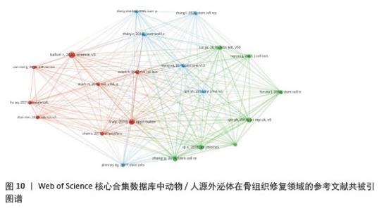
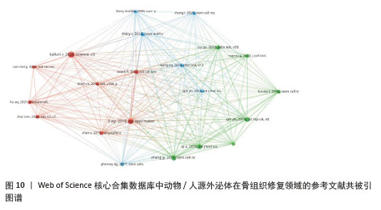
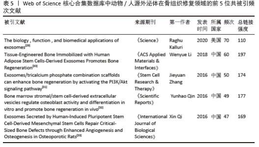
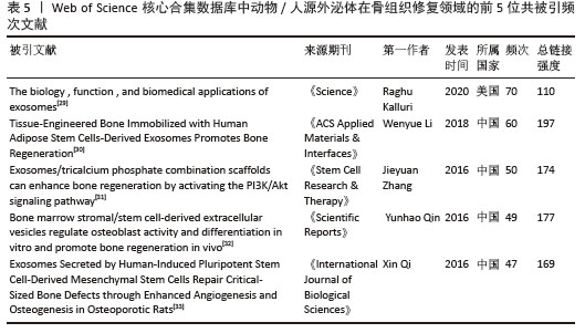
2.7 参考文献共被引结果 运用VOSviewer软件中“Co-citation-Cited references”模块对动/人源外泌体在骨组织修复领域的文献共被引情况进行可视化分析,共有17 491篇共被引的参考文献,共被引频次≥25次的有21篇,由此做出参考文献共被引共现图谱(图10)。如图所示,蓝色聚类主要代表外泌体定义和提取鉴定方式等基础理论研究;绿色聚类则聚焦于外泌体促进成骨及血管生成的具体研究;红色聚类则是聚焦于外泌体与生物材料结合的组织工程应用,这也是该领域目前的研究热点。结合表5的高共被引用文献可见[29-33],其中KALLURI等[29](2020)关于外泌体综述的被引用频次最高(70次),为该领域的研究提供了理论基础;而LI等[30](2018)关于支架负载外泌体的总链接强度最高(197),说明其研究在连接基础理论和实际应用之间起到关键作用。另外,前5篇高被引用文献中,有4篇来自中国学者,表明中国在该领域的研究中处于领先地位。 "
"
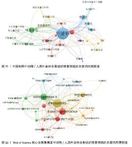
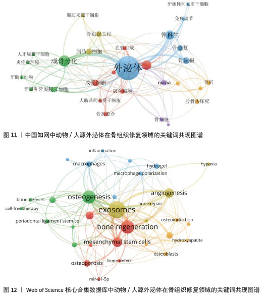
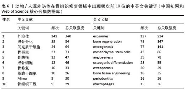
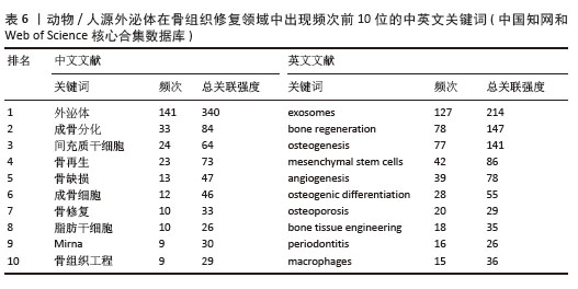
2.8 关键词 2.8.1 共现分析 运用VOSviewer软件中“Co-occurrence Author keywords”模块对动物/人源外泌体在骨组织修复领域的中英文文献关键词进行可视化分析,共现图谱每个节点代表1个关键词,节点大小代表关键词出现频率,连线越粗,共现频率越高(图11,12)。从图谱可见,英文文献的网络密度和聚类丰富度略高于中文文献,说明英文文献研究在该领域涉及的关联分支更为广泛,例如涉及了水凝胶和血管生成等方向,而中文文献的研究方向则相对集中,主要围绕干细胞和成骨分化展开。表6为中英文文献出现频次排名前10的关键词。中文文献代表关键词包括外泌体、成骨分化、间充质干细胞、骨再生等;英文文献代表关键词包括exosomes(外泌体)、bone regeneration(骨再生)、osteogenesis(成骨作用)、mesenchymal stem cells(间充质干细胞)等。无论是中文还是英文数据库,“外泌体”均为频次最高的关键词,表明其是连接各类骨组织修复研究方向的关键枢纽。而乳源外泌体关键词未显现是当前热点,乳源外泌体的研究有待进一步拓展。 "
"
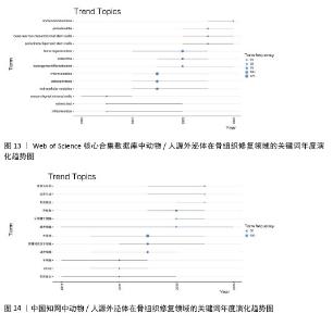
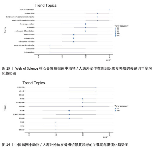
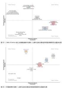
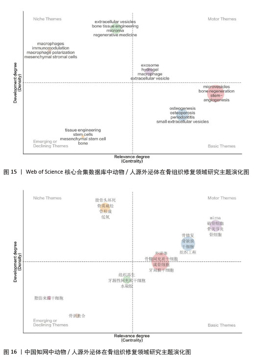
2.8.2 关键词趋势分析与主题演化 运用R语言中的Bibliometrix软件包分别对中英文数据库检索到的文献进行可视化分析,绘制出关键词年度流行趋势图(图13,14)与主题演化图谱(图15,16),分别从时间演进和空间架构两个方向来揭示该领域的研究热点和发展规律。 从时间趋势上看,中英文文献出现了相似的演变规律,并且大致可以分为3个阶段,在2019-2021年的早期阶段,英文文献中的高频词为“mesenchymal stromal cells”(间充质基质细胞)和“inflammation”(炎症),中文文献中的高频词为“骨折愈合”“破骨细胞” “miRNA”,说明该阶段研究的重心在于对细胞和机制层面的基础研究。而2022年前后,英文文献中的高频词为“extracellular vesicles” (细胞外囊泡)、“microvesicles”(微囊泡)和“osteogenesis”(成骨分化),中文文献中的高频词为“外泌体”“骨髓间充质干细胞”“成骨细胞”,说明该阶段学术界不再单纯关注干细胞本身的移植效果,而是深入研究干细胞分泌的亚细胞成分,如外泌体、细胞外囊泡等如何实现成骨功能,为后续研究奠定了基础。到了2023-2025年,研究的内容开始进一步向具体的临床应用和复杂机制延伸。英文文献中的高频词为“immunomodulation”(免疫调节)和“periodontitis”(牙周炎),中文文献中的高频词为“股骨头坏死”“血管生成”,说明当前的研究趋势已在具体的疾病模型中,通过免疫调控和血管微环境来促进骨组织再生。 从主题演化上看,该图谱的横坐标“Centrality”代表向心度,反映主题与其他领域的关联强度;纵坐标“Density”代表密度,反映主题内部的成熟度,依据象限分析可以从宏观层面揭示该领域的演化态势[34]。英文图谱所示,以“exosome”(外泌体)、“hydrogel”(水凝胶)为代表的紫色聚类位于第一象限,说明在国际上外泌体与水凝胶已经不再是单纯的基础概念或边缘性探索,而是目前最成熟且最具中心性的研究。以“extracellular vesicles”(细胞外囊泡)、“microrna”为代表的绿色聚类位于第二象限,说明该领域在机制和分子层面的研究已经较为完善。第四象限汇集了具有高向心性低密度的基础概念,如红色聚类中的“microvesicles”(微囊泡)与“bone regeneration”(骨再生),蓝色聚类中的“osteogenesis”(成骨发生)、“periodontitis”(牙周炎)和“osteoporosis”(骨质疏松)。而在第三象限中的几个关键词,反映了这些概念已经内化为学科的理论背景,不再是单一的创新点。中文图谱所示,以“骨修复”“干细胞”为代表的蓝色聚类和“mirna”“破骨细胞”为代表的紫色聚类位于第一象限,说明中文文献的研究在基础机制层面最为成熟。以“股骨头坏死”“骨质疏松”为代表的橙色聚类位于第二象限,说明国内研究者侧重于在特定的病理模型下研究骨修复的机制。第四象限则汇集了学科的基础主题,如红色聚类中的“外泌体”“骨髓间充质干细胞”“成骨细胞”,而绿色聚类中的“组织再生”“水凝胶”在第四象限出现,说明在计量学上水凝胶已经成为连接微观机制和宏观治疗的关键材料。然而,第三象限出现的内容代表边缘主题、热度平稳主题或已经衰退的主题。 "
"
| [1] ZHU G, ZHANG T, CHEN M, et al. Bone physiological microenvironment and healing mechanism: Basis for future bone-tissue engineering scaffolds. Bioact Mater. 2021;6(11):4110-4140. [2] XIE X, CHENG P, HU L, et al. Bone-targeting engineered small extracellular vesicles carrying anti-miR-6359-CGGGAGC prevent valproic acid-induced bone loss. Signal Transduct Target Ther. 2024;9(1):24. [3] CHEN X, WANG Z, DUAN N, et al. Osteoblast-osteoclast interactions. Connect Tissue Res. 2018; 59(2):99-107. [4] YU D, SHEN W, DAI J, et al. Treatment of large bone defects in load-bearing bone: traditional and novel bone grafts. J Zhejiang Univ Sci B. 2025;26(5):421-447. [5] RODHAM PL, GIANNOUDIS VP, KANAKARIS NK, et al. Biological aspects to enhance fracture healing. EFORT Open Rev. 2023;8(5):264-282. [6] MOVANIYA P, CHHATBAR R, BHATT S, et al. Comparison of Osteoinductive Potential of Autografts vs. Allografts in Mandibular Defect Models. J Pharm Bioallied Sci. 2025;17(Suppl 2): S1399-S1401. [7] HAWS L, MA B, GODFREY T, et al. Complication Rates of the Anterior Iliac Crest Bone Graft Donor Site for 426 Pediatric Patients Undergoing Alveolar Bone Grafting. J Oral Maxillofac Surg. 2025;83(9):1101-1105. [8] LU Y, MAI Z, CUI L, et al. Engineering exosomes and biomaterial-assisted exosomes as therapeutic carriers for bone regeneration. Stem Cell Res Ther. 2023;14(1):55. [9] ZOU J, YANG W, CUI W, et al. Therapeutic potential and mechanisms of mesenchymal stem cell-derived exosomes as bioactive materials in tendon-bone healing. J Nanobiotechnology. 2023;21(1):14. [10] KE W, AFONIN KA. Exosomes as natural delivery carriers for programmable therapeutic nucleic acid nanoparticles (NANPs). Adv Drug Deliv Rev. 2021;176:113835. [11] HUANG CC, KANG M, LU Y, et al. Functionally engineered extracellular vesicles improve bone regeneration. Acta Biomater. 2020;109:182-194. [12] GARCÍA-MARTÍNEZ J, SALTO R, GIRÓN MD, et al. Supplementation with a Whey Protein Concentrate Enriched in Bovine Milk Exosomes Improves Longitudinal Growth and Supports Bone Health During Catch-Up Growth in Rats. Nutrients. 2024;16(22):3814. [13] YU X, DONG M, WANG L, et al. Nanotherapy for bone repair: milk-derived small extracellular vesicles delivery of icariin. Drug Deliv. 2023;30(1): 2169414. [14] OLIVEIRA MC, PIETERS BCH, GUIMARÃES PB, et al. Bovine Milk Extracellular Vesicles Are Osteoprotective by Increasing Osteocyte Numbers and Targeting RANKL/OPG System in Experimental Models of Bone Loss. Front Bioeng Biotechnol. 2020;8:891. [15] LIU R, LIU S, WU S, et al. Milk-Derived Small Extracellular Vesicles Promote Osteogenic Differentiation and Inhibit Inflammation via microRNA-21. Int J Mol Sci. 2023;24(18):13873. [16] LEE CS, FAN J, HWANG HS, et al. Bone-Targeting Exosome Mimetics Engineered by Bioorthogonal Surface Functionalization for Bone Tissue Engineering. Nano Lett. 2023;23(4):1202-1210. [17] XU C, LI Z, KANG M, et al. Hydrogel-integrated exosome mimetics derived from osteogenically induced mesenchymal stem cells in spheroid culture enhance bone regeneration. Biomaterials. 2025;317:123088. [18] LIAN M, QIAO Z, QIAO S, et al. Nerve Growth Factor-Preconditioned Mesenchymal Stem Cell-Derived Exosome-Functionalized 3D-Printed Hierarchical Porous Scaffolds with Neuro-Promotive Properties for Enhancing Innervated Bone Regeneration. ACS Nano. 2024;18(10):7504-7520. [19] ELLEGAARD O, WALLIN JA. The bibliometric analysis of scholarly production: How great is the impact? Scientometrics. 2015;105(3):1809-1831. [20] VAN ECK NJ, WALTMAN L. Software survey: VOSviewer, a computer program for bibliometric mapping. Scientometrics. 2010;84(2):523-538. [21] CHEN CM. CiteSpace II: Detecting and Visualizing Emerging Trends and Transient Patterns in Scientific Literature. J Am Soc Inf Sci Technol. 2006;57(3):359-377. [22] 郑颖,黄珂敏.外泌体研究态势和前瞻的计量与可视化分析[J].中国组织工程研究,2024, 28(13):2126-2132. [23] 陈悦,陈超美,刘则渊,等.CiteSpace知识图谱的方法论功能[J].科学学研究,2015,33(2):242-253. [24] FURUTA T, MIYAKI S, ISHITOBI H, et al. Mesenchymal Stem Cell-Derived Exosomes Promote Fracture Healing in a Mouse Model. Stem Cells Transl Med. 2016;5(12):1620-1630. [25] ZHANG L, JIAO G, REN S, et al. Exosomes from bone marrow mesenchymal stem cells enhance fracture healing through the promotion of osteogenesis and angiogenesis in a rat model of nonunion. Stem Cell Res Ther. 2020;11(1):38. [26] CHEN CY, RAO SS, TAN YJ, et al. Extracellular vesicles from human urine-derived stem cells prevent osteoporosis by transferring CTHRC1 and OPG. Bone Res. 2019;7:18. [27] LIU W, LI L, RONG Y, et al. Hypoxic mesenchymal stem cell-derived exosomes promote bone fracture healing by the transfer of miR-126. Acta Biomater. 2020;103:196-212. [28] HU GW, LI Q, NIU X, et al. Exosomes secreted by human-induced pluripotent stem cell-derived mesenchymal stem cells attenuate limb ischemia by promoting angiogenesis in mice. Stem Cell Res Ther. 2015;6(1):10. [29] KALLURI R, LEBLEU VS. The biology, function, and biomedical applications of exosomes. Science. 2020;367(6478):eaau6977. [30] LI W, LIU Y, ZHANG P, et al. Tissue-Engineered Bone Immobilized with Human Adipose Stem Cells-Derived Exosomes Promotes Bone Regeneration. ACS Appl Mater Interfaces. 2018;10(6):5240-5254. [31] ZHANG J, LIU X, LI H, et al. Exosomes/tricalcium phosphate combination scaffolds can enhance bone regeneration by activating the PI3K/Akt signaling pathway. Stem Cell Res Ther. 2016;7(1):136. [32] QIN Y, WANG L, GAO Z, et al. Bone marrow stromal/stem cell-derived extracellular vesicles regulate osteoblast activity and differentiation in vitro and promote bone regeneration in vivo. Sci Rep. 2016;6:21961. [33] QI X, ZHANG J, YUAN H, et al. Exosomes Secreted by Human-Induced Pluripotent Stem Cell-Derived Mesenchymal Stem Cells Repair Critical-Sized Bone Defects through Enhanced Angiogenesis and Osteogenesis in Osteoporotic Rats. Int J Biol Sci. 2016;12(7):836-849. [34] 魏瑞斌.社会网络分析在关键词网络分析中的实证研究[J].情报杂志,2009,28(9):46-49. [35] YUN B, MABURUTSE BE, KANG M, et al. Short communication: Dietary bovine milk-derived exosomes improve bone health in an osteoporosis-induced mouse model. J Dairy Sci. 2020;103(9):7752-7760. [36] CHEN L, ZHU J, GE N, et al. A biodegradable magnesium alloy promotes subperiosteal osteogenesis via interleukin-10-dependent macrophage immunomodulation. Biomaterials. 2025;318:122992. [37] 张熊劲夫,陈奕达,程歆怡,等.年轻大鼠骨髓间充质干细胞来源外泌体逆转老龄大鼠骨髓间充质干细胞衰老[J].中国组织工程研究, 2025,29(36):5776-5785. [38] HU S, WANG S, YANG X, et al. Exosomes promise better bone regeneration. Regen Ther. 2025;30: 389-402. [39] TIAN L, ZHANG D, WANG Z, et al. MiR-877, an exosomal miRNA from mechanical stretch induced adipose derived stromal cells, enhances fracture healing in nonunion rats with type 2 diabetes mellitus. Stem Cell Res Ther. 2025;16(1):575. [40] LIU L, ZHOU N, FU S, et al. Endothelial cell-derived exosomes trigger a positive feedback loop in osteogenesis-angiogenesis coupling via up-regulating zinc finger and BTB domain containing 16 in bone marrow mesenchymal stem cell. J Nanobiotechnology. 2024;22(1):721. [41] SHAN J, CHENG L, LI X, et al. End-tail soaking strategy toward robust and biomimetic sandwich-layered hydrogels for full-thickness bone regeneration. Bioact Mater. 2025;49:486-501. [42] LEI T, ZHANG T, FANG T, et al. Engineering a stem cell-embedded bilayer hydrogel with biomimetic collagen mineralization for tendon-bone interface healing. Bioact Mater. 2025;49:207-217. [43] HU J, WEI J, LIU J, et al. A Novel Strategy for Fabrication of Polyamide 66/Nanohydroxyapatite Composite Bone Repair Scaffolds by Low-Temperature Three-Dimensional Printing. ACS Biomater Sci Eng. 2024;10(6):4073-4084. [44] SI Y, LI X, DONG S, et al. Hydrogel scaffold encapsulating MSC-Exos and ZIF-8 promotes bone regeneration via coordinating osteogenesis and immunomodulation. Bioact Mater. 2025;54:329-351. [45] CHEN P, WANG R, FANG S. Metabolic glycoengineered exosome-A2M nanoplatform reprograms macrophage polarization and orchestrates bone regeneration in ONFH. Cell Death Discov. 2025;11(1):510. [46] GUO Q, REN S, LIBONATI A, et al. Antler stem cell-derived exosomes restore periodontal homeostasis in a rat model with diabetic periodontitis through enhancing ROS scavenging and osteogenesis. Cell Death Discov. 2025;11(1):500. [47] WANG G, CUI Y, LENG Y, et al. Engineered three-dimensional bioactive scaffold for enhanced bone regeneration through modulating transplanted adipose derived mesenchymal stem cell and stimulating angiogenesis. Front Bioeng Biotechnol. 2024;12:1342590. [48] DU R, ZHAO B, LUO K, et al. Shape Memory Polyester Scaffold Promotes Bone Defect Repair through Enhanced Osteogenic Ability and Mechanical Stability. ACS Appl Mater Interfaces. 2023;15(36):42930-42941. [49] DONG M, SHI C, YU X, et al. Milk-derived small extracellular vesicles: nanomaterials to promote bone formation. J Nanobiotechnology. 2022; 20(1):370. [50] HUANG Q, JIANG Y, CAO Y, et al. Bone-targeting engineered milk-derived extracellular vesicles for MRI-assisted therapy of osteoporosis. Regen Biomater. 2024;11:rbae112. [51] LI J, YANG R, ZHANG J, et al. Research progress of exosome-hydrogel between 2013 and 2023 from the perspective of bibliometrics. Medicine (Baltimore). 2025;104(51):e46595. |
| [1] | Xu Canli, He Wenxing, Wang Yuping, Ba Yinying, Chi Li, Wang Wenjuan, Wang Jiajia. Research context and trend of TBK1 in autoimmunity, signaling pathways, gene expression, tumor prevention and treatment [J]. Chinese Journal of Tissue Engineering Research, 2026, 30(在线): 1-11. |
| [2] | Zhu Xiaolong, Zhang Wei, Yang Yang. Visualization analysis of research hotspots and cutting-edge information in the field of intervertebral disc regeneration and repair [J]. Chinese Journal of Tissue Engineering Research, 2026, 30(9): 2391-2402. |
| [3] | Wen Fayan, Li Yan, Qiang Tianming, Yang Chen, Shen Linming, Li Yadong, Liu Yongming. Unilateral biportal endoscopic technology for treatment of lumbar degenerative diseases: global research status and changing trends [J]. Chinese Journal of Tissue Engineering Research, 2026, 30(9): 2380-2390. |
| [4] | Lai Yu, Chen Yueping, Zhang Xiaoyun. Research hotspots and frontier trends of bioactive materials in treating bone infections [J]. Chinese Journal of Tissue Engineering Research, 2026, 30(8): 2132-2144. |
| [5] | Ye Qianqian, Pan Hang, Tian Chuan, Zhu Xiangqing, Ye Li, Zhao Xiaojuan, Shu Liping, Pan Xinghua. Effects of highly active umbilical cord mesenchymal stem cells on structure and function of thymus in elderly tree shrews [J]. Chinese Journal of Tissue Engineering Research, 2026, 30(7): 1720-1729. |
| [6] | Huang Jie, Zeng Hao, Wang Wenchi, Lyu Zhucheng, Cui Wei. Visualization analysis of literature on the effect of lipid metabolism on osteoporosis [J]. Chinese Journal of Tissue Engineering Research, 2026, 30(6): 1558-1568. |
| [7] | Yu Shiyu, Yu Sutong, Xu Yang, Zhen Xiangyan, Han Fengxuan. Advances in research and application of tissue engineering therapeutic strategies in oral submucous fibrosis [J]. Chinese Journal of Tissue Engineering Research, 2026, 30(4): 936-948. |
| [8] | Bei Chaoyong, Zhu Jialin, Yu Zhengze, Shi Niujin, Li Huarui, Peng Fenglin. Visualization analysis of research hotspots and trends in the treatment of knee osteoarthritis [J]. Chinese Journal of Tissue Engineering Research, 2026, 30(36): 9632-9639. |
| [9] | Xu Hongtao, Wang Jianping, Xu Yuehong, Li Qin, Qi Qihua, Xia Qipeng. Asiaticoside promotes osteogenic differentiation in osteoporotic rats [J]. Chinese Journal of Tissue Engineering Research, 2026, 30(35): 9113-9119. |
| [10] | Chen Yida, Cheng Xinyi, Zhao Huan, Zhou Xichao, Gu Qiaoli, Lin Xiao, Shi Qin. Effects of physiological osmotic pressure on chondrocyte differentiation and extracellular matrix metabolism [J]. Chinese Journal of Tissue Engineering Research, 2026, 30(35): 9143-9150. |
| [11] | Wang Quan, Tang Zhenglong. Visualization analysis of literature on medication-related osteonecrosis of the jaw [J]. Chinese Journal of Tissue Engineering Research, 2026, 30(35): 9327-9335. |
| [12] | Chen Xiaoxia, Zhao Lihua, Li Taowen, Qin Yimei, Liang Jinyu. Postmenopausal cognitive impairment: a bibliometric analysis of developmental context and hot trends [J]. Chinese Journal of Tissue Engineering Research, 2026, 30(35): 9336-9344. |
| [13] | He Renda, Ma Wei, Sun Yongsi, Mo Xueni. Future medical research on brain organoids: interdisciplinary training, bioengineering technologies, and optimized model maturity [J]. Chinese Journal of Tissue Engineering Research, 2026, 30(34): 9088-9094. |
| [14] | Bu Fanchen, Hua Zhen, , Li Xiaolong, Lyu Jinye, Man Hao, Wang Jianwei, . Triangular fibrocartilage complex injuries: a visualization analysis of treatment hotspots and frontiers [J]. Chinese Journal of Tissue Engineering Research, 2026, 30(34): 9095-9102. |
| [15] | Xia Jinyan, Su Meifang, Yang Wenyu, Hu Qilan, Gong Li. Application of novel materials in tissue repair of diabetic foot wounds [J]. Chinese Journal of Tissue Engineering Research, 2026, 30(32): 8563-8568. |
| Viewed | ||||||
|
Full text |
|
|||||
|
Abstract |
|
|||||