Chinese Journal of Tissue Engineering Research ›› 2026, Vol. 30 ›› Issue (9): 2391-2402.doi: 10.12307/2026.063
Previous Articles Next Articles
Visualization analysis of research hotspots and cutting-edge information in the field of intervertebral disc regeneration and repair
Zhu Xiaolong1, 2, Zhang Wei1, 3, Yang Yang1, 3
- 1Jiangxi University of Traditional Chinese Medicine, Nanchang 330000, Jiangxi Province, China; 2Fuyang Traditional Chinese Medicine Orthopedic Hospital, Hangzhou 311400, Zhejiang Province, China; 3Affiliated Hospital of Jiangxi University of Chinese Medicine, Nanchang 330000, Jiangxi Province, China
-
Received:2024-12-30Accepted:2025-04-03Online:2026-03-28Published:2025-09-29 -
Contact:Zhang Wei, PhD, Chief physician, Doctoral supervisor, Jiangxi University of Traditional Chinese Medicine, Nanchang 330000, Jiangxi Province, China; Affiliated Hospital of Jiangxi University of Chinese Medicine, Nanchang 330000, Jiangxi Province, China -
About author:Zhu Xiaolong, Doctoral candidate, Attending physician, Jiangxi University of Traditional Chinese Medicine, Nanchang 330000, Jiangxi Province, China; Fuyang Traditional Chinese Medicine Orthopedic Hospital, Hangzhou 311400, Zhejiang Province, China -
Supported by:Hangzhou Medical and Health Science and Technology Project, No. B20220070 (to ZXL)
CLC Number:
Cite this article
Zhu Xiaolong, Zhang Wei, Yang Yang. Visualization analysis of research hotspots and cutting-edge information in the field of intervertebral disc regeneration and repair[J]. Chinese Journal of Tissue Engineering Research, 2026, 30(9): 2391-2402.
share this article
Add to citation manager EndNote|Reference Manager|ProCite|BibTeX|RefWorks
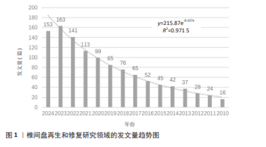
2.1 年度发文量趋势 椎间盘再生与修复研究的发文量详见图1,近15年来相关研究的发文量总体呈逐年上升趋势,发文量与时间相关关系符合指数增长模型,其方程为y=215.87e-0.157x,R2=0.971 5,符合普赖斯提出的科学文献指数增长规律,2022-2023年度发文量增加明显,2024年发文量较2023年度略下降,自2021年开始,相关发文量均保持在100篇以上,在2023年达到高峰163篇,可见近几年的研究热点较高。 "
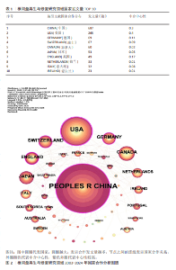
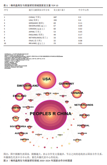
2.2 椎间盘再生与修复研究领域的国家分布情况 使用CiteSpace 6.4.R1软件进行国家分布的可视化分析,如表1,图2所示,2003-2024年,共有48个国家发表了该领域相关研究,合作图谱共得到48个节点,168条连线,各国间有良好合作关系。发文数量排名前5国家依次是:中国687篇,美国265篇,德国69篇,瑞士67篇,加拿大60篇。中介中心性前5国家依次为:美国(0.4),中国(0.3),英国(0.17),德国(0.11),瑞士(0.09),意大利(0.08)。中国的发文量最高,是美国的2倍多,但是中介中心性低于美国。节点的分析表明,中国、美国、英国及德国具有很高的中心性,他们与其他国家合作较多。"
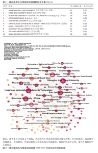
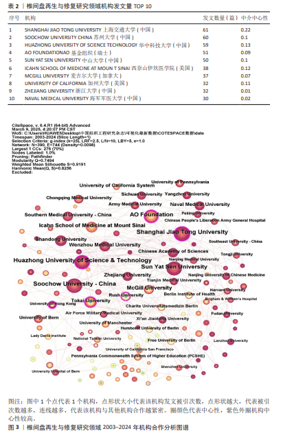
2.3 椎间盘再生与修复研究领域的机构分布情况 使用CiteSpace 6.4.R1软件进行机构分布的可视化分析,如表2,图3所示,2003-2024年,共有390个机构发表了该领域相关研究,合作图谱共得到390个节点、1 019条连线,各国间有良好合作关系。发文数量排名前5的机构依次是:上海交通大学61篇,苏州大学60篇,华中科技大学59篇,AO基金组织51篇,中山大学50篇。中介中心性前5的是上海交通大学、华中科技大学、西奈山伊坎医学院、加州大学、苏州大学。节点分析表明,中心度> 0.1的节点表示关键作用较好,从图3中可以看出上海交通大学、华中科技大学、AO基金组织(瑞士)、东海大学(日本)、拉什大学(美国)及香港大学具有较高的中心性。"
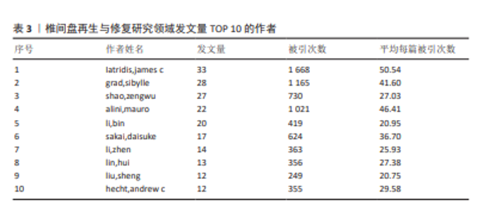
2.4 作者合作分析 根据普莱斯法则推算出核心作者的发文量,该计算公式为N=0.749×(Nmax)1/2,其中 Nmax代表发文量最多的作者的文献数量[10]。通过计算,得到N=4.29,因此,该领域核心作者的最低发文量应为5篇。通过VOSviewer 1.6.20软件的统计,发现核心作者共有235位,他们共发表996篇论文,占该领域总论文数的81.4%,这表明核心作者发表了椎间盘再生和修复领域4/5的研究成果,此次研究汇总了发文量排名前10的作者详见表3,其中来自美国西奈山伊坎医学院的Iatridis,james c学者发表33篇,总被引次数1 668次,平均每篇被引50.54次,其发文量和被引次数均排名第一,可见该学者在椎间盘再生和修复领域做出了巨大贡献。图4为作者合作分析图谱,包括该领域发文量前235的研究者,从图谱中可以发现尽管椎间盘再生与修复研究存在一定的地域性差异,但跨国合作的作者数量不容忽视,表明该领域的研究正逐步实现全球化,不同国家和地区之间的科研协作在推动创新治疗方案和新技术研发方面发挥了积极作用。"
"
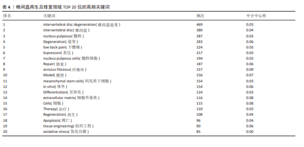
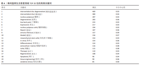
2.5 椎间盘再生与修复领域关键词共现、聚类分析 高频关键词的共现可以直观表现出该领域的研究热点,此次研究采用VOSviewer 1.6.20软件对1 229篇该领域的文献进行关键词共现分析,排名前20的高频关键词详见表4,从这些高频关键词中可以看出该领域研究方向主要聚集在椎间盘退变的发生机制及椎间盘再生和修复的策略上。关键词共现及聚类图谱详见图5,可以看出共有4个聚类方向:①椎间盘结构与组织工程(15个项目):纤维环,关节,细胞,胶原蛋白,退变,细胞外基质,基因表达,体内,椎间盘,间充质干细胞,髓核,再生,修复,组织,组织工程;②椎间盘退变的机制(13个项目):激活,凋亡,自噬,疾病,表达,炎症,椎间盘退变,下腰痛,髓核细胞,氧化应激,增生,衰老,肿瘤坏死因子α;③椎间盘退变的研究模型建立与治疗策略(7个项目):椎间盘退变,疝出,水凝胶,损伤,模型,针灸,脊柱;④干细胞与再生医学(5个项目):差异化,体外,干细胞,治疗, 移植。"
"

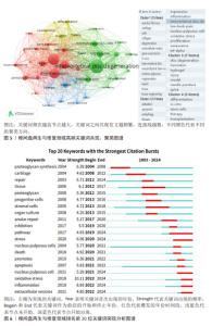
2.6 椎间盘再生与修复领域关键词突现分析、关键词时间线图 排名前20位关键词突现分析图谱详见图6,突现强度排名前20的关键词分别为:蛋白聚糖合成、软骨、修复、组织、蛋白聚糖、祖细胞、基质细胞、器官培养、纤维环修复、抑制、通路、应激、髓核细胞、死亡、促进细胞、凋亡、髓核细胞、氧化应激、炎症、细胞外囊泡。从图中可见关键词“氧化应激”展现出最高的突现强度。此外,关键词如“通路” “应激” “髓核细胞” “氧化应激” “炎症”和“细胞外囊泡”的突现尚未结束,表明这些领域仍然是近年来的研究 热点。 椎间盘再生与修复领域关键词时间线图详见图7,排序前10位聚类标签分别为:#0 氧化应激、#1 动物模型、#2 间充质干细胞、#3 髓核、#4 纤维环修复、#5 椎间盘退化、#6 增殖、#7 椎间盘细胞、#8 组织工程、#9 外泌体。"

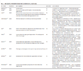
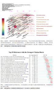
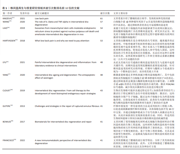
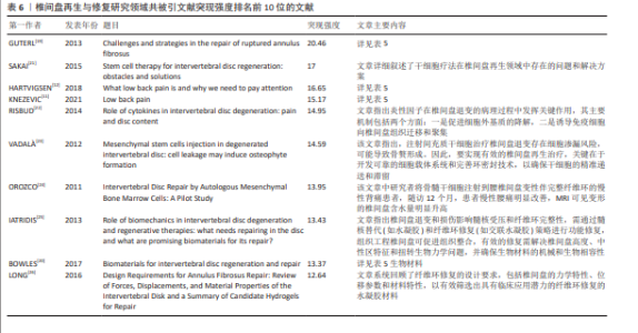
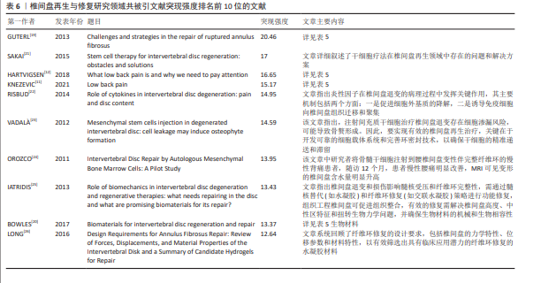
2.7 文献共被引分析 文献共被引分析揭示了椎间盘再生与修复领域的经典文献及研究前沿,在引用频次排名前10的文献中,3篇涉及椎间盘退变及腰痛的研究现状论述[11-13],1篇探讨椎间盘退变的机制[14],其余6篇聚焦于干细胞、内源性修复细胞以及组织工程等领域,探讨这些技术在促进椎间盘再生与修复方面的应用[15-20],共引用次数排名前10的文献详见表5。文献共被引次数排名第一的是Nebojsa Nick Knezevic学者于2021年在《Lancet》杂志发表的1篇文章,被引61次,文章指出了腰痛是全球生产力减退的首要原因,也是126个国家健康寿命减少的头号原因,该研究总结了腰痛的危险因素,包括遗传因素、女性性别、不良生活习惯、社会心理因素、应对机制不佳、创伤性损伤及职业危害等[11]。文献共被引次数排名第2的是王永杰研究团队在2020年在《Biomed Pharmacother》发表的1篇文章[15],该文章揭示多种细胞因子在椎间盘退变中起关键作用,如白细胞介素1β、肿瘤坏死因子α、白细胞介素6和白细胞介素17,尤其是白细胞介素1β和肿瘤坏死因子α在退行性椎间盘退变中表达显著升高,广泛研究表明白细胞介素1β和肿瘤坏死因子α在椎间盘退变中的功能作用,为其作为潜在治疗靶点的临床应用提供了新思路。共被引次数排名第3的是廖志伟团队于2019年在《Theranostics》发表的1篇文章[16],文章研究发现退行性椎间盘组织的内质网应激水平及凋亡率升高,骨髓间充质干细胞来源外泌体可通过激活蛋白激酶B(protein kinase B,AKT)和细胞外信号调节激酶(extracellular signal-regulated kinase,ERK)信号通路减轻内质网应激诱导的细胞凋亡,此外,在大鼠尾部模型中,骨髓间充质干细胞的体内递送能够调节内质网应激相关的细胞凋亡并延缓椎间盘退变。 2.8 共被引文献突现分析 突现强度可以较好地展现研究领域在一定时间范围内的研究前沿,呈现该领域的聚焦热点及趋势。2003-2024年该领域排名前20位共被引文献突发分析图谱详见图8。共被引文献突现强度排名前10位的文献详见表6。"
"
| [1] BUSER Z, ORTEGA B, D’ORO A, et al. Spine Degenerative Conditions and Their Treatments: National Trends in the United States of America. Global Spine J. 2018; 8(1):57-67. [2] CIEZA A, CAUSEY K, KAMENOV K, et al. Global estimates of the need for rehabilitation based on the Global Burden of Disease study 2019: a systematic analysis for the Global Burden of Disease Study 2019. Lancet. 2021;396(10267):2006-2017. [3] 蔡熊熊,应镒剑,张迟,等.椎间盘退变水凝胶修复策略的研究进展[J].现代实用医学,2024,36(7):974-977. [4] 柳绪超,秦雷,易伟宏.干细胞移植治疗椎间盘退变的研究现状[J].中国脊柱脊髓杂志,2023,33(12):1138-1143. [5] 刘亭亭,韩长旭,王国强.细胞移植修复椎间盘的新视角与新进展[J].中国组织工程研究,2020,24(1):154-158. [6] 宋婵婵,冉兵,宗毅,等.椎间盘退变机制及修复生物工程支架研究进展[J].中国疼痛医学杂志,2022,28(9):657-663. [7] 潘玉军,时长江,曹胜,等.负载外泌体的可注射葡聚糖/明胶复合水凝胶修复大鼠椎间盘退变[J].中国组织工程研究, 2023,27(34):5477-5482. [8] CAO S, MA Y, YANG H, et al. Long noncoding RNA HCG18 Promotes Extracellular Matrix Degradation of Nucleus Pulposus Cells in Intervertebral Disc Degeneration by Regulating the miR-4306/EPAS1 Axis. World Neurosurg. 2023;172:52-61. [9] WANG L, WANG Y, JIAO J, et al. Therapeutic Potential of Mesenchymal Stem Cell-Derived Extracellular Vesicles Carrying MicroRNAs for Modulating Autophagy and Cellular Degeneration in Intervertebral Disc Degeneration. Spine (Phila Pa 1976). 2025;50(1):7-19. [10] 赵盾,祁令臣,徐金凡,等.膝骨关节炎疼痛领域热点与前沿的可视化分析[J].中国组织工程研究,2025,29(15):3280-3289. [11] KNEZEVIC NN, CANDIDO KD, VLAEYEN J, et al. Low back pain. Lancet. 2021;398(10294): 78-92. [12] HARTVIGSEN J, HANCOCK MJ, KONGSTED A, et al. What low back pain is and why we need to pay attention. Lancet. 2018; 391(10137):2356-2367. [13] FRANCISCO V, PINO J, GONZALEZ-GAY MA, et al. A new immunometabolic perspective of intervertebral disc degeneration. Nat Rev Rheumatol. 2022;18(1):47-60. [14] LYU FJ, CUI H, PAN H, et al. Painful intervertebral disc degeneration and inflammation: from laboratory evidence to clinical interventions. Bone Res. 2021;9(1):7. [15] WANG Y, CHE M, XIN J, et al. The role of IL-1beta and TNF-alpha in intervertebral disc degeneration. Biomed Pharmacother. 2020;131:110660. [16] LIAO Z, LUO R, LI G, et al. Exosomes from mesenchymal stem cells modulate endoplasmic reticulum stress to protect against nucleus pulposus cell death and ameliorate intervertebral disc degeneration in vivo. Theranostics. 2019;9(14):4084-4100. [17] YANG S, ZHANG F, MA J, et al.Intervertebral disc ageing and degeneration: The antiapoptotic effect of oestrogen. Ageing Res Rev. 2020;57:100978. [18] CLOUET J, FUSELLIER M, CAMUS A, et al.Intervertebral disc regeneration: From cell therapy to the development of novel bioinspired endogenous repair strategies. Adv Drug Deliv Rev. 2019;146:306-324. [19] GUTERL CC, SEE EY, BLANQUER SB, et al. Challenges and strategies in the repair of ruptured annulus fibrosus. Eur Cell Mater. 2013;25:1-21. [20] BOWLES RD, SETTON LA. Biomaterials for intervertebral disc regeneration and repair. Biomaterials. 2017;129:54-67. [21] SAKAI D, ANDERSSON GB. Stem cell therapy for intervertebral disc regeneration: obstacles and solutions. Nat Rev Rheumatol. 2015; 11(4):243-256. [22] RISBUD MV, SHAPIRO IM. Role of cytokines in intervertebral disc degeneration: pain and disc content. Nat Rev Rheumatol. 2014; 10(1):44-56. [23] VADALÀ G, SOWA G, HUBERT M, et al. Mesenchymal stem cells injection in degenerated intervertebral disc: cell leakage may induce osteophyte formation. J Tissue Eng Regen Med. 2012;6(5):348-355. [24] OROZCO L, SOLER R, MORERA C, et al. Intervertebral disc repair by autologous mesenchymal bone marrow cells: a pilot study. Transplantation. 2011;92(7):822-828. [25] IATRIDIS JC, NICOLL SB, MICHALEK AJ, et al. Role of biomechanics in intervertebraldisc degeneration and regenerative therapies: what needs repairing in the disc and what are promising biomaterials for its repair? Spine J. 2013;13(3):243-262. [26] LONG RG, TORRE OM, HOM WW, et al. Design Requirements for Annulus Fibrosus Repair: Review of Forces, Displacements, and Material Properties of the Intervertebral Disk and aSummary of Candidate Hydrogels for Repair. J Biomech Eng. 2016;138(2):21007. [27] PANEBIANCO CJ, CONSTANT C, VERNENGO AJ, et al. Combining adhesive and nonadhesive injectable hydrogels for intervertebral disc repair in an ovine discectomy model. JOR Spine. 2023;6(4):1293. [28] BASATVAT S, BACH FC, BARCELLONA MN, et al. Harmonization and standardization of nucleus pulposus cell extraction and culture methods. JOR Spine. 2023; 6(1):1238. [29] DISTEFANO TJ, VASO K, PANEBIANCO CJ, et al. Hydrogel-Embedded Poly(Lactic-co-Glycolic Acid) Microspheres for the Delivery of hMSC-Derived Exosomes to Promote Bioactive Annulus Fibrosus Repair. Cartilage. 2022;13(3):788738969. [30] PANEBIANCO CJ, RAO S, HOM WW, et al. Genipin-crosslinked fibrin seeded with oxidized alginate microbeads as a novel composite biomaterial strategy for intervertebral disc cell therapy. Biomaterials. 2022;287:121641. [31] GULLBRAND SE, ASHINSKY BG, LAI A, et al. Development of a standardized histopathology scoring system for intervertebral disc degeneration and regeneration in rabbit models:An initiative of the ORS spine section. JOR Spine. 2021; 4(2):1147. [32] DISTEFANO TJ, SHMUKLER JO, DANIAS G, et al. Development of a two-part biomaterial adhesive strategy for annulus fibrosus repair and ex vivo evaluation of implant herniation risk. Biomaterials. 2020; 258:120309. [33] SARAVI B, LI Z, BASOLI V, et al.In Vitro Characterization of a Tissue Renin-Angiotensin System in Human Nucleus Pulposus Cells. Cells. 2022;11(21):3418. [34] GUO W, DOUMA L, HU MH, et al.Hyaluronic acid-based interpenetrating network hydrogel as a cell carrier for nucleus pulposus repair. Carbohydr Polym. 2022; 277:118828. [35] CUNHA C, LEITE PC, FERREIRA JR, et al.Therapeutic Strategies for IVD Regeneration through Hyaluronan/SDF-1-Based Hydrogel and Intravenous Administration of MSCs. Int J Mol Sci. 2021; 22(17):9609. [36] RUSSO F, AMBROSIO L, PEROGLIO M, et al.A Hyaluronan and Platelet-Rich Plasma Hydrogel for Mesenchymal Stem Cell Delivery in the Intervertebral Disc: An Organ Culture Study. Int J Mol Sci. 2021; 22(6):2963. [37] ZHAO K, ZHANG Y, LIAO Z, et al.Melatonin mitigates intervertebral disc degeneration by suppressing NLRP3 inflammasome activation via the EGR1/DDX3X pathway. FASEB J. 2024;38(24):70143. [38] LIAO Z, TONG B, ZHANG X, et al.Selective cargo sorting in stem cell-derived small extracellular vesicles: impact on therapeutic efficacy for intervertebral disc degeneration. Clin Transl Med. 2023;13(12):1494. [39] LIAO Z, KE W, LIU H, et al. Vasorin-containing small extracellular vesicles retard intervertebral disc degeneration utilizing an injectable thermoresponsive delivery system. J Nanobiotechnology. 2022;20(1): 420. [40] PAN C, HOU W, DENG X, et al. The Pivotal Role of Nrf2 Signal Axis in Intervertebral Disc Degeneration. J Inflamm Res. 2023; 16:5819-5833. [41] LIANG H, LUO R, LI G, et al. Lysine methylation of PPP1CA by the methyltransferase SUV39H2 disrupts TFEB-dependent autophagy and promotes intervertebral disc degeneration. Cell Death Differ. 2023;30(9):2135-2150. [42] WU ZL, LIU Y, SONG W, et al. Role of mitophagy in intervertebral disc degeneration: A narrative review. Osteoarthritis Cartilage. 2025;33(1):27-41. [43] KANG L, LIU S, LI J, et al. The mitochondria-targeted anti-oxidant MitoQ protects against intervertebral disc degeneration by ameliorating mitochondrial dysfunction and redox imbalance. Cell Prolif. 2020;53(3): 12779. [44] DENG C, LU C, WANG K, et al. Celecoxib ameliorates diabetic sarcopenia by inhibiting inflammation, stress response, mitochondrial dysfunction, and subsequent activation of the protein degradation systems. Front Pharmacol. 2024;15: 1344276. [45] 罗林钊,刘晏东,张彦军,等.炎症细胞因子及其相关通路在椎间盘退变中的作用机制[J].中国细胞生物学学报,2024, 46(10):1842-1848. [46] SONG Y, LU S, GENG W, et al. Mitochondrial quality control in intervertebral disc degeneration. Exp Mol Med. 2021;53(7): 1124-1133. [47] ZHU S, WANG J, SUO M, et al. Can extracellular vesicles be considered as a potential frontier in the treatment of intervertebral disc disease? Ageing Res Rev. 2023;92:102094. [48] PEREZ-CRUET M, BEERAVOLU N, MCKEE C, et al. Potential of Human Nucleus Pulposus-Like Cells Derived From Umbilical Cord to Treat Degenerative Disc Disease. Neurosurgery. 2019;84(1):272-283. [49] EKRAM S, KHALID S, BASHIR I, et al. Human umbilical cord-derived mesenchymal stem cells and their chondroprogenitor derivatives reduced pain and inflammation signaling and promote regeneration in a rat intervertebral disc degeneration model. Mol Cell Biochem. 2021;476(8):3191-3205. [50] SU KK, YU DC, CAO XF, et al. Bone Marrow Mesenchymal Stem Cell-Derived Exosomes Alleviate Nuclear Pulposus Cells Degeneration Through the miR-145a-5p/USP31/HIF-1alpha Signaling Pathway. Stem Cell Rev Rep. 2024;20(8):2268-2282. [51] PANG X, YANG H, PENG B.Human umbilical cord mesenchymal stem cell transplantation for the treatment of chronic discogenic low back pain. Pain Physician. 2014;17(4):525-530. [52] YUAN X, LI T, SHI L, et al. Human umbilical cord mesenchymal stem cells deliver exogenous miR-26a-5p via exosomes to inhibit nucleus pulposus cell pyroptosis through METTL14/NLRP3. Mol Med. 2021; 27(1):91. [53] PENG S, LIU X, CHANG L, et al. Exosomes Derived from Rejuvenated Stem Cells Inactivate NLRP3 Inflammasome and Pyroptosis of Nucleus Pulposus Cells via the Transfer of Antioxidants. Tissue Eng Regen Med. 2024;21(7):1061-1077. [54] JIA S, YANG T, GAO S, et al. Exosomes from umbilical cord mesenchymal stem cells ameliorate intervertebral disc degeneration via repairing mitochondrial dysfunction. J Orthop Translat. 2024;46:103-115. [55] AMBROSIO L, SCHOL J, RUIZ-FERNANDEZ C, et al. ISSLS PRIZE in Basic Science 2024: superiority of nucleus pulposus cell- versus mesenchymal stromal cell-derived extracellular vesicles in attenuating disc degeneration and alleviating pain. Eur Spine J. 2024;33(5):1713-1727. [56] MARBAN E. The Secret Life of Exosomes: What Bees Can Teach Us About Next-Generation Therapeutics. J Am Coll Cardiol. 2018;71(2):193-200. [57] 石坤,黄勇,黄雷震,等.水凝胶再生修复退变椎间盘的研究进展[J].中国修复重建外科杂志,2020,34(3):275-284. [58] CORREA S, GROSSKOPF AK, LOPEZ HH, et al. Translational Applications of Hydrogels. Chem Rev. 2021;121(18):11385-11457. [59] BIAN C, CHEN G,CHENG X, et al. Facile fabrication of nano-bioactive glass functionalized blended hydrogel with nucleus pulposus-derived MSCs to improve regeneration potential in treatment of disc degeneration by in vivo rat model. Nanomedicine. 2025;63:102790. [60] WANG X, YU L, DUAN J, et al. Anti-Stress and Anti-ROS Effects of MnOx-Functionalized Thermosensitive Nanohydrogel Protect BMSCs for Intervertebral Disc Degeneration Repair. Adv Healthc Mater. 2024;13(29): 2400343. [61] XU J, LIU S, WANG S, et al. Decellularised nucleus pulposus as a potential biologic scaffold for disc tissue engineering. Mater Sci Eng C Mater Biol Appl. 2019;99: 1213-1225. [62] BAILEY A, ARAGHI A, BLUMENTHAL S, et al. Prospective, multicenter, randomized, controlled study of anular repair in lumbar discectomy: two-year follow-up. Spine (Phila Pa 1976). 2013;38(14):1161-1169. [63] 王宇鹏, 银和平, 吴一民, 等.缝合和粘合两种方法修复山羊腰椎间盘纤维环缺损[J].中国组织工程研究,2018,22(26): 4156-4161. [64] YANG JJ, LIN YY, CHAO KH, et al. Gelatin-Poly (gamma-Glutamic Acid) Hydrogel as a Potential Adhesive for Repair of Intervertebral Disc Annulus Fibrosus: Evaluation of Cytocompatibility and Degradability. Spine (Phila Pa 1976). 2021; 46(4):243-249. [65] LI J, YUAN X, LI F, et al. A novel full endoscopic annular repair technique combined with autologous conditioned plasma intradiscal injection: a new safe serial therapeutic model for the treatment of lumbar disc herniation. Ann Palliat Med. 2021;10(1):292-301. [66] TANG G, LI Y, LIU Y, et al. Robustly Injectable Tetra-PEG Hydrogel Sealants for Annulus Fibrosus Repair. Adv Healthc Mater. 2025; 14(3):2403163. [67] JU Y, MA S, FU M, et al. Polyphenol-modified biomimetic bioadhesives for the therapy of annulus fibrosus defect and nucleus pulposus degeneration after discectomy. Acta Biomater. 2024;189:116-129. [68] ZHANG X, ZHAI H, ZHU X, et al. Polyphenol-Mediated Adhesive and Anti-Inflammatory Double-Network Hydrogels for Repairing Postoperative Intervertebral Disc Defects. ACS Appl Mater Interfaces. 2024;16(40): 53541-53554. [69] LIU M, CUI Z, XU D, et al. Chitin nanocrystal-reinforced chitin/collagen composite hydrogels for annulus fibrosus repair after discectomy. Mater Today Bio. 2025;31: 101537. |
| [1] | Xu Canli, He Wenxing, Wang Yuping, Ba Yinying, Chi Li, Wang Wenjuan, Wang Jiajia. Research context and trend of TBK1 in autoimmunity, signaling pathways, gene expression, tumor prevention and treatment [J]. Chinese Journal of Tissue Engineering Research, 2026, 30(在线): 1-11. |
| [2] | Wen Fayan, Li Yan, Qiang Tianming, Yang Chen, Shen Linming, Li Yadong, Liu Yongming. Unilateral biportal endoscopic technology for treatment of lumbar degenerative diseases: global research status and changing trends [J]. Chinese Journal of Tissue Engineering Research, 2026, 30(9): 2380-2390. |
| [3] | Lai Yu, Chen Yueping, Zhang Xiaoyun. Research hotspots and frontier trends of bioactive materials in treating bone infections [J]. Chinese Journal of Tissue Engineering Research, 2026, 30(8): 2132-2144. |
| [4] | Huang Jie, Zeng Hao, Wang Wenchi, Lyu Zhucheng, Cui Wei. Visualization analysis of literature on the effect of lipid metabolism on osteoporosis [J]. Chinese Journal of Tissue Engineering Research, 2026, 30(6): 1558-1568. |
| [5] | Jiang Kan, Alimujiang·Abudourousuli, Shalayiding·Aierxiding, Aikebaierjiang·Aisaiti, Kutiluke·Shoukeer, Aikeremujiang·Muheremu. Biomaterials and bone regeneration: research hotspots and analysis of 500 influential papers [J]. Chinese Journal of Tissue Engineering Research, 2026, 30(2): 528-536. |
| [6] | Li Kanglin, Jiang Yongdong, Wu Yufeng. Visualization analysis of piriformis syndrome: research trends and hotspots [J]. Chinese Journal of Tissue Engineering Research, 2026, 30(11): 2886-2895. |
| [7] | Wang Jiaying, Xu Chun, Mayila · Abudukelimu. Global research status, trends and hotspots of anxiety/depression in chronic obstructive pulmonary disease [J]. Chinese Journal of Tissue Engineering Research, 2026, 30(11): 2920-2932. |
| [8] | Liu Tongyan, Li Yuan, Sun Wei, Yao Bing, Fang Shanshan, Zhou Lingyun. Current status and hotspot analysis of experimental research on electroacupuncture intervention for peripheral nerve regeneration: electroacupuncture parameters, acupuncture effects and molecular mechanisms [J]. Chinese Journal of Tissue Engineering Research, 2026, 30(10): 2608-2617. |
| [9] | Huang Hailun, Wei Yatao, Liu Yongai, Wu Junzhe, Gao Heng, Sun Kui, Cao Zhenwen. Visualization analysis of lumbar spondylolisthesis treatment research from a bibliometric perspective [J]. Chinese Journal of Tissue Engineering Research, 2026, 30(10): 2618-2628. |
| [10] | Jiao Jingya, Zhang Yeting. Analysis of thematic evolution pathways in the field of physical activity and neurogenesis [J]. Chinese Journal of Tissue Engineering Research, 2026, 30(10): 2653-2661. |
| [11] | Zhao Qianwei, Sun Guangyuan . Intestinal organoids: a bibliometric analysis of the latest trends in tissue/organ biology, disease modeling, and clinical applications [J]. Chinese Journal of Tissue Engineering Research, 2026, 30(1): 238-247. |
| [12] | Liang Haobo, Wang Zeyu, Ma Wenlong, Liu Hao, Liu Youwen. Hot issues in the field of joint revision: infection, rehabilitation nursing, bone defect, and prosthesis loosening [J]. Chinese Journal of Tissue Engineering Research, 2025, 29(9): 1963-1971. |
| [13] | Xie Liugang, Cui Shuke, Guo Nannan, Li Aoyu, Zhang Jingrui. Research hotspots and frontiers of stem cells for Alzheimer’s disease [J]. Chinese Journal of Tissue Engineering Research, 2025, 29(7): 1475-1485. |
| [14] | Chang Jinxia, Liu Yufei, Niu Shaohui, Wang Chang, Cao Jianchun. Visualization analysis of macrophage polarization in tissue repair process [J]. Chinese Journal of Tissue Engineering Research, 2025, 29(7): 1486-1496. |
| [15] | Li Huijun, Li Huangyan, Zhang Yeting. Physical activity and cognition in older adults: research hotspot and topic evolution [J]. Chinese Journal of Tissue Engineering Research, 2025, 29(5): 1073-1080. |
| Viewed | ||||||
|
Full text |
|
|||||
|
Abstract |
|
|||||