Chinese Journal of Tissue Engineering Research ›› 2026, Vol. 30 ›› Issue (35): 9151-9158.doi: 10.12307/2026.432
Previous Articles Next Articles
Effects of cobimetinib versus combination of dasatinib and quercetin on stress-induced chondrocyte senescence
Zhao Minjun1, 2, Wu Xubo1, 3, Yin Jianli1, 2, Ge Yangshuo1, 2, Ding Jiaying1, 2, Huang Chunmeng1, 2, 4, Meng Tingting1, 2, 4, Wang Xuezong5, #br# Liu Zhenfeng6, Ding Daofang1, 2#br#
- 1School of Rehabilitation Medicine, 2Institute of Rehabilitation Medicine, Shanghai University of Traditional Chinese Medicine, Shanghai 201203, China; 3Pudong New Area People’s Hospital, Shanghai 201200, China; 4Shanghai Second Rehabilitation Hospital, Shanghai 200441, China; 5Shishi Orthopedic Medical Center, Shuguang Hospital Affiliated to Shanghai University of Traditional Chinese Medicine, Shanghai 200135, China; 6The Ninth People’s Hospital of Wuxi City, Wuxi 214062, Jiangsu Province, China
-
Received:2025-09-11Revised:2026-01-19Online:2026-12-18Published:2026-04-25 -
Contact:Ding Daofang, PhD, Researcher, School of Rehabilitation Medicine, Shanghai University of Traditional Chinese Medicine, Shanghai 201203, China; Institute of Rehabilitation Medicine, Shanghai University of Traditional Chinese Medicine, Shanghai 201203, China -
About author:Zhao Minjun, MS candidate, School of Rehabilitation Medicine, Shanghai University of Traditional Chinese Medicine, Shanghai 201203, China; Institute of Rehabilitation Medicine, Shanghai University of Traditional Chinese Medicine, Shanghai 201203, China -
Supported by:National Natural Science Foundation of China, Nos. 82174406 (to WXZ) and 81902306 (to DDF); the Traditional Chinese Medicine Research Project of Shanghai Municipal Health Commission, No. 2024QN012 (to HCM); the Scientific and Technological Development Project of Shanghai University of Traditional Chinese Medicine, No. 23KFL023 (to DDF); Wuxi Science and Technology Project, No. ZYYB06 (to LZF)
CLC Number:
Cite this article
Zhao Minjun, , Wu Xubo, , Yin Jianli, , Ge Yangshuo, Ding Jiaying, Huang Chunmeng, Meng Tingting, Wang Xuezong, Liu Zhenfeng, Ding Daofang. Effects of cobimetinib versus combination of dasatinib and quercetin on stress-induced chondrocyte senescence[J]. Chinese Journal of Tissue Engineering Research, 2026, 30(35): 9151-9158.
share this article
Add to citation manager EndNote|Reference Manager|ProCite|BibTeX|RefWorks
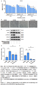
2.1 考比替尼对软骨细胞增殖活性的影响 通过检测细胞存活率来确定考比替尼的最佳干预浓度。实验结果显示,软骨细胞存活率随考比替尼作用浓度的升高(0.01-5 μmol/L)而逐渐降低,见图1A。在相同浓度条件下,细胞存活率随着干预时间的延长而下降。当干预时间达到24 h且浓度为0.1 μmol/L时细胞存活率接近90%。而药物浓度为0.5 μmol/L时,软骨细胞存活率显著低于0 μmol/L组的80%,见图1A。基于上述结果,该研究选择0.1 μmol/L作为考比替尼干预的理想浓度。 2.2 软骨细胞形态观察结果 原代SD大鼠软骨细胞培养2 d后,约70%细胞贴壁,细胞形态为类似圆形或椭圆形。白细胞介素1β处理24 h后,形态变得不规则,失去正常的圆形或椭圆形特征,呈现扁平状或伸展状,体积增大且出现纤维样改变。D+Q组处理后,细胞面积减小且趋向正常形态改变,生长速度明显放缓。考比替尼组细胞纤维样形态减少,细胞恢复卵圆形,且边界清晰,见图1B。 2.3 考比替尼对MEK-ERK1/2信号通路的调控作用 为了验证考比替尼对MEK-ERK1/2信号通路的调控作用及与D+Q组的对比,采用Western Blot检测了白细胞介素1β刺激后软骨细胞中MEK1、p-MEK1、ERK1/2及p-ERK1/2蛋白的表达情况。结果显示,与对照组相比,白细胞介素1β刺激显著上调了p-MEK1和p-ERK1/2的表达水平,而MEK和ERK总蛋白的表达无明显变化,提示白细胞介素1β可激活MEK-ERK1/2信号通路。在干预组中,发现无论是考比替尼单独处理,还是D+Q组处理,均能明显抑制p-MEK1和p-ERK1/2的表达,见图1C-E。上述结果表明,考比替尼或D+Q联合处理均可有效抑制白细胞介素1β诱导的MEK-ERK1/2信号通路激活,提示它们在缓解炎症性软骨细胞衰老中的潜在机制可能与该通路的负向调控相关。 2.4 考比替尼与D+Q对白细胞介素1β诱导的软骨细胞衰老的缓解作用对比 衰老相关β-半乳糖苷酶是衰老细胞最广泛使用的生物标志物之一[29]。两组药物分别与白细胞介素1β共同作用于"

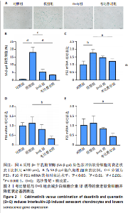
软骨细胞,处理时长为24 h,采用β-半乳糖苷酶染色法来评估两组药物对白细胞介素1β诱导的软骨细胞衰老的影响。实验结果显示,经白细胞介素1β刺激后,β-半乳糖苷酶阳性细胞比例显著上升,表明白细胞介素1β成功诱导了软骨细胞衰老,而经D+Q组药物和考比替尼处理后,β-半乳糖苷酶阳性细胞数量明显减少,见图2A,B。进一步通过实时荧光定量PCR(图2C-"


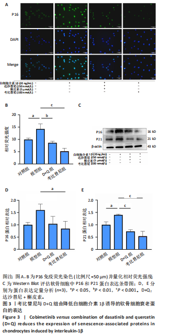
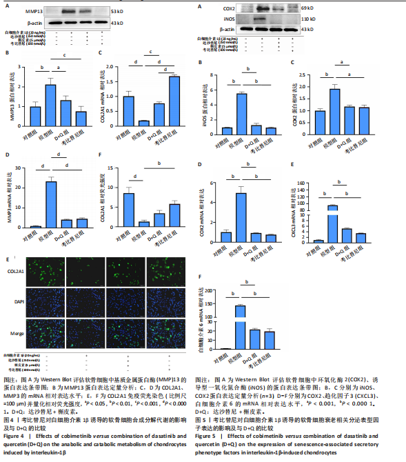
E)、免疫荧光染色(图3A,B)和Western Blot(图3C-E)实验发现,考比替尼可降低P16、P21、P53的表达水平,抑制效果优于D+Q组。综上所述,考比替尼与D+Q组合,均能有效缓解白细胞介素1β诱导的软骨细胞衰老状态。 2.5 考比替尼与D+Q对细胞外基质的影响比较 衰老软骨细胞的合成代谢能力下降,表现为细胞外基质成分的合成减少和降解增加[30]。与对照组相比,白细胞介素1β显著诱导基质金属蛋白酶13蛋白表达和基质金属蛋白酶3的mRNA表达,显著降低基质合成基因COL2A1的mRNA表达水平。考比替尼干预后,基质金属蛋白酶13蛋白表达和基质金属蛋白酶3的mRNA表达则明显降低,其中基质金属蛋白酶13蛋白表达考比替尼组低于D+Q组,但差异无显著性意义;而COL2A1的mRNA(图4C)水平显著上调,考比替尼组显著高于D+Q组(P < 0.000 1),见图4A-D。此外,免疫荧光检测结果表明COL2A1与上述分子水平检测结果相一致(图4E,F),进一步证实了考比替尼在促进软骨细胞合成代谢和抑制基质降解方面的效果优于D+Q组合。"

2.6 考比替尼抑制白细胞介素1β处理的软骨细胞中衰老相关分泌表型因子表达 衰老的软骨细胞分泌衰老相关分泌表型因子进一步促进软骨细胞的炎症以及分解代谢[31]。此外,这些衰老相关分泌表型介质促使周围健康细胞进入衰老状态。结果显示,考比替尼和D+Q组合在mRNA或蛋白质水平上能显著降低诱导型一氧化氮合酶、环氧化酶2(图5A-D)、趋化因子3以及白细胞介素6的表达,与D+Q组比较,上述指标考比替尼组降低效果更好,但差异无显著性意义(图5E,F)。这一结果表明,考比替尼和D+Q组合均能有效抑制衰老相关分泌表型因子表达。"
| [1] JIANG Y. Osteoarthritis year in review 2021: biology. Osteoarthritis Cartilage. 2022;30(2):207-215. [2] DOOLITTLE ML, SAUL D, KAUR J, et al. Multiparametric senescent cell phenotyping reveals targets of senolytic therapy in the aged murine skeleton. Nat Commun. 2023;14(1):4587. [3] VINATIER C, DOMÍNGUEZ E, GUICHEUX J, et al. Role of the Inflammation-Autophagy-Senescence Integrative Network in Osteoarthritis. Front Physiol. 2018;9:706. [4] DIEKMAN BO, LOESER RF. Aging and the emerging role of cellular senescence in osteoarthritis. Osteoarthritis Cartilage. 2024;32(4):365-371. [5] GUO Q, CHEN X, CHEN J, et al. STING promotes senescence, apoptosis, and extracellular matrix degradation in osteoarthritis via the NF-κB signaling pathway. Cell Death Dis. 2021;12(1):13. [6] YANG H, CHEN C, CHEN H, et al. Navitoclax (ABT263) reduces inflammation and promotes chondrogenic phenotype by clearing senescent osteoarthritic chondrocytes in osteoarthritis. Aging (Albany NY). 2020;12(13):12750-12770. [7] XU M, PIRTSKHALAVA T, FARR JN, et al. Senolytics improve physical function and increase lifespan in old age. Nat Med. 2018;24(8):1246-1256. [8] KIRKLAND JL, TCHKONIA T. Senolytic drugs: from discovery to translation. J Intern Med. 2020;288(5):518-536. [9] OGRODNIK M, MIWA S, TCHKONIA T, et al. Cellular senescence drives age-dependent hepatic steatosis. Nat Commun. 2017;8:15691. [10] KARNEWAR S, KARNEWAR V, SHANKMAN LS, et al. Treatment of advanced atherosclerotic mice with ABT-263 reduced indices of plaque stability and increased mortality. JCI Insight. 2024;9(2):e173863. [11] YANG C, QIAO W, XUE Q, et al. The senolytic agent ABT263 ameliorates osteoporosis caused by active vitamin D insufficiency through selective clearance of senescent skeletal cells. J Orthop Translat. 2024;49:107-118. [12] ZHAO J, ZHENG L, DAI G, et al. Senolytics cocktail dasatinib and quercetin alleviate chondrocyte senescence and facet joint osteoarthritis in mice. Spine J. 2025;25(1):184-198. [13] WU W, WU X, QIU L, et al. Quercetin influences intestinal dysbacteriosis and delays alveolar epithelial cell senescence by regulating PTEN/PI3K/AKT signaling in pulmonary fibrosis. Naunyn Schmiedebergs Arch Pharmacol. 2024;397(7): 4809-4822. [14] KRZYSTYNIAK A, WESIERSKA M, PETRAZZO G, et al. Combination of dasatinib and quercetin improves cognitive abilities in aged male Wistar rats, alleviates inflammation and changes hippocampal synaptic plasticity and histone H3 methylation profile. Aging (Albany NY). 2022;14(2):572-595. [15] KANG C. Senolytics and Senostatics: A Two-Pronged Approach to Target Cellular Senescence for Delaying Aging and Age-Related Diseases. Mol Cells. 2019;42(12): 821-827. [16] HERNANDEZ-SEGURA A, DE JONG TV, MELOV S, et al. Unmasking Transcriptional Heterogeneity in Senescent Cells. Curr Biol. 2017;27(17):2652-2660. [17] SHARMA A K, ROBERTS R L, BENSON R D J, et al. The Senolytic Drug Navitoclax (ABT-263) Causes Trabecular Bone Loss and Impaired Osteoprogenitor Function in Aged Mice. Front Cell Dev Biol. 2020;8:354. [18] LARKIN J, ASCIERTO P A, DRÉNO B, et al. Combined vemurafenib and cobimetinib in BRAF-mutated melanoma. N Engl J Med. 2014;371(20):1867-1876. [19] ZOU J, LEI T, GUO P, et al. Mechanisms shaping the role of ERK1/2 in cellular senescence (Review). Mol Med Rep. 2019;19(2):759-770. [20] CHAPPELL WH, STEELMAN LS, LONG JM, et al. Ras/Raf/MEK/ERK and PI3K/PTEN/Akt/mTOR inhibitors: rationale and importance to inhibiting these pathways in human health. Oncotarget. 2011;2(3):135-164. [21] CHEN H, CHEN H, LIANG J, et al. TGF-β1/IL-11/MEK/ERK signaling mediates senescence-associated pulmonary fibrosis in a stress-induced premature senescence model of Bmi-1 deficiency. Exp Mol Med. 2020;52(1):130-151. [22] BLAGOSKLONNY MV. Cellular senescence: when growth stimulation meets cell cycle arrest. Aging (Albany NY). 2023;15(4):905-913. [23] WANG Z, MA L, SU M, et al. Baicalin induces cellular senescence in human colon cancer cells via upregulation of DEPP and the activation of Ras/Raf/MEK/ERK signaling. Cell Death Dis. 2018;9(2):217. [24] CHAPPELL WH, CANDIDO S, ABRAMS SL, et al. Influences of TP53 and the anti-aging DDR1 receptor in controlling Raf/MEK/ERK and PI3K/Akt expression and chemotherapeutic drug sensitivity in prostate cancer cell lines. Aging (Albany NY). 2020;12(11):10194-10210. [25] JIANG E, DINESH A, JADHAV S, et al. Canagliflozin shares common mTOR and MAPK signaling mechanisms with other lifespan extension treatments. Life Sci. 2023;328:121904. [26] GKIONI L, NESPITAL T, BAGHDADI M, et al. The geroprotectors trametinib and rapamycin combine additively to extend mouse healthspan and lifespan. Nat Aging. 2025;5(7):1249-1265. [27] CHEN Y, GUO H, LI L, et al. Long Non-Coding RNA (lncRNA) Small Nucleolar RNA Host Gene 15 (SNHG15) Alleviates Osteoarthritis Progression by Regulation of Extracellular Matrix Homeostasis. Med Sci Monit. 2020;26: e923868. [28] MAURER S, KIRSCH V, RUTHS L, et al. Senolytic therapy combining Dasatinib and Quercetin restores the chondrogenic phenotype of human osteoarthritic chondrocytes by the release of pro-anabolic mediators. Aging Cell. 2025;24(1):e14361. [29] COPP ME, FLANDERS MC, GAGLIARDI R, et al. The combination of mitogenic stimulation and DNA damage induces chondrocyte senescence. Osteoarthritis Cartilage. 2021;29(3):402-412. [30] HAO W, CHANG M, SHI D, et al. Therapeutic targets in aging-related osteoarthritis: A focus on the extracellular matrix homeostasis. Life Sci. 2025;368:123487. [31] MYAKISHEVA SN, LINKOVA NS, KOZHEVNIKOVA EO, et al. Chondrocytes secretory phenotype associated with aging: role in the pathogenesis of osteoarthritis and prospects for peptide bioregulation. Adv Gerontol. 2023;36(3):313-323. [32] RIM YA, NAM Y, JU JH. The Role of Chondrocyte Hypertrophy and Senescence in Osteoarthritis Initiation and Progression. Int J Mol Sci. 2020;21(7):2358. [33] OGRODNIK M, CARLOS ACOSTA J, ADAMS PD, et al. Guidelines for minimal information on cellular senescence experimentation in vivo. Cell. 2024;187(16): 4150-4175. [34] YAN J, CHEN S, YI Z, et al. The role of p21 in cellular senescence and aging-related diseases. Mol Cells. 2024;47(11):100113. [35] LI X, LI C, ZHANG W, et al. Inflammation and aging: signaling pathways and intervention therapies. Signal Transduct Target Ther. 2023;8(1):239. [36] TONUTTI A, GRANATA V, MARRELLA V, et al. The role of WNT and IL-1 signaling in osteoarthritis: therapeutic implications for platelet-rich plasma therapy. Front Aging. 2023;4:1201019. [37] HUANG X, YOU Y, XI Y, et al. p-Coumaric Acid Attenuates IL-1β-Induced Inflammatory Responses and Cellular Senescence in Rat Chondrocytes. Inflammation. 2020;43(2):619-628. [38] SHENG J, LIU K, SUN D, et al. Association of RDM1 with osteosarcoma progression via cell cycle and MEK/ERK signalling pathway regulation. J Cell Mol Med. 2021; 25(16):8039-8046. [39] YU Y, ZHANG X, LIU F, et al. A stress-induced miR-31-CLOCK-ERK pathway is a key driver and therapeutic target for skin aging. Nat Aging. 2021;1(9):795-809. [40] GROGAN L, SHAPIRO P. Progress in the development of ERK1/2 inhibitors for treating cancer and other diseases. Adv Pharmacol. 2024;100:181-207. [41] GUÉGAN J, EZAN F, GAILHOUSTE L, et al. MEK1/2 overactivation can promote growth arrest by mediating ERK1/2-dependent phosphorylation of p70S6K. J Cell Physiol. 2014;229(7):903-915. [42] EL-HOSS J, KOLIND M, JACKSON MT, et al. Modulation of endochondral ossification by MEK inhibitors PD0325901 and AZD6244 (Selumetinib). Bone. 2014;59:151-161. [43] WANG X, XUE Y, YE W, et al. The MEK-ERK1/2 signaling pathway regulates hyaline cartilage formation and the redifferentiation of dedifferentiated chondrocytes in vitro. Am J Transl Res. 2018,10(10):3068-3085. [44] PRASADAM I, MAO X, SHI W, et al. Combination of MEK-ERK inhibitor and hyaluronic acid has a synergistic effect on anti-hypertrophic and pro-chondrogenic activities in osteoarthritis treatment. J Mol Med (Berl). 2013;91(3):369-380. [45] FAN S, CHANG Y, XIONG X, et al. Reversible SAHH inhibitor ameliorates MIA-induced osteoarthritis of rats through suppressing MEK/ERK pathway. Biomed Pharmacother. 2024;170:115975. [46] WANG M, YUANG-CHI CHANG A. Molecular mechanism of action and potential biomarkers of growth inhibition of synergistic combination of afatinib and dasatinib against gefitinib-resistant non-small cell lung cancer cells. Oncotarget. 2018;9(23):16533-16546. [47] HASINOFF BB, PATEL D. Mechanisms of the Cardiac Myocyte-Damaging Effects of Dasatinib. Cardiovasc Toxicol. 2020;20(4):380-389. [48] LI Y, HE Y, HAN X, et al. Dasatinib suppresses invasion and induces apoptosis in nasopharyngeal carcinoma. Int J Clin Exp Pathol. 2015;8(7):7818-7824. [49] MILANOVIC M, FAN DNY, BELENKI D, et al. Senescence-associated reprogramming promotes cancer stemness. Nature. 2018;553(7686):96-100. [50] SI H, YANG T, LI L, et al. miR-140 Attenuates the Progression of Early-Stage Osteoarthritis by Retarding Chondrocyte Senescence. Mol Ther Nucleic Acids. 2020;19:15-30. [51] WU X, ZHOU X, WANG S, et al. DNA damage response (DDR): a link between cellular senescence and human cytomegalovirus. Virol J. 2023;20(1):250. [52] ZHU Y, TCHKONIA T, PIRTSKHALAVA T, et al. The Achilles’ heel of senescent cells: from transcriptome to senolytic drugs. Aging Cell. 2015;14(4):644-658. [53] UR RASHID H, XU Y, AHMAD N, et al. Promising anti-inflammatory effects of chalcones via inhibition of cyclooxygenase, prostaglandin E (2), inducible NO synthase and nuclear factor κb activities. Bioorg Chem. 2019;87:335-365. [54] HAN Z, WANG K, DING S, et al. Cross-talk of inflammation and cellular senescence: a new insight into the occurrence and progression of osteoarthritis. Bone Res. 2024;12(1):69. [55] FANG C, LIU B, WAN M. “Bone-SASP” in Skeletal Aging. Calcif Tissue Int. 2023; 113(1):68-82. [56] JEON OH, WILSON DR, CLEMENT CC, et al. Senescence cell-associated extracellular vesicles serve as osteoarthritis disease and therapeutic markers. JCI Insight. 2019;4(7):e125019. |
| [1] | Lyu Guoqing, Aizimaitijiang·Rouzi, Xiong Daohai. Irisin inhibits ferroptosis in human articular chondrocytes: roles and mechanisms [J]. Chinese Journal of Tissue Engineering Research, 2026, 30(6): 1359-1367. |
| [2] | Zhang Haiwen, Zhang Xian, Xu Taichuan, Li Chao. Bibliometric and visual analysis of the research status and trends of senescence in osteoporosis [J]. Chinese Journal of Tissue Engineering Research, 2026, 30(6): 1580-1591. |
| [3] | Lai Jiaming, , Song Yuling, Chen Zixi, Wei Jinghuan, Cai Hao, , Li Guoquan, . Screening of diagnostic markers for endothelial cell Senescence in mice with radiation-induced heart disease and analysis of immune infiltration [J]. Chinese Journal of Tissue Engineering Research, 2026, 30(6): 1450-1463. |
| [4] | Bu Yangyang, Ning Xinli, Zhao Chen. Intra-articular injections for the treatment of osteoarthritis of the temporomandibular joint: different drugs with multiple combined treatment options [J]. Chinese Journal of Tissue Engineering Research, 2026, 30(5): 1215-1224. |
| [5] | Zhang Qian, Huang Dongfeng. Weighted gene co-expression network analysis combined with machine learning to screen and validate biomarkers for osteoarthritis [J]. Chinese Journal of Tissue Engineering Research, 2026, 30(5): 1096-1105. |
| [6] | Chen Yida, Cheng Xinyi, Zhao Huan, Zhou Xichao, Gu Qiaoli, Lin Xiao, Shi Qin. Effects of physiological osmotic pressure on chondrocyte differentiation and extracellular matrix metabolism [J]. Chinese Journal of Tissue Engineering Research, 2026, 30(35): 9143-9150. |
| [7] | Yang Huaqun, Abudouainijiang·Abulimiti, Wang Fazheng, Maimaitishawutiaji·Maimaiti, Li Simi, Muhetaer·Maimaitirexiati. Weighted gene co-expression network analysis combined with machine learning identifies autophagy and senescence signature genes in osteoarthritis chondrocytes [J]. Chinese Journal of Tissue Engineering Research, 2026, 30(34): 8889-8898. |
| [8] | Wang Yanfei, Jin Lianhai, Li Qingya, Fu Yuanfei, Tan Huangsheng, Deng Pengwei, Gao Kun. Synovial fluid exosome-mediated crosstalk between synoviocytes and chondrocytes in development and progression of knee osteoarthritis [J]. Chinese Journal of Tissue Engineering Research, 2026, 30(34): 9032-9040. |
| [9] | Liu Bangding, Tang Yongliang, Li Ni, Ren Bo. Quercetin-loaded hydrogel materials for treatment of infected bone defects [J]. Chinese Journal of Tissue Engineering Research, 2026, 30(32): 8427-8435. |
| [10] | Chen Shichao, Deng Yunyi, Zhao Renshengjie, Yu Ke, Li Guangwen. Antibacterial properties of photocrosslinkable hydrogel loaded with quercetin-silver nanoparticles for infected wounds [J]. Chinese Journal of Tissue Engineering Research, 2026, 30(32): 8436-8442. |
| [11] | Wei Bingqi, Zhang Xinyue, Ren Xingyue, Sun Jiahui, Chen Liu, Li Yijing, Qi Yifan, Wang Shangzeng. Zinc finger DHHC-type containing 2 emerges as a novel therapeutic target in osteoarthritis pathogenesis: genome-wide data analysis in European populations [J]. Chinese Journal of Tissue Engineering Research, 2026, 30(29): 7715-7723. |
| [12] | Li Zhengpeng, Shao Weigang, Zeng Hao, Xiang Kelin, Zhang Botao, Zou Shunyi, Chen Sheng, Qi Wen. Osteoarthritis characteristic genes and prediction of targeted food-medicine homology traditional Chinese medicine: bioinformatics analysis and kinetic simulation [J]. Chinese Journal of Tissue Engineering Research, 2026, 30(29): 7739-7748. |
| [13] | Guo Shanshan, Ma Ding, Dong Bingchen. Metabolic dysregulation in osteoarthritis: mechanisms and targeted therapeutic strategies [J]. Chinese Journal of Tissue Engineering Research, 2026, 30(29): 7654-7662. |
| [14] | Yan Yuge, Wang Yanxi, Qi Xiang, Cao Shan, Zou Xiaoyan, Liu Yujuan. Screening biomarkers for premature ovarian insufficiency based on cellular senescence and endoplasmic reticulum stress with experimental validation [J]. Chinese Journal of Tissue Engineering Research, 2026, 30(28): 7447-7455. |
| [15] | Su Jiemao, Qi Yansong, Kong Keyu, Zhai Zanjing, Xu Yongsheng. Role of chondrocyte ferroptosis in the pathogenesis of osteoarthritis [J]. Chinese Journal of Tissue Engineering Research, 2026, 30(24): 6282-6288. |
| Viewed | ||||||
|
Full text |
|
|||||
|
Abstract |
|
|||||