Chinese Journal of Tissue Engineering Research ›› 2024, Vol. 28 ›› Issue (34): 5508-5515.doi: 10.12307/2024.841
Previous Articles Next Articles
Aerobic exercise upregulates the thioredoxin system and inhibits cardiomyocyte apoptosis in aging rats
Xu Zheng1, Zhao Xiaoqin1, Chen Xiaodan1, Wang Jiapu2, Bao Fenmiao1, Yu Liang3, Li Junping3, Wei Yan2
- 1School of Physical Education, 2School of Biomedical Engineering, Taiyuan University of Technology, Taiyuan 030024, Shanxi Province, China; 3School of Sports Human Science, Beijing Sport University, Beijing 100084, China
-
Received:2023-11-20Accepted:2024-01-16Online:2024-12-08Published:2024-03-14 -
Contact:Zhao Xiaoqin, Associate professor, School of Physical Education, Taiyuan University of Technology, Taiyuan 030024, Shanxi Province, China -
About author:Xu Zheng, Master candidate, School of Physical Education, Taiyuan University of Technology, Taiyuan 030024, Shanxi Province, China -
Supported by:the National Natural Science Foundation of China, No. 32071168 (to YL [project participant]); the Fundamental Research Funds for the Central Universities, No. 2022YB019 (to YL); the Natural Science Foundation of Shanxi Province (General Program), No. 201901D111079 (to ZXQ)
CLC Number:
Cite this article
Xu Zheng, Zhao Xiaoqin, Chen Xiaodan, Wang Jiapu, Bao Fenmiao, Yu Liang, Li Junping, Wei Yan. Aerobic exercise upregulates the thioredoxin system and inhibits cardiomyocyte apoptosis in aging rats[J]. Chinese Journal of Tissue Engineering Research, 2024, 28(34): 5508-5515.
share this article
Add to citation manager EndNote|Reference Manager|ProCite|BibTeX|RefWorks
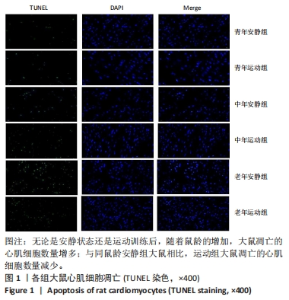
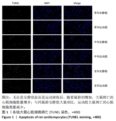
2.1 实验动物数量分析 36只大鼠全部进入结果分析。 2.2 各组大鼠心肌细胞凋亡 TUNEL染色显示,无论是安静状态还是运动训练后,随着鼠龄的增加,大鼠凋亡的心肌细胞数量增多;与同鼠龄安静组大鼠相比,运动组大鼠凋亡的心肌细胞数量减少,见图1。以上结果表明,10周的有氧运动能够明显抑制衰老大鼠心肌细胞的凋亡。"
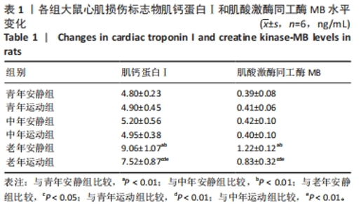
2.3 各组大鼠血清肌钙蛋白Ⅰ和肌酸激酶同工酶MB水平 见表1。 "
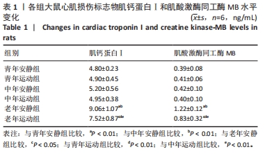
肌钙蛋白Ⅰ和肌酸激酶同工酶MB为心肌损伤标志物水平。 安静组间对比:与青年安静组相比,中年安静组大鼠血清肌钙蛋白Ⅰ和肌酸激酶同工酶MB水平无明显变化(P > 0.05),老年安静组大鼠血清肌钙蛋白Ⅰ和肌酸激酶同工酶MB水平升高(P < 0.01)。与中年安静组相比,老年安静组大鼠血清肌钙蛋白Ⅰ和肌酸激酶同工酶MB水平升高(P < 0.01)。 运动前后对比:与同龄段安静组相比,青年运动组、中年运动组大鼠血清肌钙蛋白Ⅰ和肌酸激酶同工酶MB水平均无明显变化(P > 0.05),老年运动组大鼠血清肌钙蛋白Ⅰ和肌酸激酶同工酶MB水平降低(P < 0.01)。 运动组间对比:与青年运动组对比,中年运动组大鼠血清肌钙蛋白Ⅰ和肌酸激酶同工酶MB水平无明显变化(P > 0.05),老年运动组大鼠血清肌钙蛋白Ⅰ和肌酸激酶同工酶MB水平升高(P < 0.01)。与中年运动组相比,老年运动组大鼠血清肌钙蛋白Ⅰ和肌酸激酶同工酶MB水平升高(P < 0.01)。 2.4 各组大鼠心肌组织Bax、Bcl-2、Caspase 3蛋白表达 见图2。"
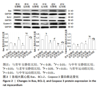
心肌细胞凋亡检测后,对大鼠心肌组织的凋亡相关蛋白表达进行分析。无论是安静状态还是运动训练后,大鼠心肌Bax和Caspase 3蛋白表达呈升高趋势,而Bcl-2蛋白表达呈降低趋势。 安静组间对比:与青年安静组相比,中年安静组大鼠心肌Bax蛋白表达升高(P < 0.01),Bcl-2蛋白表达降低(P > 0.01),Caspase 3蛋白表达无明显变化(P > 0.05);老年安静组大鼠心肌Bax和Caspase 3蛋白表达升高(P < 0.01),Bcl-2蛋白表达降低(P < 0.01)。与中年安静组相比,老年安静组大鼠心肌Bax和Caspase 3蛋白表达升高(P < 0.01),Bcl-2蛋白表达降低(P < 0.01)。 运动前后对比:与青年安静组相比,青年运动组大鼠心肌Bax和Caspase 3蛋白表达降低(P < 0.01,P < 0.05),Bcl-2蛋白表达升高(P < 0.01)。与中年安静组相比,中年运动组大鼠心肌Bax蛋白表达降低(P < 0.01),Bcl-2蛋白表达升高(P < 0.01),Caspase 3蛋白表达无明显变化(P > 0.05)。与老年安静组相比,老年运动组大鼠心肌Bax和Caspase 3蛋白表达降低(P < 0.01,P < 0.05),Bcl-2蛋白表达升高(P < 0.01)。 运动组间对比:与青年运动组大鼠比,中年运动组大鼠心肌Bax蛋白表达无明显变化(P > 0.05),Caspase 3蛋白表达升高(P < 0.01),Bcl-2蛋白表达降低(P < 0.01);老年运动大鼠心肌Caspase 3、Bax蛋白表达升高(P < 0.01),Bcl-2蛋白表达降低(P < 0.01)。与中年运动组大鼠比,老年运动组大鼠心肌Bax和Caspase 3蛋白表达均升高(P < 0.01),Bcl-2蛋白表达降低(P < 0.01)。 2.5 各组大鼠心肌组织硫氧还蛋白系统蛋白表达 见图3。"

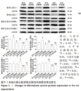
安静组间对比:与青年安静组相比,中年安静组大鼠心肌硫氧还蛋白1和硫氧还蛋白2蛋白表达降低(P < 0.01,P < 0.05),硫氧还蛋白还原酶1和硫氧还蛋白还原酶2蛋白表达无明显变化(P > 0.05);老年安静组大鼠心肌组织硫氧还蛋白1、硫氧还蛋白2、硫氧还蛋白还原酶1、硫氧还蛋白还原酶2蛋白表达均降低(P < 0.01,P < 0.05)。与中年安静组相比,老年安静组大鼠心肌硫氧还蛋白1、硫氧还蛋白2、硫氧还蛋白还原酶和硫氧还蛋白还原酶2蛋白表达均降低(P < 0.01,P < 0.05)。 运动前后对比:与青年安静组相比,青年运动组大鼠心肌硫氧还蛋白1和硫氧还蛋白2蛋白表达升高(P < 0.05),硫氧还蛋白还原酶1和硫氧还蛋白还原酶2蛋白表达无明显变化(P > 0.05);与中年安静组相比,中年运动组大鼠心肌组织硫氧还蛋白1、硫氧还蛋白2、硫氧还蛋白还原酶1、硫氧还蛋白还原酶2蛋白表达均升高(P < 0.01,P < 0.05)。与老年安静组相比,老年运动组大鼠心肌组织硫氧还蛋白1、硫氧还蛋白2、硫氧还蛋白还原酶1、硫氧还蛋白还原酶2蛋白表达均升高(P < 0.01,P < 0.05)。 运动组间对比:与青年运动组大鼠比,中年运动组大鼠心肌硫氧还蛋白1和硫氧还蛋白2蛋白表达均降低(P < 0.01,P < 0.05),硫氧还蛋白还原酶1蛋白表达无明显变化(P > 0.05),硫氧还蛋白还原酶2蛋白表达升高(P < 0.05);老年运动组大鼠心肌组织硫氧还蛋白1、硫氧还蛋白2和硫氧还蛋白还原酶2蛋白表达降低(P < 0.01,P < 0.05),硫氧还蛋白还原酶1蛋白表达无明显变化(P > 0.05)。与中年运动组大鼠比,老年运动组大鼠心肌组织硫氧还蛋白1和硫氧还蛋白2蛋白表达均无明显变化(P > 0.05),硫氧还蛋白还原酶1和硫氧还蛋白还原酶2蛋白表达均显著降低(P < 0.01)。 2.6 各组大鼠心肌组织硫氧还蛋白系统下游蛋白表达 见图4。"
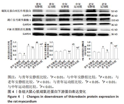

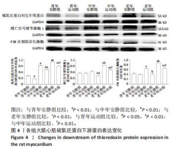
安静状态下,随着鼠龄的增加,大鼠心肌组织硫氧还蛋白相互作用蛋白、凋亡信号调节激酶1和P38丝裂原活化激酶的蛋白表达水平呈升高趋势。 安静组间对比:与青年安静组相比,中年安静组大鼠心肌硫氧还蛋白相互作用蛋白和P38丝裂原活化激酶蛋白表达升高(P < 0.01),凋亡信号调节激酶1蛋白表达无明显变化(P < 0.05);老年安静组大鼠心肌硫氧还蛋白相互作用蛋白、凋亡信号调节激酶1和P38丝裂原活化激酶蛋白表达均升高(P < 0.01)。与中年安静组相比,老年安静组大鼠心肌组织硫氧还蛋白相互作用蛋白、凋亡信号调节激酶1和P38丝裂原活化激酶蛋白表达均升高(P < 0.01)。 运动前后对比:与青年安静组对比,青年运动组大鼠心肌硫氧还蛋白相互作用蛋白表达降低(P < 0.01),凋亡信号调节激酶1和P38丝裂原活化激酶蛋白均无明显变化(P > 0.05)。与同龄段安静组对比,中年运动组、老年运动组大鼠心肌硫氧还蛋白相互作用蛋白、凋亡信号调节激酶1和P38丝裂原活化激酶蛋白表达均降低(P < 0.01)。 运动组间对比:与青年运动组比,中年运动组大鼠心肌硫氧还蛋白相互作用蛋白表达升高(P < 0.01),凋亡信号调节激酶1蛋白表达降低(P < 0.05),P38丝裂原活化激酶蛋白无明显变化(P > 0.05);老年运动组大鼠心肌组织硫氧还蛋白相互作用蛋白、凋亡信号调节激酶1和P38丝裂原活化激酶蛋白表达均升高(P < 0.01)。与中年运动组比,老年运动组大鼠心肌组织硫氧还蛋白相互作用蛋白、凋亡信号调节激酶1、P38丝裂原活化激酶蛋白表达均升高(P < 0.01)。"

| [1] MORAN A, GU D, ZHAO D, et al. Future cardiovascular disease in china: markov model and risk factor scenario projections from the coronary heart disease policy model-china.Circ Cardiovasc Qual Outcomes. 2010;3(3):243-252. [2] Gude NA, Broughton KM, Firouzi F, et al. Cardiac ageing: extrinsic and intrinsic factors in cellular renewal and senescence. Nat Rev Cardiol. 2018;15(9):523-542. [3] Costantino S, Paneni F, Cosentino F. Ageing, metabolism and cardiovascular disease. J Physiol. 2016;594(8):2061-2073. [4] Liao PH, Hsieh DJ, Kuo CH, et al. Moderate exercise training attenuates aging-induced cardiac inflammation, hypertrophy and fibrosis injuries of rat hearts. Oncotarget. 2015;6(34):35383-35394. [5] Shen T, Liu X, Zhuang B, et al. Efficacy and Safety of Different Aerobic Exercise Intensities in Patients With Heart Failure With Reduced Ejection Fraction: Design of a Multicenter Randomized Controlled Trial (HF-EI Trial). Front Cardiovasc Med. 2021;8:705972. [6] Guo M, Lu B, Gan J, et al. Apoptosis detection: a purpose-dependent approach selection. Cell Cycle. 2021;20(11):1033-1040. [7] Liu Y, Xue N, Zhang B, et al. Role of Thioredoxin-1 and its inducers in human health and diseases. Eur J Pharmacol. 2022;919:174756. [8] Murata R, Watanabe H, Nosaki H, et al. Long-Acting Thioredoxin Ameliorates Doxorubicin-Induced Cardiomyopathy via Its Anti-Oxidative and Anti-Inflammatory Action. Pharmaceutics. 2022;14(3):562. [9] 张丽红,李海涛,马建林,等.血清Trx1、FGL2与急性心肌梗死后心力衰竭患者预后的关系[J].现代生物医学进展,2023,23(11): 2102-2107. [10] Kaplán P, Tatarková Z, Lichardusová L, et al. Age-Associated Changes in Antioxidants and Redox Proteins of Rat Heart. Physiol Res. 2019;68(6):883-892. [11] BEDFORD TG, TIPTON CM, WILSON NC, et al. Maximum oxygen consumption of rats and its changes with various experimental procedures. J Appl Physiol Respir Environ Exerc Physiol. 1979;47(6):1278-1283. [12] 刘文锋,刘少鹏,傅让,等. 耐力运动对增龄大鼠脑皮层突触可塑性的影响及相关调控机制[J].中国应用生理学杂志,2019,35(4): 339-345,350. [13] 刘吉焕,王鹏,袁顺灵,等.有氧耐力运动调控大鼠心肌细胞自噬的机制[J].中国组织工程研究,2023,27(23):3714-3720. [14] LI W, LIU F. Myocardial Cell Aging in the Elderly. Aging Pathobiol Ther. 2020; 2(3):134-142. [15] Eng J, McClelland RL, Gomes AS, et al. Adverse Left Ventricular Remodeling and Age Assessed with Cardiac MR Imaging: The Multi-Ethnic Study of Atherosclerosis. Radiology. 2016;278(3):714-722. [16] Frangogiannis NG. Cardiac fibrosis: Cell biological mechanisms, molecular pathways and therapeutic opportunities. Mol Aspects Med. 2019;65:70-99. [17] Beliveau P, Cheriet F, Anderson SA, et al. Quantitative assessment of myocardial fibrosis in an age-related rat model by ex vivo late gadolinium enhancement magnetic resonance imaging with histopathological correlation. Comput Biol Med. 2015;65:103-113. [18] Lefferts WK, Davis MM, Valentine RJ. Exercise as an Aging Mimetic: A New Perspective on the Mechanisms Behind Exercise as Preventive Medicine Against Age-Related Chronic Disease. Front Physiol. 2022;13:866792. [19] Darband SG, Sadighparvar S, Yousefi B, et al. Combination of exercise training and L-arginine reverses aging process through suppression of oxidative stress, inflammation, and apoptosis in the rat heart. Pflugers Arch. 2020;472(2):169-178. [20] Fatahi A, Zarrinkalam E, Azizbeigi K, et al. Cardioprotective effects of exercise preconditioning on ischemia-reperfusion injury and ventricular ectopy in young and senescent rats. Exp Gerontol. 2022;162:111758. [21] 李欣,丁树哲,卢健.耐力训练对衰老小鼠心肌Akt/mTOR信号通路的影响[J].中国运动医学杂志,2010,29(1): 38-41. [22] SAVITSKAYA MA, ONISHCHENKO GE. Mechanisms of Apoptosis. Biochemistry (Mosc). 2015;80(11):1393-1405. [23] Moldoveanu T, Follis AV, Kriwacki RW, et al. Many players in BCL-2 family affairs. Trends Biochem Sci. 2014;39(3):101-111. [24] Ludwig LM, Maxcy KL, LaBelle JL. Flow Cytometry-Based Detection and Analysis of BCL-2 Family Proteins 4and Mitochondrial Outer Membrane Permeabilization (MOMP). Methods Mol Biol. 2019; 1877:77-91. [25] Pereira RM, Mekary RA, da Cruz Rodrigues KC, et al. Protective molecular mechanisms of clusterin against apoptosis in cardiomyocytes. Heart Fail Rev. 2018;23(1):123-129. [26] DeBalsi KL, Hoff KE, Copeland WC. Role of the mitochondrial DNA replication machinery in mitochondrial DNA mutagenesis, aging and age-related diseases. Ageing Res Rev. 2017;33:89-104. [27] Czabotar PE, Lessene G, Strasser A, et al. Control of apoptosis by the BCL-2 protein family: implications for physiology and therapy. Nat Rev Mol Cell Biol. 2014;15(1):49-63. [28] No MH, Heo JW, Yoo SZ, et al. Effects of aging and exercise training on mitochondrial function and apoptosis in the rat heart. Pflugers Arch. 2020;472(2):179-193. [29] Kwak HB, Song W, Lawler JM. Exercise training attenuates age-induced elevation in Bax/Bcl-2 ratio, apoptosis, and remodeling in the rat heart. FASEB J. 2006;20(6):791-793. [30] Pei Z, Yang C, Guo Y, et al. Effect of different exercise training intensities on age-related cardiac damage in male mice. Aging (Albany NY). 2021;13(17):21700-21711. [31] Kwak HB, Lee Y, Kim JH, et al. MnSOD overexpression reduces fibrosis and pro-apoptotic signaling in the aging mouse heart. J Gerontol A Biol Sci Med Sci. 2015;70(5):533-544. [32] Lu J, Holmgren A. Thioredoxin system in cell death progression. Antioxid Redox Signal. 2012;17(12):1738-1747. [33] Bjørklund G, Zou L, Peana M, et al. The Role of the Thioredoxin System in Brain Diseases. Antioxidants (Basel). 2022;11(11):2161. [34] McCarver AC, Lessner DJ. Molecular characterization of the thioredoxin system from Methanosarcina acetivorans. FEBS J. 2014;281(20):4598-4611. [35] Yu Y, Xing K, Badamas R, et al. Overexpression of thioredoxin-binding protein 2 increases oxidation sensitivity and apoptosis in human lens epithelial cells. Free Radic Biol Med. 2013;57:92-104. [36] 黄云霞,时杜娟,蒙玉娜,等.弥漫大B细胞淋巴瘤中Trx、TrxR-1及TXNIP的表达及临床意义[J]. 临床与实验病理学杂志,2022,38(4): 443-447. [37] Mitchell DA, Morton SU, Fernhoff NB, et al. Thioredoxin is required for S-nitrosation of procaspase-3 and the inhibition of apoptosis in Jurkat cells. Proc Natl Acad Sci U S A. 2007;104(28):11609-11614. [38] Mitchell DA, Marletta MA. Thioredoxin catalyzes the S-nitrosation of the caspase-3 active site cysteine. Nat Chem Biol. 2005;1(3):154-158. [39] Mahmood DF, Abderrazak A, El Hadri K, et al. The thioredoxin system as a therapeutic target in human health and disease. Antioxid Redox Signal. 2013;19(11):1266-1303. [40] Pérez VI, Lew CM, Cortez LA, et al. Thioredoxin 2 haploinsufficiency in mice results in impaired mitochondrial function and increased oxidative stress. Free Radic Biol Med. 2008;44(5):882-892. [41] Li Y, Xu P, Wang Y, et al. Different Intensity Exercise Preconditions Affect Cardiac Function of Exhausted Rats through Regulating TXNIP/TRX/NF-ĸBp65/NLRP3 Inflammatory Pathways. Evid Based Complement Alternat Med. 2020;2020:5809298. [42] Marschner RA, Banda P, Wajner SM, et al. Short-term exercise training improves cardiac function associated to a better antioxidant response and lower type 3 iodothyronine deiodinase activity after myocardial infarction. PLoS One. 2019;14(9):e0222334. [43] 黄海高,李悦山.Trx-ASK1在多柔比星诱导的乳鼠心肌细胞凋亡中的作用[J].中国病理生理杂志,2012,28(6):1028-1033. [44] 赵晓琴,赵俊杰,李晓宇,等.2型糖尿病大鼠心肌损伤时心肌组织中硫氧还蛋白系统的变化[J]. 生理学报,2010,62(3): 261-268. [45] Liu Q, Sargent MA, York AJ, et al. ASK1 regulates cardiomyocyte death but not hypertrophy in transgenic mice. Circ Res. 2009;105(11): 1110-1117. |
| [1] | Yang Yifeng, Ye Nan, Wang Lin, Guo Shuaicheng, Huang Jian. Signaling pathway of dexmedetomidine against ischemia-reperfusion injury [J]. Chinese Journal of Tissue Engineering Research, 2024, 28(9): 1464-1469. |
| [2] | Yue Yun, Wang Peipei, Yuan Zhaohe, He Shengcun, Jia Xusheng, Liu Qian, Li Zhantao, Fu Huiling, Song Fei, Jia Menghui. Effects of croton cream on JNK/p38 MAPK signaling pathway and neuronal apoptosis in cerebral ischemia-reperfusion injury rats [J]. Chinese Journal of Tissue Engineering Research, 2024, 28(8): 1186-1192. |
| [3] | Wang Ji, Zhang Min, Li Wenbo, Yang Zhongya, Zhang Long. Effect of aerobic exercise on glycolipid metabolism, skeletal muscle inflammation and autophagy in type 2 diabetic rats [J]. Chinese Journal of Tissue Engineering Research, 2024, 28(8): 1200-1205. |
| [4] | Shen Feiyan, Yao Jixiang, Su Shanshan, Zhao Zhongmin, Tang Weidong. Knockdown of circRNA WD repeat containing protein 1 inhibits proliferation and induces apoptosis of chondrocytes in knee osteoarthritis [J]. Chinese Journal of Tissue Engineering Research, 2024, 28(4): 499-504. |
| [5] | Chen Zepeng, Hou Yonghui, Chen Shudong, Hou Yu, Lin Dingkun. Tauroursodeoxycholic acid treats spinal cord injury by reducing apoptosis of spinal cord neurons under glucose and oxygen deprivation [J]. Chinese Journal of Tissue Engineering Research, 2024, 28(4): 528-534. |
| [6] | Yi Zhi, Zhan Hongwei, Wang Yaobin, Liang Xiaoyuan, Niu Yongkang, Xiang Dejian, Geng Bin, Xia Yayi. Caveolin-1 mediated fluid shear stress regulates proliferation and apoptosis of MC3T3-E1 osteoblasts [J]. Chinese Journal of Tissue Engineering Research, 2024, 28(34): 5440-5445. |
| [7] | Hou Zengtao, Dong Zhiwei, Zhang Jinfeng, Yang Xiaohui, Fan Xiao. Platelet-rich fibrin regulates apoptosis to promote cartilage repair in rats with knee osteoarthritis [J]. Chinese Journal of Tissue Engineering Research, 2024, 28(32): 5167-5171. |
| [8] | Wang Xi, Yu Li, Jia Qiyu, Huang Jinyong, Liu Zebiao, Zhang Jun, Jiayidaer•Dilimulati, Xie Zengru, Ma Hairong. Effects of filament B knockdown on proliferation, migration and apoptosis of mouse MC3T3-E1 cells [J]. Chinese Journal of Tissue Engineering Research, 2024, 28(32): 5177-5181. |
| [9] | Lu Mengya, Wu Xian, She Zeyu, Xia Shuai, Lu Man, Yang Yonghui. Acupotomy prevents knee osteoarthritis in rats by modulating chondrocyte apoptosis in the mitochondrial pathway [J]. Chinese Journal of Tissue Engineering Research, 2024, 28(32): 5190-5195. |
| [10] | Zhang Jinpu, Wang Junli, Zhang Siqi, Chen Jiahao, Yang Qiushi. Effectiveness of exercise interventions for fibromyalgia syndrome: a Meta-analysis [J]. Chinese Journal of Tissue Engineering Research, 2024, 28(32): 5210-5216. |
| [11] | Gao Jie, , , Zou Xingxing, Wen Banghong, Li Yuandi, Su Min, , , Hu Rong, , . Effect of Pax6 gene expression on hydrogen peroxide-induced aging in bone marrow mesenchymal stem cells [J]. Chinese Journal of Tissue Engineering Research, 2024, 28(31): 4921-4925. |
| [12] | Li Huijun, Li Chuikun, Wei Cuilan, Zhang Yeting. Notch1-mediated aerobic exercise promotes hippocampal nerve cell proliferation in Alzheimer’s disease mice [J]. Chinese Journal of Tissue Engineering Research, 2024, 28(31): 4951-4957. |
| [13] | Qian Longjie, Su Wenli, Zhu Wenxian, Wang Yixin. SRT1720, an activator of silent information regulator 1, alleviates acute traumatic brain injury in a rat model [J]. Chinese Journal of Tissue Engineering Research, 2024, 28(28): 4447-4454. |
| [14] | Jiang Yu, Xu Lin, Zhao Yalin, Liu Gang, Zhang Yaqi, Bai Huizhong, Ren Jingpei, Zeng Jie, Mu Xiaohong. Effects of Shujin Jiannao Prescription on cell apoptosis in rats with hypoxic-ischemic brain injury [J]. Chinese Journal of Tissue Engineering Research, 2024, 28(28): 4477-4483. |
| [15] | Zhou Minghan, Zhang Hui, Zheng Xianbo, Xu Wuji. Significance of PI3K/Akt/HIF-1α signaling pathway expression in nucleus pulposus cells at different oxygen concentrations in delaying intervertebral disc degeneration [J]. Chinese Journal of Tissue Engineering Research, 2024, 28(28): 4491-4497. |
| Viewed | ||||||
|
Full text |
|
|||||
|
Abstract |
|
|||||