Chinese Journal of Tissue Engineering Research ›› 2024, Vol. 28 ›› Issue (8): 1161-1167.doi: 10.12307/2023.871
Previous Articles Next Articles
Functional exercise for tendon adhesion in a model of deep flexor tendon II injury of the third toe
Cheng Jie1, Wang Jihong2, Zhang Pei3
- 1Department of Trauma Area C, 2Department of Hand and Foot Microsurgery Area B, 3Department of Spinal Surgery Area A, the Second Affiliated Hospital of Inner Mongolia Medical University, Hohhot 010030, Inner Mongolia Autonomous Region, China
-
Received:2022-10-26Accepted:2022-12-24Online:2024-03-18Published:2023-07-18 -
Contact:Zhang Pei, Master, Chief physician, Professor, Department of Spinal Surgery Area A, the Second Affiliated Hospital of Inner Mongolia Medical University, Hohhot 010030, Inner Mongolia Autonomous Region, China -
About author:Cheng Jie, Master, Associate chief physician, Department of Trauma Area C, the Second Affiliated Hospital of Inner Mongolia Medical University, Hohhot 010030, Inner Mongolia Autonomous Region, China -
Supported by:Science and Technology Million Joint Project of Inner Mongolia Medical University, No. YKD2018KJBW(LH)012 (to CJ); Scientific Research Project of Higher Education Institutions in Inner Mongolia Autonomous Region, No. NJZY22673 (to CJ)
CLC Number:
Cite this article
Cheng Jie, Wang Jihong, Zhang Pei. Functional exercise for tendon adhesion in a model of deep flexor tendon II injury of the third toe[J]. Chinese Journal of Tissue Engineering Research, 2024, 28(8): 1161-1167.
share this article
Add to citation manager EndNote|Reference Manager|ProCite|BibTeX|RefWorks

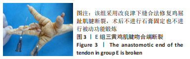
2.1 实验动物数量分析 术后5-7天发现,E组所有鸡右足第三趾伸直畸形,不能屈曲,通过解剖发现所有标本屈趾深肌腱断裂(图3),而鸡等实验动物不能配合,在日常生活中鸡有拔地的习惯,不能主动制动,导致了标本肌腱吻合端的断裂,说明肌腱修复术后合理制动的必要性,E组不给予其余指标的检测。在对鸡进行被动功能锻炼时,一定要尽可能地减少鸡的恐惧,如:平时多到鸡舍、让鸡不感到陌生感;在进行功能锻炼之前根据鸡的状态进行一定的安抚,待鸡的情绪平稳后再行功能锻炼;对于特别不配合的鸡,可给予镇静药物等。其余各组把与同组相比屈伸功能差异较大的样本给予剔除,后期切开发现这些剔除的样本屈肌腱断裂,最终A组17份、B 组19份、C组14份、D组20份标本进行指标的检测。"
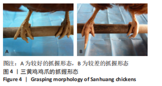
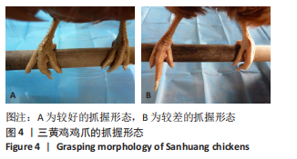
2.2 各组鸡爪的抓握形态 术后3周取下石膏,可见A、B、C组鸡部分右足第三趾有轻度屈曲挛缩,整体主动屈曲形态较好,鸡爪有较好抓握形态,这3组鸡爪抓握形态较好的是B、C组;D组鸡所有第三趾因屈曲挛缩明显而不能主动抓握木棍,无抓握形态;A组鸡爪抓握形态位于B、C组与D组之间,见图4。"
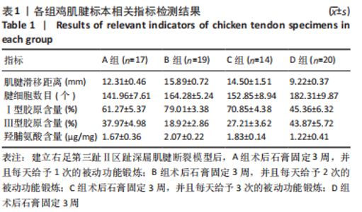
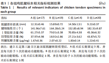
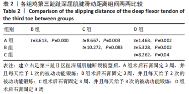
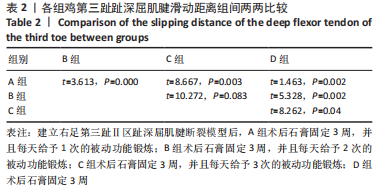
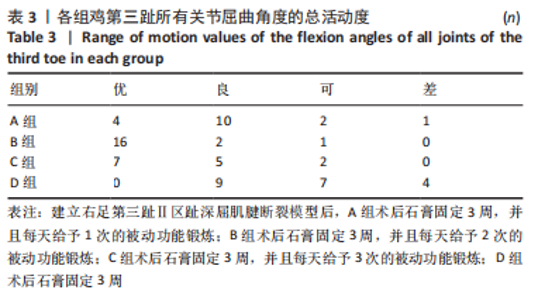
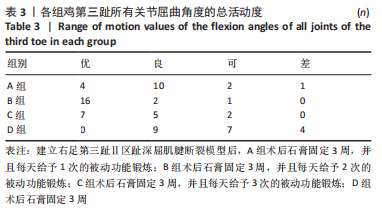
2.3 各组鸡第三趾趾深屈肌腱滑移距离和所有关节屈曲角度 经SNK法分析得出,B、C组鸡第三趾趾深屈肌腱滑移距离比较差异无显著性意义(P > 0.05),B、C组鸡第三趾趾深屈肌腱滑移距离大于A、D组(P < 0.05),A组鸡第三趾趾深屈肌腱滑移距离大于D组(P < 0.05),见表1,2。第三趾所有关节屈曲角度的总活动度,A组的优良率为82.4%,B组的优良率为94.7%,C组的优良率为85.7%,D组的优良率为42.9%,见表3,此指标经χ2检验分析得出各组间优良率比较差异均有显著性意义(P < 0.05)。"
"
"
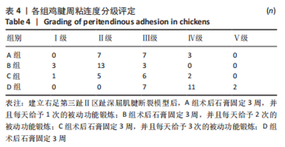
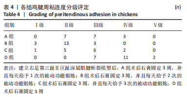
2.4 各组鸡肌腱吻合端形态及腱周粘连度分级评定 B、C组吻合端膨大不明显,质地和正常腱性组织差别不大,腱周组织瘢痕不多,相对于C组,B组的粘连程度相对轻;C组有5份标本的吻合端有分离,断端间是瘢痕组织连接;D组吻合端膨大明显,质地较硬,腱周组织瘢痕明显,粘连程度重;A组的表现介于B、C组和D组之间,见表4。此指标经χ2分析得出,各组间腱周粘连度分级结果比较差异有显著性意义(P < 0.05)。"

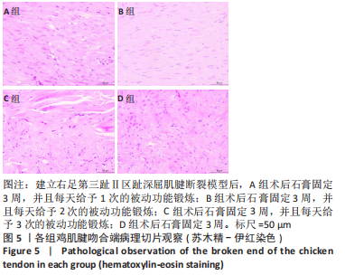
2.5 各组鸡肌腱吻合端苏木精-伊红染色和天狼星红染色的观察 苏木精-伊红染色可见,A、C、B组的腱细胞排列顺序逐渐好转,数量逐渐接近正常,细胞核的形态逐渐向梭形发展、染色逐渐浅染,C组可见正常的肌腱组织和瘢痕组织有明显的交界区;D组的腱细胞排列较紊乱,细胞核的大小和染色差异较大,见图5。"

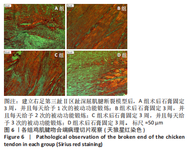
天狼星红染色可以很好地显示Ⅰ型和Ⅲ型胶原纤维。天狼星红染色染色可见,A、C、B组胶原纤维排列逐渐有方向性,鲜红粗大的Ⅰ型胶原纤维逐渐增多,绿色细小的Ⅲ型胶原纤维逐渐减少;E组胶原纤维排列方向性差,Ⅰ型和Ⅲ型胶原纤维交叉分布,见图6。"
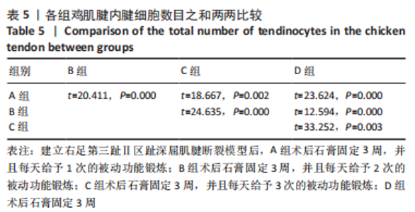
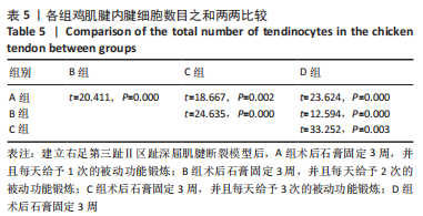
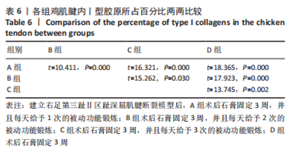
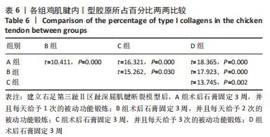
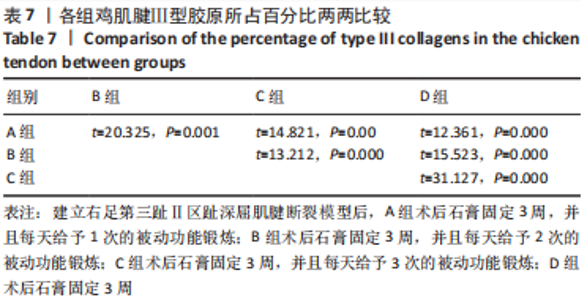
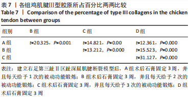
各组鸡肌腱吻合端腱细胞数量及Ⅰ型和Ⅲ型胶原含量,见表1。经方差分析及SNK法分析,4组间腱细胞数量及Ⅰ型和Ⅲ型胶原含量两两比较差异均有显著性意义(P < 0.05),见表5-7。"
"
"
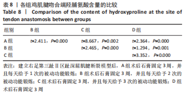
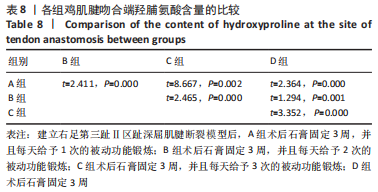
2.6 各组鸡肌腱吻合端羟脯氨酸含量测定 各组鸡肌腱吻合端羟脯氨酸含量见表1。经方差及SNK法分析,4组间羟脯氨酸含量两两比较差异均有显著性意义(P < 0.05),见表8。"
| [1] LEE JS, KIM YH. Factors associated with limited hand motion after hand trauma. Medicine (Baltimore). 2019;98(3):e14183. [2] THANGAVELU M, VEERASAMY N, KANTHAN A. Study on evaluation and management of hand injuries. J Evol Med Dent Sci. 2016;5(66):4703-4707. [3] POLINDER S, IORDENS GI, PANNEMAN MJ, et al. Trends in incidence and costs of injuries to the shoulder, arm and wrist in The Netherlands between 1986 and 2008. BMC Public Health. 2013;13:531. [4] 强力,薛明宇,卜凡玉,等.游离腕横纹嵌合皮瓣修复手指复合组织缺损八例[J].中华显微外科杂志,2017,40(4):371-373. [5] KRITIOTIS C, PHILLIPS A, MUIR L, et al. Practice in wide-awake hand surgery:differences between United Kingdom and Cyprus. Hand Clin. 2019;35(1):43-50. [6] KARJALAINEN T, JOKINEN K, SEBASTIN SJ, et al. Correlations among objectively measured impairment,outcome classification systems,and subjectively perceived disability after flexor tendon repair. J Hand Surg Am. 2019;44(5):361-365. [7] 高君,王维,那磊,等.肌腱粘连的预防:现状和进展[J].中国组织工程研究,2014,18(46):7515-7519. [8] STAUBER T, BLACHE U, SNEDEKER JG. Tendon tissue microdamage and the limits of intrinsic repair. Matrix Biol. 2020;85-86:68-79. [9] 张家乐,梁远,王静成.肌腱粘连的研究进展[J].实用临床医药杂志,2021,25(12):119-123. [10] 路明宽,蔡传栋,王伟,等.防肌腱粘连膜制备材料的研究进展[J].上海交通大学学报(医学版),2021,41(4):550-553. [11] 王骏,刘振峰,沈霞,等.指屈肌腱Ⅱ区修复术后早期活动方式对指功能恢复的影响[J].中华手外科杂志,2021,37(1):64-67. [12] MICHEL PA, KRONENBERG D, NEU G, et al. Microsurgical reconstruction affects the outcome in a translational mouse model for Achilles tendon healing. J Orthop Translat. 2020;24:1-11. [13] ACKERMAN JE, BEST KT, MUSCAT SN, et al. Metabolic Regulation of Tendon Inflammation and Healing Following Injury. Curr Rheumatol Rep. 2021;23(3):1-9. [14] 庄加川,李敏姣,曾锦浩,等.单根肌腱缝线埋线修复肌腱损伤的临床应用[J].中华手外科杂志,2020,36(4):300-301. [15] 陈东生,贾麒钰,阿卜杜萨拉木·阿力木江,等.手部肌腱损伤与修复:737例流行病学因素分析[J].中国组织工程研究,2022,26(35): 5676-5684. [16] 庄加川,李敏姣,曾锦浩,等.单根肌腱缝线埋线修复肌腱损伤的临床应用[J].中华手外科杂志,2020,36(4):300-301. [17] 杨占宇,朱海波,王旼娴.动力型支具防治手指屈肌腱松解术后肌腱粘连的临床研究[J].中华手外科杂志,2020,36(4):298-299. [18] 程杰,王继宏,郝增涛,等.改良津下缝合法即刻生物力学实验和在动物模型中的应用[J].临床骨科杂志,2018,21(6):749-752. [19] 顾江江,赵凤朝,程琪,等.酒精灼烧致青脚麻鸡股骨头坏死造模方法的可行性分析[J]. 中国组织工程研究,2020,24(17):2700-2705. [20] 汤锦波,侍德,石井清一.各种伤情下屈肌腱的愈合及肌腱粘连形成[J].中华手外科杂志,1992,8(1):31-35. [21] 王继宏,温树正.天狼星红染色法在肌腱胶原形态分析中的应用[J].中华实验外科杂志,2011,28(7):1096-1098. [22] 杨秀颖,杜冠华.组织羟脯氨酸含量微量测定方法及应用[J].中国药理学通报,2004,20(7):836-837. [23] PETERS SE, JHA B, ROSS M. Rehabilitation following surgery for flexor tendon injuries of the hand. Cochrane Database Syst Rev. 2021;1(1): CD012479. [24] LIU C, BAI J, YU K, et al. Biological Amnion Prevents Flexor Tendon Adhesion in Zone II:A Controlled, Multicentre Clinical Trial. Biomed Res Int. 2019;2019(8):1-9. [25] PRAKASH S, KALRA P, DHAL A. Flexor tendon repair with amniotic membrane. Int Orthop. 2020;44(10):2037-2045. [26] NEUWIRTH M, BÜRGER H, PALLE W, et al. One-stage reconstruction of isolated and combined tendon defects with the vascularized adductor magnus tendon graft:Surgical technique and preliminary results. J Plast Reconstr Aesthet Surg. 2016;69(7):928-935. [27] KREY D, BORCHERS J, MCCAMEY K. Tendon needling for treatment of tendinopathy:A systematic review. Phys Sportsmed. 2015;43(1):80-86. [28] FUJIHARA Y, OTA H, WATANABE K. Utility of early active motion for flexor tendon repair with concomitant injuries: A multivariate analysis. Injury. 2018;49(12):2248-2251. [29] BIGORRE N, DELAQUAIZE F, DEGEZ F, et al. Primary flexor tendons repair in zone 2: Current trends with GEMMSOR survey results. Hand Surg Rehabil. 2018;37(5):281-288. [30] STAUBER T, BLACHE U, SNEDEKER JG. Tendon tissue microdamage and the limits of intrinsic repair. Matrix Biol. 2020;85-86:68-79. [31] KOEHLER SV, SAUERBIER M, TERZIS A. Rupture Rate,Functional Outcome and Patient Satisfaction after Primary Flexor Tendon Repair with the Modified 4-Strand Core Suture Technique by Tsuge and Using the Arthrex FiberLoop((R)) with Early Motion Rehabilitation. J Clin Med. 2021;10(19):4538. [32] MANNINEN M, KARJALAINEN T, MÄÄTTÄ J, et al. Epidemiology of Flexor Tendon Injuries of the Hand in a Northern Finnish Population. Scand J Surg. 2017;106(3):278-282. [33] SHEN H, YONEDA S, SAKIYAMA-ELBERT SE, et al. Flexor Tendon Injury and Repair.The Influence of Synovial Environment on the Early Healing Response in a Canine Model. J Bone Joint Surg Am. 2021;103(9):e36. [34] 高君,王维,那磊,等.构建趾深屈肌腱横断模型:移植腱周膜预防肌腱粘连[J].中国组织工程研究,2015,19(18):2896-2900. [35] MICHEL PA, KRONENBERG D, NEU G, et al. Microsurgical reconstruction affects the outcome in a translational mouse model for Achilles tendon healing. J Orthop Translat. 2020;24:1-11. [36] GHAVAMIAN A, MOUSAVI SJ, AVRIL S. Computational Studv of Growth and RemodeIing in Ascending Thoracic Aortic Aneurysms Considering variations of smooth Muscle cell Basal Tone. Front Bioeng Biotechnol. 2020;8:587376. [37] LEONG NL, KATOR JL, CLEMENS TL, et al. Tendon and ligament healing and current approaches to tendon and ligament regeneration. J Orthop Res. 2020;38(1):7-12. [38] JACKSON JE, KOPECKI Z, ANDERSON PJ, et al. In vitro analysis of the effect of Flightless I on murine tenocyte cellular functions. J Orthop Surg Res. 2020;15(1):170. [39] CONNIZZO BK, PIET JM, SHEFELBINE SJ, Et al. Age-associated changes in the response of tendon explants to stress deprivation is sex-dependent. Connect Tissue Res. 2020;61(1):48-62. [40] SEREYSKY JB, FLATOW EL, ANDARAWIS-PURI N. Musculoskeletal regeneration and its implications for the treatment of tendinopathy. Int J Exp Pathol. 2013;94(4):293-303. [41] SUN YL, THORESON AR, CHA SS, et al. Temporal response of canine flexor tendon to limb uspension. J Appl Physiol (1985). 2010;109(6): 1762-1768. [42] SZCZODRY M, ZHANG J, LIM C, et al. Treadmill running exercise results in the presence of numerous myofibroblasts in mouse patellar tendons. J Orthop Res. 2009;27(10):1373-1378. [43] 郭文,王继宏,温树正,等.应力在肌腱愈合中的作用[J].中国组织工程研究,2015,19(29):4715-4720. [44] 陈建海,姜保国,傅中国,等.肌腱损伤修复后早期最大抗拉力与功耗变化的实验研究明[J].中华手外科杂志,2004,20(2):117-119. [45] 范家强,郝云甲,张在轶,等.改良Giftbox缝合法联合束状捆扎技术治疗急性跟腱断裂的疗效分析[J].中华解剖与临床杂志, 2022,27(1):30-34. [46] 何家雄,吴家盛,朱俊德,等.超声引导下改良Bunnell法经皮缝合术治疗急性闭合性跟腱断裂的疗效[J].川北医学院学报,2022, 37(3):345-348. [47] 叶建兴,梁会平,刘强,等.彩超引导下微创改良Krackow缝合法治疗跟腱断裂临床疗效研究[J].医学理论与实践,2022,35(19): 3306-3308. |
| [1] | Hu Guangzhi, Lu Hongyan. Changes in pulmonary pericytes and tube formation of pulmonary vascular endothelial cells in mouse models of broncho-pulmonary dysplasia [J]. Chinese Journal of Tissue Engineering Research, 2024, 28(4): 522-527. |
| [2] | Pan Yujun, Shi Changjiang, Cao Sheng, Mu Huaizhao, Zhang Yilong, Wang Kaihua. An injectable dextran/gelatin composite hydrogel loaded with exosomes for repair of rat intervertebral disc degeneration [J]. Chinese Journal of Tissue Engineering Research, 2023, 27(34): 5477-5482. |
| [3] | Liu Huan, Li Han, Ma Yunhao, Zhong Weijian, Ma Guowu. Osteogenic capacity of partially demineralized dentin particles in the maxillary sinus lift [J]. Chinese Journal of Tissue Engineering Research, 2023, 27(3): 354-359. |
| [4] | Dong Shiwu, Zhou Lanxi, Shao Lu, Yu Zhengwen. Effect of magnesium alloy biomaterial degradation on endothelialized cells [J]. Chinese Journal of Tissue Engineering Research, 2023, 27(21): 3398-3406. |
| [5] | Liang Hanying, Ma Yunhao, Li Han, Li Dongyang, Zhong Weijian, Ma Guowu. Osteogenic effects of partially demineralized autogenous dentin particles in implant site preservation [J]. Chinese Journal of Tissue Engineering Research, 2023, 27(16): 2488-2492. |
| [6] | Li Wei, Lin Meina, Lu Yongping, Sui Yu, Jiang Miao. Feasibility and advantages of mesenchymal stem cells for repairing Achilles tendon injury [J]. Chinese Journal of Tissue Engineering Research, 2023, 27(15): 2404-2411. |
| [7] | Chen Dongsheng, Jia Qiyu, Abudusalamu·Alimujiang, Guo Jian, Feng Dongwei, Wu Tong, Ma Chuang. Tendon injury and repair of the hand: analysis of epidemiological factors in 737 cases [J]. Chinese Journal of Tissue Engineering Research, 2022, 26(35): 5676-5684. |
| [8] | Li Chuanhong, Yu Xing, Yang Yongdong, Yang Kaitan, Zhao He. Subarachnoid injection via the posterior atlanto-occipital interspace is an alternative way of administration in a rat model of spinal cord injury [J]. Chinese Journal of Tissue Engineering Research, 2022, 26(33): 5376-5383. |
| [9] | Guo Yangyan, Yu Zhengwen, Zhang Jian. Research hotspots of magnesium alloy biomaterials in an in vivo animal [J]. Chinese Journal of Tissue Engineering Research, 2022, 26(22): 3556-3565. |
| [10] | Gou Yongsheng, Ding Keyuan, Xu Shengqian, Li Haibo, Zhong Gang, Li Liang, Teng Lin. Comparison of double Kirschner wire with double screw fixation in the treatment of avulsion fracture at the base of the distal phalanx [J]. Chinese Journal of Tissue Engineering Research, 2022, 26(18): 2849-2853. |
| [11] | Wang Shuyun, Xie Junhui, Yu Xuefeng. Effect and mechanism of mesenchymal stem cells in the treatment of diabetic nephropathy [J]. Chinese Journal of Tissue Engineering Research, 2022, 26(1): 148-152. |
| [12] | Zhang Chao, Lü Xin. Heterotopic ossification after acetabular fracture fixation: risk factors, prevention and treatment progress [J]. Chinese Journal of Tissue Engineering Research, 2021, 25(9): 1434-1439. |
| [13] | Zhao Binbin, Zhong Weijian, Ma Guowu, Li Yongqi, Wang Ning. Comparison of the osteogenic effect of three different bone graft materials [J]. Chinese Journal of Tissue Engineering Research, 2021, 25(10): 1507-1510. |
| [14] | Feng Yong, Zhao Yanxu, Zhang Minze. Human amniotic membrane, drugs, and growth factors prevent adhesion after repair of tendon injury [J]. Chinese Journal of Tissue Engineering Research, 2020, 24(4): 625-630. |
| [15] | Huo Shaochuan, Wang Haibin, Tang Hongyu, Wang Yueqi, Chen Qunqun, Feng Ziyu, Li Yikai. Treatment of knee osteoarthritis by tonifying kidney and spleen and activating blood circulation herbs via chondrocyte IL-1beta/ERR-alpha/SOX9/Col2alpha1 signaling pathway in a rat model [J]. Chinese Journal of Tissue Engineering Research, 2020, 24(35): 5577-5581. |
| Viewed | ||||||
|
Full text |
|
|||||
|
Abstract |
|
|||||