Chinese Journal of Tissue Engineering Research ›› 2023, Vol. 27 ›› Issue (30): 4824-4829.doi: 10.12307/2023.833
Previous Articles Next Articles
Three-dimensional finite element analysis of different restorative methods for ultrashort implants
Reyila·Kuerban, Xiaheida·Yilaerjiang, Chen Xin, Zilala·Julaiti, Baibujiafu·Yellisi, Nijati·Turson
- Department of Stomatology, Second Affiliated Hospital of Xinjiang Medical University, Urumqi 830063, Xinjiang Uygur Autonomous Region, China
-
Received:2022-08-30Accepted:2022-11-30Online:2023-10-28Published:2023-04-03 -
Contact:Nijati·Turson, Master, Chief physician, Associate professor, Department of Stomatology, Second Affiliated Hospital of Xinjiang Medical University, Urumqi 830063, Xinjiang Uygur Autonomous Region, China -
About author:Reyila·Kuerban, Master candidate, Department of Stomatology, Second Affiliated Hospital of Xinjiang Medical University, Urumqi 830063, Xinjiang Uygur Autonomous Region, China -
Supported by:Natural Science Foundation of Xinjiang Uygur Autonomous Region, No. 2016D01C192 (to NT)
CLC Number:
Cite this article
Reyila·Kuerban, Xiaheida·Yilaerjiang, Chen Xin, Zilala·Julaiti, Baibujiafu·Yellisi, Nijati·Turson. Three-dimensional finite element analysis of different restorative methods for ultrashort implants[J]. Chinese Journal of Tissue Engineering Research, 2023, 27(30): 4824-4829.
share this article
Add to citation manager EndNote|Reference Manager|ProCite|BibTeX|RefWorks

2.1 不同修复方式对应同一方向力时种植修复体及骨组织应力分布 施加垂直载荷:当2枚植体采用单冠方式进行修复时,牙冠的应力集中均位于冠的内侧壁与基台的连接处,但第一磨牙区稍偏向近中、第二磨牙区稍偏向远中,各组件应力峰值集中于中央螺丝上,2枚种植体的单冠应力值无明显差异;当2枚植体采用联冠修复方式时,牙冠的应力峰值集中于联冠连接体靠近龈缘处,各部件应力峰值集中于联冠上;垂直向载荷时,应力峰值则随着修复方式的不同而随之改变。采用单冠修复时,各部件受力均匀无明显差异,主要集中于中央螺丝、基台及种植体上,单冠修复各部件的应力峰值均高于联冠修复,骨组织应力峰值也明显大于联冠修复,见图4。"

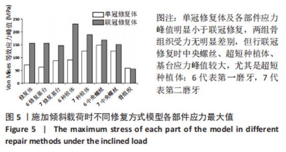
施加斜向载荷:当2枚植体采用单冠修复方式时,牙冠的应力集中区位于冠的内侧壁与基台的连接处,各组件应力峰值集中于中央螺丝上,其余各个接触组件之间应力峰值无明显差值,变化均匀;当2枚植体采用联冠修复时,牙冠的应力峰值集中于联冠连接体靠近龈缘处,冠、中央螺丝、基台、超短种植体应力分布较集中;单冠修复体及各部件应力峰值明显小于联冠修复,两组骨组织受力无明显差别,但行联冠修复时中央螺丝、超短种植体、基台应力峰值较大,尤其是超短种植体,见图5。"

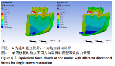
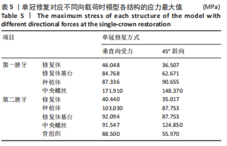
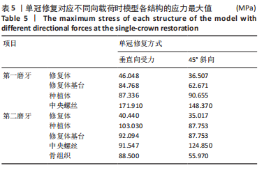
2.2 同一修复方式对应不同方向力时种植修复体及骨组织应力分布 采用单冠修复时:施加垂直与斜向载荷时,应力分布大小无明显差异,各部件之间应力值较均匀,应力峰值位于中央螺丝及修复体基台、皮质骨上,不管从哪一种方向施加力,力都大致随着超短种植体的长轴向骨组织方向分散,斜向受力时骨组织的应力分布较小,见图6,表5。"
"
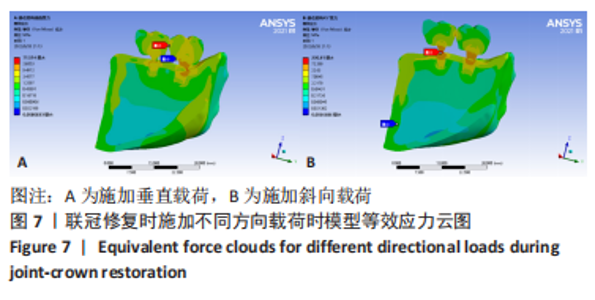
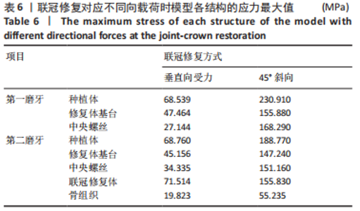
采用联冠修复时:施加垂直与斜向载荷时,种植修复体的应力主要分布在联冠中间的连接体上;但施加斜向载荷时,应力峰值主要集中于中央螺丝及与其接近的超短种植体、基台上,超短种植体、种植修复体、基台、中央螺丝的应力峰值也明显大于施加垂直载荷时,施加垂直载荷时各组件应力峰值小且均匀,见图7,表6。"

"
| [1] 胡佩男,刘洋,李阳,等.口腔微生态与龋病防治的研究进展[J].现代医药卫生,2022,38(9):1531-1535. [2] 马学荣,张华林,岳进,等.牙列缺损的修复方式对口腔健康相关生活质量的影响[J].宁夏医科大学学报,2020,42(7):749-752. [3] 安辉.口腔种植修复和常规修复在牙列缺损治疗中的效果对比[J].中国实用医药,2021,16(20):100-102. [4] 吴佩玲,张晓倩,尼加提·吐尔逊,等.上颌窦外提升中黏膜穿孔成因与修复后的种植(英文)[J].中国组织工程研究,2014,18(51):8223-8227. [5] 齐昱晗,石前会,安哲庆,等.环状植骨修复牙列缺损伴骨量不足[J].中国组织工程研究,2023,27(17):2738-2744. [6] 付丽,孙悦,翟婧捷,等.3D打印及CGF在双侧下牙槽神经移位同期牙种植术中的应用[J].口腔医学研究,2016,32(10):1003-1005. [7] SRINIVASAN M, VAZQUEZ L, RIEDER P, et al. Survival rates of short (6 mm) micro-rough surface implants: a review of literature and meta-analysis. Clin Oral Implants Res. 2014;25(5):539-545. [8] 安尼卡尔·安尼瓦尔,帕丽黛姆·图尔迪,阿迪力·麦木提敏,等.上颌前牙区不同种植修复体在不同咬合受力下的三维有限元分析[J].中国组织工程研究,2020,24(16):2531-2536. [9] 吴越琳,刘加荣.三维有限元分析在口腔医学领域的研究进展[J].口腔医学, 2021,41(7):659-663. [10] AROSIO P, GRECO GB, ZANIOL T, et al. Sinus augmentation and concomitant implant placement in low bone-density sites. A retrospective study on an undersized drilling protocol and primary stability. Clin Implant Dent Relat Res. 2018;20(2):151-159. [11] LIU C, XING Y, LI Y, et al. Bone quality effect on short implants in the edentulous mandible: a finite element study. BMC Oral Health. 2022;22(1):139. [12] 白布加甫·叶力思,热娜古丽·买合木提,艾孜买提江·赛依提,等.上颌中切牙联冠种植修复体在不同咬合方式中的应力分析[J].中国组织工程研究, 2022,26(4):567-572. [13] 孙江伟,王俊祥,白布加甫·叶力思,等.不同光滑颈圈种植体修复时应力分布的三维有限元分析[J].中国组织工程研究,2023,27(7):1004-1011. [14] ERCAL P, TAYSI AE, AYVALIOGLU DC, et al. Impact of peri-implant bone resorption, prosthetic materials, and crown to implant ratio on the stress distribution of short implants: a finite element analysis. Med Biol Eng Comput. 2021;59(4):813-824. [15] 施梦汝,谢伟丽,施武阁,等.柱形锥形种植体在不同种植深度的三维有限元研究[J].口腔医学,2019,39(7):577-580. [16] HUSSEIN FA, SALLOOMI KN, ABDULRAHMAN BY, et al. Effect of thread depth and implant shape on stress distribution in anterior and posterior regions of mandible bone: A finite element analysis. Dent Res J (Isfahan). 2019;16(3):200-207. [17] OZAN O, KURTULMUS-YILMAZ S. Biomechanical Comparison of Different Implant Inclinations and Cantilever Lengths in All-on-4 Treatment Concept by Three-Dimensional Finite Element Analysis. Int J Oral Maxillofac Implants. 2018;33(1): 64-71. [18] DU L, LI Z, CHANG X, et al. Effects of the Screw-Access Hole Diameter on the Biomechanical Behaviors of 4 Types of Cement-Retained Implant Prosthodontic Systems and Their Surrounding Cortical Bones: A 3D Finite Element Analysis. Implant Dent. 2018;27(5):555-563. [19] 黄耀文.不同材料修复下颌第一前磨牙楔状缺损的三维有限元分析[D].南昌:南昌大学,2020. [20] 李希光,郅克谦,高岭,等.基于三维有限元评价种植体不同倾斜角度在上颌后牙区骨量不足的应力分析[J].口腔医学研究,2019,35(7):671-675. [21] QIN S, GAO Z. Comparative evaluation of short or standard implants with different prosthetic designs in the posterior mandibular region: a three-dimensional finite element analysis study. Comput Methods Biomech Biomed Engin. 2022:1-11. doi: 10.1080/10255842.2022.2124859. [22] DE MATOS JDM, LOPES GDRS, NAKANO LJN, et al. Biomechanical evaluation of 3-unit fixed partial dentures on monotype and two-piece zirconia dental implants. Comput Methods Biomech Biomed Engin. 2022;25(3):239-246. [23] XIE B, CHEN J, ZHAO T, et al. Three-dimensional finite element analysis of anterior fixed partial denture supported by implants with different materials. Ann Anat. 2022;243:151943. [24] 李春.口腔种植与传统固定义齿修复牙列缺损的疗效对比[J].临床医学工程, 2021,28(4):481-482. [25] 安维康,张薇,郑亚飞,等.影响短种植体成功率的因素探讨[J].中华口腔医学研究杂志(电子版),2021,15(3):129-134. [26] SLOTTE C, GRØNNINGSAETER A, HALMØY AM, et al. Four-Millimeter-Long Posterior-Mandible Implants: 5-Year Outcomes of a Prospective Multicenter Study. Clin Implant Dent Relat Res. 2015;17 Suppl 2:e385-395. [27] 叶芷君,徐普.超短种植体在萎缩后牙区的临床研究进展[J].成都医学院学报,2021,16(5):673-675. [28] NISAND D, RENOUARD F. Short implant in limited bone volume. Periodontol 2000. 2014;66(1): 72-96. [29] LOMBARDO G, MARINCOLA M, SIGNORIELLO A, et al. Single-crown, short and ultra-short implants, in association with simultaneous internal sinus lift in the atrophic posterior maxilla: a three-year retrospective study. Materials(Basel). 2020;13(9): E2208. [30] NIZAM N, GÜRLEK Ö, KAVAL ME. Extra-Short Implants with Osteotome Sinus Floor Elevation: A Prospective Clinical Study. Int J Oral Maxillofac Implants. 2020;35(2):415-422. [31] PELLIZZER EP, MARCELA DE LUNA GOMES J, ARAÚJO LEMOS CA, et al. The influence of crown-to-implant ratio in single crowns on clinical outcomes: A systematic review and meta-analysis. J Prosthet Dent. 2021;126(4):497-502. [32] TANG Y, YU H, WANG J, et al. Influence of crown-to-implant ratio and different prosthetic designs on the clinical conditions of short implants in posterior regions: A 4-year retrospective clinical and radiographic study. Clin Implant Dent Relat Res. 2020;22(1):119-127. [33] 夏勋,胡常琦,黄江琴,等.短种植体冠根比对骨改建的影响:5年临床观察[J].口腔医学研究,2020,36(7):683-687. [34] MEIJER HJA, BOVEN C, DELLI K, et al. Is there an effect of crown-to-implant ratio on implant treatment outcomes? A systematic review. Clin Oral Implants Res. 2018;29 Suppl 18(Suppl Suppl 18):243-252. [35] HINGSAMMER L, WATZEK G, POMMER B. The influence of crown-to-implant ratio on marginal bone levels around splinted short dental implants: A radiological and clincial short term analysis. Clin Implant Dent Relat Res. 2017;19(6):1090-1098. [36] RAVIDÀ A, BAROOTCHI S, ALKANDERI A, et al. The Effect of Crown-to-Implant Ratio on the Clinical Outcomes of Dental Implants: A Systematic Review. Int J Oral Maxillofac Implants. 2019;34(5):1121-1131. [37] GUARNIERI R, DI NARDO D, GAIMARI G, et al. Short vs. Standard Laser-Microgrooved Implants Supporting Single and Splinted Crowns: A Prospective Study with 3 Years Follow-Up. J Prosthodont. 2019;28(2):e771-e779. [38] ROMERO-MILLÁN JJ, AIZCORBE-VICENTE J, PEÑARROCHA-DIAGO M, et al. Implants in the Posterior Maxilla: Open Sinus Lift Versus Conventional Implant Placement. A Systematic Review. Int J Oral Maxillofac Implants. 2019;34(4):e65-e76. [39] 胡常琦,魏振宇,魏洪武,等.短种植体(≤6 mm)在萎缩后牙区5~7年疗效的回顾性研究[J].口腔医学研究,2021,37(1):58-62. [40] CALVO-GUIRADO JL, LÓPEZ TORRES JA, DARD M, et al. Evaluation of extrashort 4-mm implants in mandibular edentulous patients with reduced bone height in comparison with standard implants: a 12-month results. Clin Oral Implants Res. 2016;27(7):867-874. [41] PAPASPYRIDAKOS P, DE SOUZA A, VAZOURAS K, et al. Survival rates of short dental implants (≤6 mm) compared with implants longer than 6 mm in posterior jaw areas: A meta-analysis. Clin Oral Implants Res. 2018;29 Suppl 16:8-20. [42] 马新扬,王方,唐成芳,等.基台角度对种植体周围骨组织应力分布的影响[J].山西医科大学学报,2021,52(1):97-101. [43] 刘世员.不同角度基台Straumann种植系统行前牙种植修复的疗效及并发症分析[J].中国口腔种植学杂志,2017,22(1):36-39. [44] ROSSI F, BOTTICELLI D, CESARETTI G, et al. Use of short implants(6 mm) in a single -tooth replacement: a 5 -year follow -up prospective randomized controlled multicenter clinical study. Clin Oral Implants Res. 2016;27(4): 458-464. [45] 邹立东,韩劼,吴敏节,等.后牙连续缺失后短种植体联冠修复早期负重的临床研究[J].中华口腔医学杂志,2018,53(10):653-658. [46] 徐淑兰,郭泽鸿,宁颖圆,等.种植义齿冠根比与临床并发症[J].口腔疾病防治,2020,28(9):545-550. [47] NAENNI N, SAHRMANN P, SCHMIDLIN PR, et al. Five-Year Survival of Short Single-Tooth Implants (6 mm): A Randomized Controlled Clinical Trial. J Dent Res. 2018; 97:887-892. [48] 莫嘉骥,乔士冲,张枭,等.后牙区短种植体(≤6 mm)单冠修复3-5年临床效果评价[J].中国口腔颌面外科杂志,2019,17(3):235-239. [49] SUMI T, BRAIAN M, SHIMADA A, et al. Characteristics of implant-CAD/CAM abutment connections of two different internal connection systems. J Oral Rehabil. 2012;39(5):391-398. [50] 邹丽妍.种植体成品基台和个性化基台的机械性能及适合性的对比研究[D].沈阳:中国医科大学,2021. [51] MITSIAS M, KOUTAYAS SO, WOLFART S, et al. Influence of zirconia abutment preparation on the fracture strength of single implant lithium disilicate crowns after chewing simulation. Clin Oral Implants Res. 2014;25(6):675-682. |
| [1] | Fang Xingyan, Tian Zhenli, Zhao Zheyi, Wen Ping, Xie Tingting. Effects of sodium arsenite on human umbilical vein endothelial cell injury and sphingosine kinases 1/sphingosine 1-phosphate signaling axis [J]. Chinese Journal of Tissue Engineering Research, 2023, 27(在线): 1-7. |
| [2] | Guo Shuhui, Yang Ye, Jiang Yangyang, Xu Jianwen. Screening and validation of neurogenic bladder miRNA-mRNA regulatory network [J]. Chinese Journal of Tissue Engineering Research, 2023, 27(在线): 1-8. |
| [3] | Zhong Yizheng, Huang Peizhen, Cai Qunbin, Zheng Liqin, He Xingpeng, Dong Hang. Microstructural indexes that determine the trabecular bone maximum stress of micro-finite element models [J]. Chinese Journal of Tissue Engineering Research, 2023, 27(9): 1313-1318. |
| [4] | Wu Taoguang, Nie Shaobo, Chen Hua, Zhu Zhengguo, Qi Lin, Tang Peifu. Biomechanical characteristics of a new multi-dimensional cross locking plate in the treatment of subtrochanteric nonunion [J]. Chinese Journal of Tissue Engineering Research, 2023, 27(9): 1330-1334. |
| [5] | Tang Huiyu, Hou Biao, Xia Xiaodan, Xiang Wei, Xie Songlin. Effect of mechanical tension stress on arterial vessels after limb osteotomy in rabbits [J]. Chinese Journal of Tissue Engineering Research, 2023, 27(9): 1422-1426. |
| [6] | Peng Zhixin, Yan Wengang, Wang Kun, Zhang Zhenjiang. Finite element analysis and structural optimization design of 3D printed forearm braces [J]. Chinese Journal of Tissue Engineering Research, 2023, 27(9): 1340-1345. |
| [7] | Liu Jinyu, Zhang Hanshuo, Cui Hongpeng, Pan Lingzhi, Zhao Boran, Li Fei, Ding Yu. Finite element biomechanical analysis of minimally invasive treatment of cervical spondylotic myelopathy and accurate exercise rehabilitation [J]. Chinese Journal of Tissue Engineering Research, 2023, 27(9): 1359-1364. |
| [8] | Wen Xinghua, Ding Huanwen, Cheng Kai, Yan Xiaonan, Peng Yuanhao, Wang Yuning, Liu Kang, Zhang Huiwu. Three-dimensional finite element model analysis of intramedullary nailing fixation design for large femoral defects in Beagle dogs [J]. Chinese Journal of Tissue Engineering Research, 2023, 27(9): 1371-1376. |
| [9] | Dang Yi, Du Chengyan, Yao Honglin, Yuan Nenghua, Cao Jin, Xiong Shan, Zhang Dingmei, Wang Xin. Hormonal osteonecrosis and oxidative stress [J]. Chinese Journal of Tissue Engineering Research, 2023, 27(9): 1469-1476. |
| [10] | Ruan Ling, Wang Guanghua, Wu Rongping, Jin Zhan, Lyu Zhenqing, Zhang Nan, Li Shoubang. Correlation between exercise intensity and lipid metabolism disorder and oxidative stress in a high-diet rat model [J]. Chinese Journal of Tissue Engineering Research, 2023, 27(8): 1149-1155. |
| [11] | Wu Chuang, Muhetaer · Kelimu, Maimaitili · Aisha, Yang Hang. Influence of blood components on blood flow characteristics of individualized communicating aneurysm [J]. Chinese Journal of Tissue Engineering Research, 2023, 27(8): 1219-1223. |
| [12] | Nie Chenchen, Su Kaiqi, Gao Jing, Fan Yongfu, Ruan Xiaodi, Yuan Jie, Duan Zhaoyuan, Feng Xiaodong. The regulatory role of circular RNAs in cerebral ischemia-reperfusion injury [J]. Chinese Journal of Tissue Engineering Research, 2023, 27(8): 1286-1291. |
| [13] | Liang Jiaqi, Liu Hengxu, Yang Jinxin, Yang Yi, Deng Xuhui, Tan Mingjian, Luo Jiong. Health benefit relationship between exercise and intestinal bacteria [J]. Chinese Journal of Tissue Engineering Research, 2023, 27(8): 1292-1299. |
| [14] | Liu Jiaxin, Jia Peng, Men Yutao, Liu Lu, Wang Yeming, Ye Jinduo. Design and optimization of bone trabecular structure with triply periodic minimal surfaces [J]. Chinese Journal of Tissue Engineering Research, 2023, 27(7): 992-997. |
| [15] | Zhu Lin, Gu Weiping, Wang Can, Chen Gang. Biomechanical analysis of All-on-Four and pterygomaxillary implants under different maxillary bone conditions [J]. Chinese Journal of Tissue Engineering Research, 2023, 27(7): 985-991. |
| Viewed | ||||||
|
Full text |
|
|||||
|
Abstract |
|
|||||