Chinese Journal of Tissue Engineering Research ›› 2023, Vol. 27 ›› Issue (20): 3236-3241.doi: 10.12307/2023.433
Previous Articles Next Articles
Effects of icariin on NRG1-ErbB signaling pathways in hippocampus of schizophrenia rats
Liu Yunqin1, Lin Li2, Xiao Wenhao1, Ji Qiuming1, Liu Yanqin3
- 1Department of Psychiatry, 3Operating Room, Wuhan Wudong Hospital, Wuhan 430084, Hubei Province, China; 2Laboratory of Molecular Cell Biology, School of Basic Medicine, Hubei University of Chinese Medicine, Wuhan 430065, Hubei Province, China
-
Received:2022-04-24Accepted:2022-06-15Online:2023-07-18Published:2022-11-19 -
Contact:Liu Yunqin, Associate chief physician, Department of Psychiatry, Wuhan Wudong Hospital, Wuhan 430084, Hubei Province, China -
About author:Liu Yanqin, Associate chief nurse, Operating Room, Wuhan Wudong Hospital, Wuhan 430084, Hubei Province, China -
Supported by:The 59th-Batch General Project of China Postdoctoral Science Foundation, No. 2016M592319 (to LL); Medical and Health Research Project of Wuhan Municipal Health and Family Planning Commission, No. wx14c70 (to XWH)
CLC Number:
Cite this article
Liu Yunqin, Lin Li, Xiao Wenhao, Ji Qiuming, Liu Yanqin. Effects of icariin on NRG1-ErbB signaling pathways in hippocampus of schizophrenia rats[J]. Chinese Journal of Tissue Engineering Research, 2023, 27(20): 3236-3241.
share this article
Add to citation manager EndNote|Reference Manager|ProCite|BibTeX|RefWorks
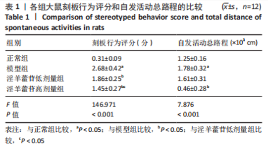
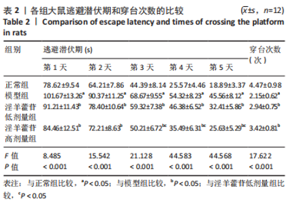
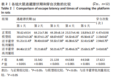
2.1 实验动物数量分析 48只大鼠全部进入结果分析。 2.2 各组大鼠行为学表现 逃避潜伏期采用重复测量资料方差分析,组间和时间效应显著(F=97.991,378.088,P < 0.001),进一步采用多重分析。与正常组比较,模型组大鼠刻板行为评分、自发活动总路程和逃避潜伏期明显增加(P < 0.05),穿台次数明显减少(P < 0.05)。与模型组比较,淫羊藿苷低剂量组大鼠刻板行为评分和逃避潜伏期明显减少(P < 0.05),穿台次数明显增加(P < 0.05);淫羊藿苷高剂量组刻板行为评分、自发活动总路程和逃避潜伏期明显减少(P < 0.05),穿台次数明显增加(P < 0.05)。淫羊藿苷高剂量组大鼠刻板行为评分、逃避潜伏期(第3-5天)明显低于淫羊藿苷低剂量组(P < 0.05),见表1,2。"

"
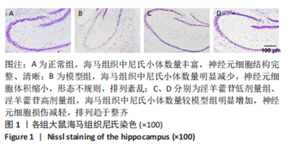
2.3 各组大鼠海马组织病理变化 各组大鼠海马组织尼氏染色见图1。正常组海马组织中尼氏小体数量丰富,神经元细胞结构完整、清晰;与正常组比较,模型组海马组织中尼氏小体数量明显减少,神经元细胞体积缩小,形态不规则,排列紊乱;与模型组比较,淫羊藿苷低、高剂量组海马组织中尼氏小体数量明显增加,神经元细胞损伤减轻,排列趋于整齐。"

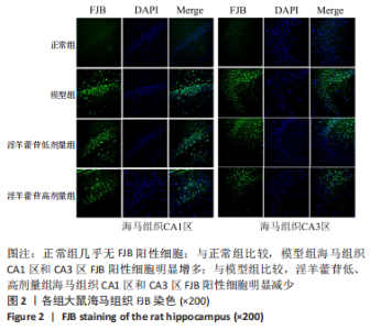
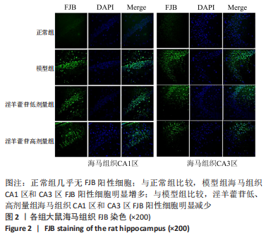
各组大鼠海马组织FJB染色见图2。正常组几乎无FJB阳性细胞;与正常组比较,模型组海马组织CA1区和CA3区FJB阳性细胞明显增多;与模型组比较,淫羊藿苷低、高剂量组海马组织CA1区和CA3区FJB阳性细胞明显减少。"
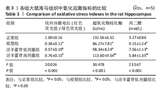
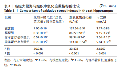
2.4 各组大鼠海马组织内氧化应激水平 与正常行组比较,模型组大鼠海马组织线粒体膜电位、超氧化物歧化酶活性降低(P < 0.05),丙二醛水平升高(P < 0.05);与模型组比较,淫羊藿苷低、高剂量组海马组织线粒体膜电位和超氧化物歧化酶活性升高(P < 0.05),丙二醛水平降低(P < 0.05);淫羊藿苷高剂量组海马组织超氧化物歧化酶活性高于淫羊藿苷低剂量组,丙二醛水平低于淫羊藿苷低剂量组(P < 0.05),见表3。"
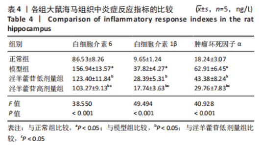
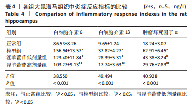
2.5 各组大鼠海马组织内炎症反应水平 与正常组比较,模型组大鼠海马组织中白细胞介素6、白细胞介素1β及肿瘤坏死因子α水平明显增加(P < 0.05);与模型组比较,淫羊藿苷低、高剂量组海马组织中白细胞介素6、白细胞介素1β及肿瘤坏死因子α水平明显减少(P < 0.05),且淫羊藿苷高剂量组海马组织中白细胞介素6、白细胞介素1β及肿瘤坏死因子α水平低于淫羊藿苷低剂量组(P < 0.05),见表4。"

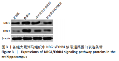
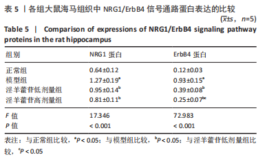
2.6 各组大鼠海马组织中NRG1/ErbB4信号通路蛋白表达 蛋白印迹法检测海马组织中相关蛋白表达,见图3。与正常组比较,模型组大鼠海马组织中NRG1和ErbB4蛋白表达升高(P < 0.05);与模型组比较,淫羊藿苷低、高剂量组海马组织中NRG1和ErbB4蛋白表达降低(P < 0.05),淫羊藿苷高剂量组海马组织中ErbB4蛋白表达低于淫羊藿苷低剂量组(P < 0.05),见表5。"

"
| [1] McCUTCHEON RA, MARQUES TR, HOWES OD. Schizophrenia-an overview. JAMA Psychiatry. 2020;77(2):201-210. [2] SRIVASTAVA A, DADA O, QIAN J, et al. Epigenetics of schizophrenia. Psychiatry Res. 2021;305:114218. [3] RICHETTO J, MEYER U. Epigenetic modifications in schizophrenia and related disorders: molecular scars of environmental exposures and source of phenotypic variability. Biol Psychiatry. 2021;89(3):215-226. [4] 彭兴,马辛,任艳萍,等. 精神分裂症患者语义距离与认知功能的相关性研究[J].中华行为医学与脑科学杂志,2020,29(9):797-801. [5] GAMA MARQUES J, OUAKININ S. Schizophrenia–schizoaffective–bipolar spectra: an epistemological perspective. CNS Spectr. 2021;26(3):197-201. [6] 陈彦霖,陈诚,李泽平,等. NRG1/ErbB4信号通路与精神疾病关联的研究进展[J].中国现代医学杂志,2020,30(15):44-49. [7] HASAN A, ROEH A, LEUCHT S, et al. Add-on spironolactone as antagonist of the NRG1-ERBB4 signaling pathway for the treatment of schizophrenia: Study design and methodology of a multicenter randomized, placebo-controlled trial. Contemp Clin Trials Commun. 2020;17:100537. [8] LIN E, LIN CH, LANE HY. Logistic ridge regression to predict bipolar disorder using mRNA expression levels in the N-methyl-D-aspartate receptor genes. J Affect Disord. 2022;297:309-313. [9] WANG M, GAO H, LI W, et al. Icariin and its metabolites regulate lipid metabolism: From effects to molecular mechanisms. Biomed Pharmacother. 2020;131:110675. [10] 路宇仁,陈昳冰,崔元璐,等.淫羊藿苷药理作用研究进展[J]. 中国实验方剂学杂志,2018,24(17):209-220. [11] 陈静,高瑜,夏海建,等.淫羊藿苷调节大鼠脑内N-甲基-D-天冬氨酸受体分子机制研究[J].中国药业,2020,29(11):18-21. [12] WANG S, MA J, ZENG Y, et al. Icariin, an up-and-coming bioactive compound against neurological diseases: network pharmacology-based study and literature review. Drug Des Devel Ther. 2021;15:3619-3641. [13] ZHOU D, LV D, WANG Z, et al. GLYX-13 ameliorates schizophrenia-like phenotype induced by MK-801 in mice: role of hippocampal NR2B and DISC1. Front Mol Neurosci. 2018;11:121. [14] 万争艳,李宁,向玲玲,等.精神分裂症模型大鼠前额叶皮质PKA和内皮细胞趋化因子-5的表达变化[J]. 中国比较医学杂志,2019,29(10):92-97. [15] JAARO-PELED H, SAWA A. Neurodevelopmental factors in schizophrenia. Psychiatr Clin North Am. 2020,43(2):263-274. [16] PARK HJ, CHOI I, LEEM KH. Decreased brain pH and pathophysiology in schizophrenia. Int J Mol Sci. 2021;22(16):8358. [17] MATSUDA Y, MAKINODAN M, MORIMOTO T, et al. Neural changes following cognitive remediation therapy for schizophrenia. Psychiatry Clin Neurosci. 2019; 73(11):676-684. [18] WRIGHT AC, BROWNE J, SKIEST H, et al. The relationship between conventional clinical assessments and momentary assessments of symptoms and functioning in schizophrenia spectrum disorders: A systematic review. Schizophr Res. 2021; 232(13):11-27. [19] HE C, WANG Z, SHI J. Pharmacological effects of icariin. Adv Pharmacol. 2020;87: 179-203. [20] 陈溪,谷洪顺,张兰,等.淫羊藿苷对MK-801致精神分裂症小鼠模型的影响[J].中国康复理论与实践,2016,22(4):395-398. [21] 刘运琴,刘燕芹,戢汉斌,等.淫羊藿苷对精神分裂症模型大鼠认知功能的改善作用及机制[J].中国药房,2021,32(7):812-818. [22] WINSHIP IR, DURSUN SM, BAKER GB, et al. An overview of animal models related to schizophrenia. Can J Psychiatry. 2020;64(1):5-17. [23] 陈溪,马登磊,李林.淫羊藿苷对cuprizone诱导精神分裂症样小鼠模型行为学变化,髓鞘脱失及神经炎性反应的影响[J].首都医科大学学报,2021, 42(5):761-767. [24] KÖŞGER F, YİĞİTASLAN S, EŞSİZOĞLU A, et al. Inflammation and oxidative stress in deficit schizophrenia. Noro Psikiyatr Ars. 2020;57(4):303-307. [25] UPTHEGGROVE R, KHANDAKER GM. Cytokines, oxidative stress and cellular markers of inflammation in schizophrenia. Curr Top Behav Neurosci. 2020;44: 49-66. [26] PRESTWOOD TR, ASGARIROOZBEHANI R, WU S, et al. Roles of inflammation in intrinsic pathophysiology and antipsychotic drug-induced metabolic disturbances of schizophrenia. Behav Brain Res. 2021;402:113101. [27] CJIEN YL, HWU HG, HANG TJ, et al. Clinical implications of oxidative stress in schizophrenia: Acute relapse and chronic stable phase. Prog Neuropsychopharmacol Biol Psychiatry. 2020;99:109868. [28] 范瑜,吴嘉,林静.集体心理干预对精神分裂症患者氧化应激、细胞凋亡及炎症反应的影响[J].海南医学院学报,2017,23(16):2299-2302. [29] JING X, DU T, CHEN K, et al. Icariin protects against iron overload-induced bone loss via suppressing oxidative stress. J Cell Physiol. 2019;234(7):10123-10137. [30] WANG P, MENG Q, WANG W, et al. Icariin inhibits the inflammation through down-regulating NF-κB/HIF-2α signal pathways in chondrocytes. Biosci Rep. 2020;40(11):BR20203107. [31] RAJASEKARAN A, SHIVAKUMAR V, KALMAADY SV, et al. Impact of NRG1 HapICE gene variants on digit ratio and dermatoglyphic measures in schizophrenia. Asian J Psychiatry. 2020;54:102363. [32] LI C, TAO H, YANG X, et al. Assessment of a combination of serum proteins as potential biomarkers to clinically predict Schizophrenia. Int J Med Sci. 2018;15(9): 900-906. [33] ZHANG Z, LI Y, HE F, et al. Sex differences in circulating neuregulin1-beta1 and beta-secretase 1 expression in childhood-onset schizophrenia. Compr Psychiatry. 2020;100:152176. [34] ZIEBA J, MPRRIS MJ, WEICKERT CS, et al. Behavioural effects of high fat diet in adult Nrg1 type III transgenic mice. Behav Brain Res. 2020;377:112217. [35] JAHN K, HEESE A, KEBIR O, et al. Differential methylation pattern of schizophrenia candidate genes in tetrahydrocannabinol-consuming treatment-resistant schizophrenic patients compared to non-consumer patients and healthy controls. Neuropsychobiology. 2021;80(1):36-44. [36] XIE R, HONG S, YE Y, et al. Ketamine affects the expression of ErbB4 in the hippocampus and prefrontal cortex of rats. J Mol Neuroscie. 2020;70(6):962-967. [37] SKIRZEWSKI M, CRONIN ME, MURPHY R, et al. ErbB4 null mice display altered mesocorticolimbic and nigrostriatal dopamine Levels, as well as deficits in cognitive and motivational behaviors. eNeuro. 2020;7(3):ENEURO.0395-19.2020. [38] YANG JZ, KANG CY, WU C, et al. Pharmacogenetic associations of NRG1 polymorphisms with neurocognitive performance and clinical symptom response to risperidone in the untreated schizophrenia. Schizophr Res. 2021;231(1-3):67-69. [39] ZHANG C, NI P, LIU Y, et al. GABAergic abnormalities associated with sensorimotor corticostriatal community structural deficits in ErbB4 knockout mice and first-episode treatment-naive patients with schizophrenia. Neurosci Bull. 2020;36(2): 97-109. [40] DABBAH-ASSADI F, ALON D, GOLANI I, et al. The influence of immune activation at early vs late gestation on fetal NRG1-ErbB4 expression and behavior in juvenile and adult mice offspring. Brain Behav Immun. 2019;79:207-215. |
| [1] | Guo Shuhui, Yang Ye, Jiang Yangyang, Xu Jianwen. Screening and validation of neurogenic bladder miRNA-mRNA regulatory network [J]. Chinese Journal of Tissue Engineering Research, 2023, 27(在线): 1-8. |
| [2] | Fang Xingyan, Tian Zhenli, Zhao Zheyi, Wen Ping, Xie Tingting. Effects of sodium arsenite on human umbilical vein endothelial cell injury and sphingosine kinases 1/sphingosine 1-phosphate signaling axis [J]. Chinese Journal of Tissue Engineering Research, 2023, 27(在线): 1-7. |
| [3] | He Xi, Wan Yu, Tang Yuting, Yang Anning, Wu Kai, Jiao Yun, Bai Zhigang, Jiang Yideng, Shen Jiangyong. Erastin inhibits proliferation of hypertrophic scar fibroblasts [J]. Chinese Journal of Tissue Engineering Research, 2023, 27(在线): 1-. |
| [4] | Zhong Yizheng, Huang Peizhen, Cai Qunbin, Zheng Liqin, He Xingpeng, Dong Hang. Microstructural indexes that determine the trabecular bone maximum stress of micro-finite element models [J]. Chinese Journal of Tissue Engineering Research, 2023, 27(9): 1313-1318. |
| [5] | Cao Sheng, Kong Lingwei, Xu Kun, Sun Zhijie. Correlation of cervical sagittal force line parameters with degenerative segment and Pfirrmann classification in patients with cervical intervertebral disc degeneration [J]. Chinese Journal of Tissue Engineering Research, 2023, 27(9): 1319-1324. |
| [6] | Ke Yuqi, Chen Changjian, Wu Hao, Zheng Lianjie. Comparison of 12-month follow-up results of primary total hip arthroplasty between modified direct anterior approach and direct anterior approach [J]. Chinese Journal of Tissue Engineering Research, 2023, 27(9): 1377-1382. |
| [7] | Zhang Lichuang, Gao Huali, Wang Jingchao, Lin Huijun, Wu Chonggui, Ma Yinghui, Huang Yunfei, Fang Xue, Zhai Weitao. Effect of tendon manipulation with equal emphasis on muscles and bones on accelerating the functional rehabilitation of quadriceps femoris after total knee arthroplasty [J]. Chinese Journal of Tissue Engineering Research, 2023, 27(9): 1383-1389. |
| [8] | Du Xueting, Zhang Xiaodong, Chen Yanjun, Wang Mei, Chen Wubiao, Huang Wenhua. Application of compressed sensing technology in two-dimensional magnetic resonance imaging of the ankle joint [J]. Chinese Journal of Tissue Engineering Research, 2023, 27(9): 1396-1402. |
| [9] | You Zhengqiu, Zhang Zhongzu, Wang Qunbo. Early symptomatic intervertebral disc pseudocysts after discectomy detected on MRI [J]. Chinese Journal of Tissue Engineering Research, 2023, 27(9): 1403-1409. |
| [10] | Li Chao, Zhang Peipei, Xu Mengting, Li Linlin, Ding Jiangtao, Liu Xihua, Bi Hongyan. Respiratory training improves morphological changes of the multifidus muscle in patients with chronic nonspecific lower back pain assessed by musculoskeletal ultrasound [J]. Chinese Journal of Tissue Engineering Research, 2023, 27(9): 1417-1421. |
| [11] | He Yinhao, Li Xiaosheng, Chen Hongwen, Chen Tiezhu. 3D printed porous tantalum metal in the treatment of developmental dysplasia of the hip: current status and application prospect [J]. Chinese Journal of Tissue Engineering Research, 2023, 27(9): 1455-1461. |
| [12] | Dang Yi, Du Chengyan, Yao Honglin, Yuan Nenghua, Cao Jin, Xiong Shan, Zhang Dingmei, Wang Xin. Hormonal osteonecrosis and oxidative stress [J]. Chinese Journal of Tissue Engineering Research, 2023, 27(9): 1469-1476. |
| [13] | Jiang Xiaocheng, Shi Lu, Wang Yinbin, Li Qiujiang, Xi Chuangzhen, Ma Zefeng, Cai Lijun. Systematical evaluation of bone fusion rate after interbody fusion in patients with osteoporosis and lumbar degenerative disease treated with teriparatide [J]. Chinese Journal of Tissue Engineering Research, 2023, 27(9): 1427-1433. |
| [14] | Lian Shilin, Zhang Yan, Jiang Qiang, Zhang Hanshuo, Li Tusheng, Ding Yu. Interventional effects of whole blood and platelet-rich plasma with different preparation methods on nucleus pulposus cells [J]. Chinese Journal of Tissue Engineering Research, 2023, 27(8): 1199-1204. |
| [15] | Wu Dongzhe, Gao Xiaolin, Li Chuangtao, Wang Hao. Constructing the prediction model of maximal oxygen uptake by back-propagation neural network based on the cardiorespiratory optimal point [J]. Chinese Journal of Tissue Engineering Research, 2023, 27(8): 1224-1231. |
| Viewed | ||||||
|
Full text |
|
|||||
|
Abstract |
|
|||||