Chinese Journal of Tissue Engineering Research ›› 2023, Vol. 27 ›› Issue (22): 3580-3586.doi: 10.12307/2023.359
Previous Articles Next Articles
Aging morphological characteristics of uncinate process of cervical vertebra and its clinical significance
Wang Xing1, Ageru2, Jiregelegen2, Zhang Chi2, Zhao Lei2, Zhang Yuanyuan2, Bu Jiaqi2, Keerqin3, Zhao Xueting3, Yang yuanhuizi2, #br# Wang Chaoqun4, Li Kun1, Zhang Shaojie1, Shi Jun5, Li Zhijun1
- 1Department of Anatomy of Basic Medical College (Digital Medical Center), 2First Clinical Medical College, 3Ordos Clinical Medical College, 4Imaging Department of Affiliated Hospital, 5Department of Physiology of Basic Medical College, Inner Mongolia Medical University, Hohhot 010010, Inner Mongolia Autonomous Region, China
-
Received:2022-04-15Accepted:2022-06-22Online:2023-08-08Published:2022-11-02 -
Contact:Li Zhijun, Professor, Doctoral supervisor, Department of Anatomy of Basic Medical College (Digital Medical Center), Inner Mongolia Medical University, Hohhot 010010, Inner Mongolia Autonomous Region, China Shi Jun, Associate professor, Department of Physiology of Basic Medical College, Inner Mongolia Medical University, Hohhot 010010, Inner Mongolia Autonomous Region, China -
About author:Wang Xing, MD, Associate professor, Department of Anatomy of Basic Medical College (Digital Medical Center), Inner Mongolia Medical University, Hohhot 010010, Inner Mongolia Autonomous Region, China Ageru, First Clinical Medical College, Inner Mongolia Medical University, Hohhot 010010, Inner Mongolia Autonomous Region, China -
Supported by:the National Natural Science Foundation of China, No. 81860382 (to WX); Natural Science Foundation Project of Inner Mongolia Autonomous Region, No. 2020MS03061 (to WX); Youth Science and Technology Talents Support Program of Inner Mongolia Autonomous Region Colleges and Universities, No. NJYT22009 (to WX); Science and Technology Plan Project of Inner Mongolia Autonomous Region, No. 2019GG158 (to WX); Key Scientific Research Project of Inner Mongolia Medical University, No. YKD2021ZD011 (to WX); Health Commission Medical and Health Technology Project of Inner Mongolia Autonomous Region, No. 202201217, 202201188 (to WX); National Natural Science Foundation of China, No. 81860383 (to LZJ); Natural Science Foundation of Inner Mongolia Autonomous Region, No. 2020LH08021 (to LZJ); Natural Science Foundation of Inner Mongolia Autonomous Region, No. 2019MS08017 (to ZSJ)
CLC Number:
Cite this article
Wang Xing, Ageru, Jiregelegen, Zhang Chi, Zhao Lei, Zhang Yuanyuan, Bu Jiaqi, Keerqin, Zhao Xueting, Yang yuanhuizi, Wang Chaoqun, Li Kun, Zhang Shaojie, Shi Jun, Li Zhijun. Aging morphological characteristics of uncinate process of cervical vertebra and its clinical significance[J]. Chinese Journal of Tissue Engineering Research, 2023, 27(22): 3580-3586.
share this article
Add to citation manager EndNote|Reference Manager|ProCite|BibTeX|RefWorks
2.1 钩突相关径线在性别与侧别间的比较 见图2。"
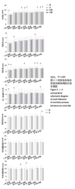
2.1.1 钩突长 在性别比较中,< 10岁组、30-39岁组和≥60岁组间差异有显著性意义(P < 0.05);在左右侧别比较中,20-29岁组、30-39岁组、50-59岁组和≥60岁组间差异有显著性意义(P < 0.05)。 2.1.2 钩突高 在性别比较中,仅10-19岁组和50-59岁组差异有显著性意义(P < 0.05);左右侧别间比较均差异无显著性意义(P > 0.05)。 2.1.3 钩突宽 在性别比较中,< 10岁组、30-39岁组、50-59岁组和≥60岁组间差异有显著性意义(P < 0.05);在左右侧别比较中,仅在50-59岁组内差异有显著性意义(P < 0.05)。 2.1.4 钩-横间距 在性别比较中,< 10岁组、10-19岁组和≥60岁组间差异有显著性意义(P < 0.05),左右侧别间比较均差异无显著性意义(P > 0.05)。 2.1.5 钩突间距 在性别比较中,仅在≥60岁组内差异有显著性意义(P < 0.05)。 2.1.6 钩突前脚间距 在性别比较中均差异无显著性意义(P > 0.05)。 2.1.7 钩突后脚间距 在性别比较中仅在20-29岁组和30-39岁组差异有显著性意义(P < 0.05)。 2.2 钩突相关径线椎序增加的变化规律 见图3。"
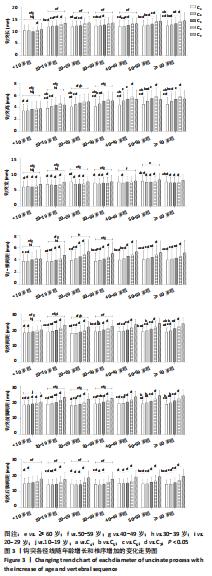

2.2.1 钩突长 总体随年龄的增长呈缓慢递增趋势,< 10岁组与10-19岁组至≥60岁组间比较均差异有显著性意义(P < 0.05),10-19岁组与40-49岁组和≥60岁组间比较均差异有显著性意义(P < 0.05);椎序间的比较中除了在< 10岁组外,其余在各年龄组中总体随椎序的增加呈递增趋势,仅在C6与C7间差异有显著性意义(P < 0.05)外,其余均差异无显著性意义(P > 0.05);10-19岁组中C3与C4-7、C4,5与 C6,7、C6与C7;20-29岁组中C3-5与C6,7;30-39岁组中C3,4与C6,7、C5与C7;40-49岁组中C3-5与C6,7;50-59岁组中C3,4与C5-7、C5与C6,7;50-59岁组中C3与C4-7、C4与C5-7、C5与C6,7、C6与C7间均差异有显著性意义(P < 0.05),其余均差异无显著性意义(P > 0.05)。 2.2.2 钩突高 随年龄的增长呈缓慢递增趋势,< 10岁组至40-49岁组分别与随后递增年龄组间比较均差异有显著性意义(P < 0.05);椎序间的比较中,在< 10岁组和20-29岁组中随椎序的增加呈递增趋势,其余各年龄组中均随椎序的增加呈递增趋势,但其峰值位于C6处,在< 10岁组中C3,4与C7,10-19岁组和20-29岁中C3与C4-7、C4与C6,7,30-39岁组中C3与C4-7、C3与C3-5,40-49岁组中C3与C4-7、C4,5与C6、C6与C7,50-59岁组和≥60岁组中C3与C4-7、C4与C5-7、C5与C6、C6与C7间均差异有显著性意义(P < 0.05),其余均差异无显著性意义(P > 0.05)。 2.2.3 钩突宽 总体随年龄的增长呈缓慢递增趋势,在< 10岁组与10-19岁组至≥60岁组间比较均差异有显著性意义(P < 0.05),10-19岁组与20-29岁组至≥60岁组间比较均差异有显著性意义(P < 0.05),20-29岁组和30-39岁组分别与40-49岁组至≥60岁组间比较均差异有显著性意义(P < 0.05),40-49岁组与50-59岁组、50-59岁组与≥60岁组间比较均差异有显著性意义(P < 0.05);在各年龄组中总体随椎序的增加呈“V”字形,两峰值位于C3和C7处,在< 10岁组、10-19岁组和30-39岁组中均是C3-6与C7间差异有显著性意义(P < 0.05),在20-29岁组、50-59岁和≥60岁组中C3与C5-7、C4-6与C7间差异有显著性意义(P < 0.05),其余均差异无显著性意义(P > 0.05)。 2.2.4 钩-横间距 总体随年龄增长呈波浪状,在< 10岁组和10-19岁组分别与20-29岁组至≥60岁组间比较均差异有显著性意义(P < 0.05),20-29岁组与30-39岁组间、30-39岁组与40-49岁组至≥60岁组间比较均差异有显著性意义(P < 0.05)。在各年龄组中总体随椎序的增加呈递增趋势,在< 10岁组中C3与C6、C4与C6,7,10-19岁组中C3,4与C6,7、C4,5与C7,20-29岁组和30-39岁组中C3与C5-7、C4与C6,7, C5,6与C7,40-49岁组中C3,4与C5-7,C5,6与C7,50-59岁组和≥60岁组中C3与C4,6,7、C4,5与C6,7、C6与C7间比均差异有显著性意义(P < 0.05),其余均差异无显著性意义(P > 0.05)。钩突间距在各年龄组中总体随椎序的增加呈递增趋势,在< 10岁组中C3,4与C7,10-19岁组中C3与C4,6,7、C4,5与C6,7、C6与C7,20-29岁组中C3,4与C6,7、C4,5与C7,30-39岁组中C3,4与C5-7、C5,6与C7,40-49岁组中C3-5与C5-7,C4与C6,7、C6与C7,50-59岁组中C3,4与C5-7,C5与C6,7、C6与C7,≥60岁组中C3与C4-7、C4与C5-7、C5与C6,7、C6与C7间均差异有显著性意义(P < 0.05),其余均无差异显著性意义(P > 0.05)。 2.2.5 钩突间距 随年龄增长呈波浪状,在年龄增长中< 10岁组与10-19岁组和30-39岁组至≥60岁组间比较均差异有显著性意义(P < 0.05),10-19岁组与20-29岁组和50-59岁组至≥60岁组间比较均差异有显著性意义(P < 0.05),20-29岁组与30-39岁组至≥60岁组、30-39岁组与50-59岁组和≥60岁组间比较均差异有显著性意义(P < 0.05)。椎序间的比较中,增加呈递增趋势,在< 10岁组中C3、4与C7,10-19岁组中C3与C4,6,7,20-29岁组中C3,4与C6,7、C5,6与C7,30-39岁组中均是C3,4与C5-7,C5,6与C7,40-49岁组中C3-5与C6,7,C6与C7,50-59岁C3,4与C5-7、C5与C6,7、C6与C7,≥60岁组中C3与C4-7、C4与C5-7、C5与C6,7、C6与C7间差异有显著性意义(P < 0.05),其余均差异无显著性意义(P > 0.05)。 2.2.6 钩突前脚间距 总体随年龄增长呈波浪状,在年龄增长中< 10岁组仅与10-19岁组间比较均差异有显著性意义(P < 0.05),10-19岁组、20-29岁组和30-39岁组分别与40-49岁组至≥60岁组间比较均差异有显著性意义(P < 0.05)。在各年龄组中总体随椎序的增加呈递增趋势,在< 10岁组中C3与C6,7、C4-6与C7,10-19岁组、30-39岁组和40-49岁组中C3-5与C6,7、C6与C7,20-29岁组中C3,6与C7、C4,5与C6,7, 50-59岁组和≥60岁组中C3,4与C5-7、C5与C6,7、C6与C7间差异有显著性意义(P < 0.05),其余均无差异显著性意义(P > 0.05)。 2.2.7 钩突后脚间距 总体随年龄增长也呈波浪状,在年龄增长中< 10岁组至40-49岁组分别与50-59岁组和≥60岁组间比较均差异有显著性意义(P < 0.05)。在各年龄组中总体随椎序的增加递增趋势,在< 10岁组中C3,4与C7,10-19岁组中C3,4与C6,7、C5,6与C7,20-29岁组和40-49岁组中C3-6与C7,30-39岁组中C3与C6,7、C4,5与C7,50-59岁组中C3与C5-7、C4与C6,7、C5,6与C7,≥60岁组中C3与C5-7、C4,5与C6,7、C6与C7间有差异显著性意义(P < 0.05),其余均差异无显著性意义(P > 0.05)。"

| [1] CAGNIE B, BARBAIX E, VINCK E, et al. Extrinsic risk factors for compromised blood flow in the vertebral artery: anatomical observations of the transverse foramina from C3 to C7. Surg Radiol Anat. 2005;27(4):312-316. [2] NOURBAKHSH A, YANG J, GALLAGHER S, et al. A safe approach to explore/identify the V2 segment of the vertebral artery during anterior approaches to cervical spine and/or arterial repairs: anatomical study. J Neurosurg Spine. 2010;12(1):25-32. [3] GILES LG. Mechanisms of neurovascular compression within the spinal and intervertebral canals. J Manipulative Physiol Ther. 2000;23(2):107-111. [4] 宋明, 万业达, 何岸苇, 等.应用CTA图像分析颈椎钩突及上关节突对椎动脉的影响[J].天津医科大学学报,2014,20(2):150-153. [5] BULUT MD, ALPAYCI M, ŞENKöY E, et al. Decreased Vertebral Artery Hemodynamics in Patients with Loss of Cervical Lordosis. Med Sci Monit. 2016;22:495-500. [6] JÓNSSON H JR, BRING G, RAUSCHNING W, et al. Hidden cervical spine injuries in traffic accident victims with skull fractures. J Spinal Disord. 1991;4(3):251-263. [7] 唐冲, 孙宇, 潘胜发.平山病患者与非平山病患者钩椎关节在CT上的形态学差异[J].中国脊柱脊髓杂志,2014,24(1):13-19. [8] 李亚梅, 贾功伟, 郑元义, 等.肌肉骨骼超声探测颈椎钩椎关节方法的初步建立[J].中华物理医学与康复杂志,2014,36(2):95-99. [9] 王星. 青少年Luschca关节增龄变化的影像解剖学研究[D].呼和浩特:内蒙古医学院,2010. [10] 康小燕. 7-12岁儿童“Luschka joint”的数字化三维形态测量与有限元分析[D].呼和浩特:内蒙古医科大学,2016. [11] 朱建兵, 龚建平, 钱铭辉.螺旋CT研究颈椎钩突的大小及其相关因素[J].颈腰痛杂志,2006,27(2):88-92. [12] BLAND JH, BOUSHEY DR. Anatomy and physiology of the cervical spine. Semin Arthritis Rheum. 1990;20(1):1-20. [13] LU J, EBRAHEIM NA, YANG H, et al. Cervical uncinate process: an anatomic study for anterior decompression of the cervical spine. Surg Radiol Anat. 1998;20(4):249-252. [14] ERBULUT DU, ZAFARPARANDEH I, LAZOGLU I, et al. Application of an asymmetric finite element model of the C2-T1 cervical spine for evaluating the role of soft tissues in stability. Med Eng Phys. 2014; 36(7):915-921. [15] HARTMAN J. Anatomy and clinical significance of the uncinate process and uncovertebral joint: A comprehensive review. Clin Anat. 2014; 27(3):431-440. [16] WANG Z, ZHAO H, LIU JM, et al. Resection or degeneration of uncovertebral joints altered the segmental kinematics and load-sharing pattern of subaxial cervical spine: A biomechanical investigation using a C2-T1 finite element model. J Biomech. 2016;49(13):2854-2862. [17] RAVEENDRANATH V, KAVITHA T, UMAMAGESWARI A. Morphometry of the Uncinate Process, Vertebral Body, and Lamina of the C3-7 Vertebrae Relevant to Cervical Spine Surgery. Neurospine. 2019;16(4):748-755. [18] SILBERSTEIN CE. The evolution of degenerative changes in the cervical spine and an investigation into the “joints of luschka”. Clin Orthop Relat Res. 1965;40:184-204. [19] 王星, 康小燕, 史君, 等.学龄前期儿童钩椎关节部位的组织学观察及意义[J].局解手术学杂志,2016,25(12):859-862. [20] 李梓瑜, 刘宇航, 史君, 等.基于硬组织切片染色的颈椎钩椎关节的解剖研究[J].局解手术学杂志,2022,31(1):15-18. [21] 王星, 史君, 张少杰, 等.三维图像测量青少年颈椎钩突的形态特征[J].中国组织工程研究与临床康复,2011,15(30):5587-5590. [22] 王星, 王威, 史君, 等.青少年颈椎钩突-横突孔间距的增龄变化及临床意义[J].局解手术学杂志,2014,23(2):144-145. [23] 王星, 史君, 张少杰, 等.青少年颈椎钩突与横突孔的相关性研究及临床意义[J].局解手术学杂志,2016,25(10):728-731. [24] 王星, 张少杰, 史君, 等.CT三维重建青少年颈椎钩突与椎体各结构的相关性[J].中国组织工程研究,2017,21(3):412-417. [25] 瞿东滨, 金大地, 钟世镇.颈椎钩突的解剖学测量及临床意义[J].中国矫形外科杂志,2002,9(1):49-51. [26] KLAASSEN Z, TUBBS RS, APAYDIN N, et al. Vertebral spinal osteophytes. Anat Sci Int. 2011;86(1):1-9. [27] KOCABIYIK N, ERCIKTI N, TUNALI S. Morphometric analysis of the uncinate processes of the cervical vertebrae. Folia Morphol (Warsz). 2017;76(3):440-445. [28] SUN JC, YANG HS, SHI JG, et al. Morphometric Analysis of the Uncinate Process as a Landmark for Anterior Controllable Antedisplacement and Fusion Surgery: A Study of Radiologic Anatomy. World Neurosurg. 2018;113:e101-e107. [29] PAIT TG, KILLEFER JA, ARNAUTOVIC KI. Surgical anatomy of the anterior cervical spine: the disc space, vertebral artery, and associated bony structures. Neurosurgery. 1996;39(4):769-776. [30] CIVELEK E, KIRIS T, HEPGUL K, et al. Anterolateral approach to the cervical spine: major anatomical structures and landmarks. Technical note. J Neurosurg Spine. 2007;7(6):669-678. [31] MILNE N. The role of zygapophysial joint orientation and uncinate processes in controlling motion in the cervical spine. J Anat. 1991;178: 189-201. [32] SARINGER WF, REDDY B, NöBAUER-HUHMANN I, et al. Endoscopic anterior cervical foraminotomy for unilateral radiculopathy: anatomical morphometric analysis and preliminary clinical experience. J Neurosurg. 2003;98(2 Suppl):171-180. [33] RUSSO VM, GRAZIANO F, PERIS-CELDA M, et al. The V <sub/>2</sub> segment of the vertebral artery: anatomical considerations and surgical implications. J Neurosurg Spine. 2011;15(6):610-619. [34] TUBBS RS, ROMPALA OJ, VERMA K, et al. Analysis of the uncinate processes of the cervical spine: an anatomical study. J Neurosurg Spine. 2012;16(4):402-407. [35] UğUR HC, UZ A, ATTAR A, et al. Anatomical projection of the cervical uncinate process in ventral, ventrolateral, and posterior decompressive surgery. J Neurosurg. 2000;93(2 Suppl):248-251. [36] YILMAZLAR S, KOCAELI H, UZ A, et al. Clinical importance of ligamentous and osseous structures in the cervical uncovertebral foraminal region. Clin Anat. 2003;16(5):404-410. [37] 胡明华, 陈傲, 唐顺胜, 等.解剖学测量在颈椎前路减压手术中的应用[J].广西医学,2015,37(7):935-937. [38] 孙明元, 裴守明.颈椎横突孔的临床应用解剖[J].中国解剖与临床, 2000,5(3):164. [39] 李敏才, 李应续, 胡振武.椎动脉横突段的应用解剖学研究[J].咸宁学院学报(医学版),2005,19(6):479-480. [40] YANG XL, SUN JM. Effect of cervical spine instability on cervical spondylosis of vertebral artery. Zhongguo Gu Shang. 2009;22(5):352-353. |
| [1] | Yang Zhishan, Tang Zhenglong. YAP/TAZ, a core factor of the Hippo signaling pathway, is involved in bone formation [J]. Chinese Journal of Tissue Engineering Research, 2023, 27(8): 1264-1271. |
| [2] | Feng Minshan, Han Changxiao, Liang Dongzhu, Zhao Weidong, Yin Xunlu, Liu Guangwei, Zhu Liguo. In vitro biomechanical characteristics of the effect of Rotation-Traction Manipulation on lower cervical vertebral displacement [J]. Chinese Journal of Tissue Engineering Research, 2023, 27(18): 2820-2823. |
| [3] | Wang Xing, Li Kun, Ma Yuan, Zhang Shaojie, Wang Chaoqun, Gao Mingjie, Zheng Bingwu, Chen Jie, Li Xiaohe, Zhang Zhifeng, Zheng Leigang, Shi Jun, Li Zhijun . Comparison of finite element model establishment and validity verification of total cervical vertebrae between human and adult rhesus monkeys [J]. Chinese Journal of Tissue Engineering Research, 2023, 27(13): 2022-2027. |
| [4] | Niu Jingwei, Zhang Qingtao, Wang Na, Guan Shuo, Wang Ying. Correlation analysis between thyroid function-related Thrβ and Thrsp genes and mechanical properties of Achilles tendon development in rats [J]. Chinese Journal of Tissue Engineering Research, 2023, 27(11): 1653-1658. |
| [5] | Kou Bowen, Han Ye, Hao Yiguang, Xu Haoxiang, Wen Wangqiang, Zhang Zepei, Lan Jie, Miao Jun. Morphological changes and strain characteristics of lumbar intervertebral disc during sitting in forward flexion [J]. Chinese Journal of Tissue Engineering Research, 2022, 26(36): 5792-5797. |
| [6] | Liu Zhichao, Zhang Fan, Sun Qi, Kang Xiaole, Yuan Qiaomei, Liu Genzhe, Chen Jiang. Morphology and activity of human nucleus pulposus cells under different hydrostatic pressures [J]. Chinese Journal of Tissue Engineering Research, 2021, 25(8): 1172-1176. |
| [7] | Guan Qian, Luan Zuo, Ye Dou, Yang Yinxiang, Wang Zhaoyan, Wang Qian, Yao Ruiqin. Morphological changes in human oligodendrocyte progenitor cells during passage [J]. Chinese Journal of Tissue Engineering Research, 2021, 25(7): 1045-1049. |
| [8] | Tang Shuo, Hou Decai. Correlation between traditional Chinese medicine syndrome types of femoral head necrosis and hip joint morphology [J]. Chinese Journal of Tissue Engineering Research, 2021, 25(36): 5867-5871. |
| [9] | Wang Xing, Xu Xuebin, Zhang Shaojie, Zheng Bingwu, Yang Xi, Wang Chaoqun, Li Ping, Ma Yuan, Li Kun, Chen Jie, Li Xiaohe, Shi Jun, Li Zhijun. Establishment and validation of C0-T1 finite element model of cervical vertebrae in adult rhesus monkeys [J]. Chinese Journal of Tissue Engineering Research, 2021, 25(35): 5632-5637. |
| [10] | Zhu Feilong, Zhang Ming, Wu Yu, Wang Bin, Guo Xiaoqi, Cao Jiangang, Zhu Qian, Chen Wei. Foot posture and gait in adolescent idiopathic scoliosis patients: three-dimensional morphological analysis and biomechanics evaluation [J]. Chinese Journal of Tissue Engineering Research, 2021, 25(33): 5294-5300. |
| [11] | Wang Shuguang, Cai Tongchuan, Feng Xinmin, Nan Liping, Wang Feng, Zhu Lei, Chen Dong, Zhang Liang. Meta-analysis of adverse events between anterior and posterior fusion surgery for multiple-level cervical spondylosis [J]. Chinese Journal of Tissue Engineering Research, 2021, 25(30): 4907-4914. |
| [12] | Zhang Xinming, Liu Zhihua, Zhang Xinmin, Shen Yankui, Wang Chunli. Establishment of a total cervical spine model and characteristics of traction force and traction angles under different traction orientations [J]. Chinese Journal of Tissue Engineering Research, 2021, 25(30): 4805-4811. |
| [13] | You Bin, Huang Xiuzhu, Lin Yu, Huang Xi. Changes in bone morphology, bone mineral density and bone metabolism in different parts of natural aging rats [J]. Chinese Journal of Tissue Engineering Research, 2021, 25(26): 4118-4122. |
| [14] | Li Ping, Lin Yu, Chen Xiang, Liu Zhentao, Xiao Lili, Lin Xueyi, Hua Peng . Characteristics of bone remodeling in female ovariectomized rat models of osteoporosis undergoing Erzhi Pill extract intervention [J]. Chinese Journal of Tissue Engineering Research, 2021, 25(2): 191-195. |
| [15] | Pei Tianlong, Wang Yongkang, Wu Wenjie, Zhao Hong, Wang Longsheng . Anatomical and morphological characteristics of knee joint in hemophilic arthritis patients with three-dimensional CT and X-ray films [J]. Chinese Journal of Tissue Engineering Research, 2021, 25(12): 1886-1890. |
| Viewed | ||||||
|
Full text |
|
|||||
|
Abstract |
|
|||||