Chinese Journal of Tissue Engineering Research ›› 2023, Vol. 27 ›› Issue (22): 3531-3536.doi: 10.12307/2023.349
Previous Articles Next Articles
High tibial osteotomy combined with arthroscopy to treat degenerative tear in the posterior horn of medial meniscus combined with varus deformity of the knee
Zhong Hehe, Jin Ying, Liu Xiuqi, Xiang Kuan, Wu Shuhong, Peng Jiachen, Liu Yi
- Department of Orthopedics, Affiliated Hospital of Zunyi Medical University, Zunyi 563000, Guizhou Province, China
-
Received:2022-05-05Accepted:2022-06-13Online:2023-08-08Published:2022-11-02 -
Contact:Wu Shuhong, Chief physician, Department of Orthopedics, Affiliated Hospital of Zunyi Medical University, Zunyi 563000, Guizhou Province, China -
About author:Zhong Hehe, Master, Attending physician, Department of Orthopedics, Affiliated Hospital of Zunyi Medical University, Zunyi 563000, Guizhou Province, China -
Supported by:Zunyi Science and Technology Plan Project, No. 2020-216 (to ZHH); Guizhou Provincial Health Commission Fund Project, No. gzwkj2021-257 (to ZHH)
CLC Number:
Cite this article
Zhong Hehe, Jin Ying, Liu Xiuqi, Xiang Kuan, Wu Shuhong, Peng Jiachen, Liu Yi. High tibial osteotomy combined with arthroscopy to treat degenerative tear in the posterior horn of medial meniscus combined with varus deformity of the knee[J]. Chinese Journal of Tissue Engineering Research, 2023, 27(22): 3531-3536.
share this article
Add to citation manager EndNote|Reference Manager|ProCite|BibTeX|RefWorks
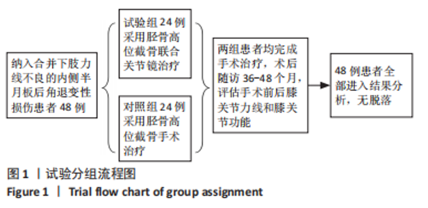
2.1 参与者数量分析 48例患者全部进入结果分析。 2.2 试验分组流程图 见图1。"
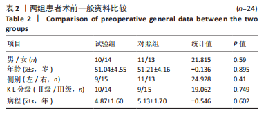
2.3 两组患者一般资料比较 两组患者的性别、年龄、侧别、骨关节炎Kellgren-Lawrence分级及病程等术前一般资料比较差异均无显著性意义(P > 0.05),见表2,具有可比性。"
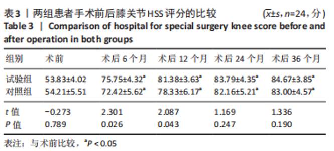
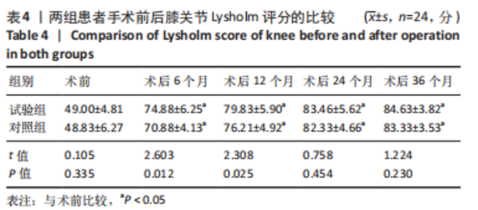
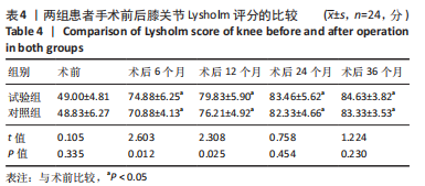
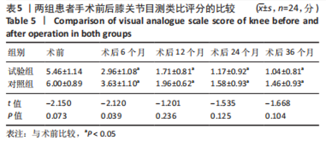
2.4 两组并发症发生情况 两组患者均获得36-48个月随访。所有患者术中未发生血管、神经损伤并发症,术后对照组1例患者切口延迟愈合,经换药后痊愈,所有患者无截骨部位不愈合及延迟愈合、无植入物相关并发症。 2.5 两组患者膝关节功能比较 见表3-5。"
"
"
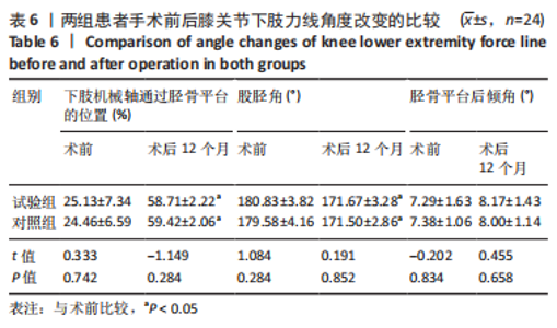
两组患者术前的膝关节HSS评分、Lysholm评分、目测类比评分比较差异均无显著性意义(P > 0.05),两组术后不同时间点的膝关节HSS评分、Lysholm评分、目测类比评分均较术前明显改善(P < 0.05)。试验组术后6,12个月的膝关节HSS评分高于对照组(P < 0.05),术后6,12个月的膝关节Lysholm评分高于对照组(P < 0.05),术后6个月的目测类比评分低于对照组(P < 0.05)。 2.6 两组患者影像学检查结果比较 见表6。 "
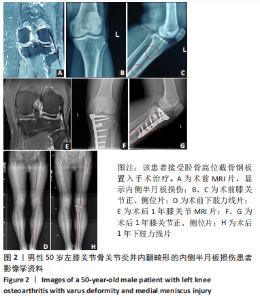
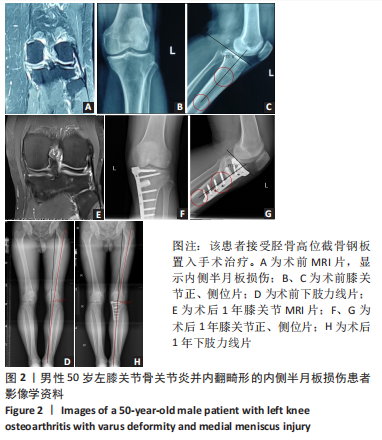
术前,两组间下肢机械轴通过胫骨平台的位置、股胫角、胫骨平台后倾角比较差异无显著性意义(P > 0.05);术后12个月,两组间下肢机械轴通过胫骨平台的位置、股胫角、胫骨平台后倾角比较差异无显著性意义(P > 0.05)。两组术后12个月的下肢机械轴通过胫骨平台的位置、股胫角均较术前明显改善(P < 0.05)。 2.7 典型病例 病例1: 50岁男性患者,左膝关节疼痛5年,入院后诊断为:左膝骨性关节炎并内翻畸形、左膝内侧半月板损伤,K-L分级Ⅱ级,接受关节镜下内侧半月板清理+胫骨高位截骨手术治疗,其治疗前后的影像学资料,见图2。"
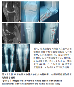
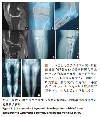
病例2:53岁女性患者,入院后诊断为:左膝关节骨性关节炎并内翻畸形、左膝内侧半月板损伤,接受胫骨高位截骨手术治疗,其治疗前后的影像学资料,见图3。"
| [1] KAPILA R, SHARMA PK, CHUGH A, et al. Management of osteoarthritis knee by graduated open wedge high tibial osteotomy in 40-60 years age group using limb reconstruction system: A clinical study. J Clin Diagn Res. 2015;9(10):RC09-11. [2] ZHAO X, MENG F, HU S, et al.The synovium attenuates cartilage degeneration in KOA through activation of the Smad2/3-Runx1 cascade and chondrogenesis-related miRNAs. Mol Ther Nucleic Acids. 2020; 22(10):832-845. [3] XIE K, JIANG X, HAN X, et al. Association between knee malalignment and ankle degeneration in patients with end-stage knee osteoarthritis. J Arthroplasty. 2018;33(12):3694-3698. [4] KIM KI, SEO MC, SONG SJ, et al. Change of chondral lesions and predictive factors after medial open-wedge high tibial osteotomy with a locked plate system. Am J Sports Med. 2017;45(7):1615-1621. [5] BONASIA DE, PELLEGRINO P, D’AMELIO A, et al. Meniscal Root Tear Repair: Why, When and How. Orthop Rev (Pavia). 2015;7(2):34-39. [6] YIM JH, SEON JK, SONG EK, et al. A comparative study of meniscectomy and nonoperative treatment for degenerative horizontaltears of the medial meniscus. Am J sports Med. 2013;41(7):1565-1570. [7] 李儒军,钟群杰,倪磊,等.内侧半月板退变性损伤的关节镜下分型[J].中华骨科杂志,2014,34(3):293-297. [8] SCHUSTER P, RICHTER J. Editorial commentary: High tibial osteotomy is effective, even in patients with severe osteoarthritis: Contradiction of another dogma from the past. Arthroscopy. 2021;37(2):645-646. [9] LIU X, CHEN Z, GAO Y, et al. High tibial osteotomy: Review of techniques and biomechanics. J Healthc Eng. 2019;2019:8363128. [10] LOIA MC, VANNI S, ROSSO F, et al. High tibial osteotomy in varus knees: indications and limits. Joints. 2016;4(2):98-110. [11] YAO RZ, LIU WQ, SUN LZ, et al. Effectiveness of high tibial osteotomy with or without other procedures for medial compartment osteoarthritis of knee: an update meta-analysis. J Knee Surg. 2020; 34(9):952-961. [12] 恽常军,钱文杰,王岩峰,等.胫骨高位截骨联合关节镜手术治疗膝关节内侧骨关节炎[J].中华创伤骨科杂志,2020,22(9):808-812. [13] EL GHAZALY SA, RAHMAN AAA, YUSRY AH, et al. Arthroscopic partial meniscectomy is superior to physical rehabilitation in the management of symptomatic unstable meniscal tears. Int Orthop. 2015;39(4): 769-775. [14] EMMANUEL K, QUINN E, NIU J, et al. Quantitative measures of meniscus extrusion predict incident radiographic knee osteoarthritis data from the Osteoarthritis Initiative. Osteoarthritis Cartilage. 2015; 24(2):262-269. [15] KIM TW, KIM BK, KIM DW, et al. The SPECT/CT evaluation of compartmental changes after open wedge high tibial osteotomy. Knee Surg Relat Res. 2016;28(4):263-269. [16] 李沼,关振鹏.单髁置换术治疗膝关节内侧间室骨关节炎[J].中华骨科杂志,2019,39(14):902-908. [17] ZHANG FJ, LI HC, BA ZC, et al. Robotic arm-assisted vs conventional unicompartmental knee arthroplasty: A meta-analysis of the effects on clinical outcomes. Medicine (Baltimore). 2019;98(35):e16968. [18] HANSEN EN, ONG KL, LAU E, et al. Unicondylar knee arthroplasty has fewer complications but higher revision rates than total knee arthroplasty in a study of large United States databases. J Arthroplasty. 2019;34:1617-1625. [19] HUNT LP, BLOM AW, MATHARU GS, et al. Patients receiving a primary unicompartmental knee replacement have a higher risk of revision but a lower risk of mortality than predicted had they received a total knee replacement: data from the National Joint Registry for England, Wales, Northern Ireland. J Arthroplasty. 2021;36(2):471-477. [20] 裴征,丁镇涛,李沼,等.基于倾向性评分匹配的单髁与全膝置换术治疗膝内侧骨关节炎早期效果比较[J].中华外科杂志,2020, 58(6):452-456. [21] Liu JN, Agarwalla A, Garcia G, et al. Return to sport following isolated opening wedge high tibial osteotomy. Knee. 2019;26(6): 1306-1312. [22] JIN C, SONG EK, SANTOSO A, et al.Survival and risk factor analysis of medial open wedge high tibial osteotomy for unicompartment knee osteoarthritis. Arthroscopy. 2020;36(2):535-543. [23] ALY AS, ABDELHAMID ALSABIR AR, FAHMY HA, et al. Modified oblique high tibial osteomy with minimal fixation for correction of adolescent tibia vara:a prospective case series study. J Child Orthop. 2021;15(1): 6-11. [24] 黄野,柳剑,王兴山.等.胫骨高位截骨术适应证解析[J].中华外科杂志,2020,58(6):420-423. [25] HOHLOCH L, KIM S, MEHL J, et al. Customized post-operative alignment improves clinical outcome following medial open-wedge osteotomy. Knee Surg Sports Traumatol Arthrosc. 2018;26(9):2766-2773. [26] NHA KW, KIM HJ, AHN HS, et al. Change in posterior tibial slope after open-wedge and closed-wedge high tibial osteotomy: a Meta-analysis. Am J Sports Med. 2016;44(11):3006-3013. [27] NOYES FR, GOEBEL SX, WEST J. Opening wedge tibial osteotomy: the 3-triangle method to correct axial alignment and tibial slope. Am J Sports Med. 2005;33(3):378-387. [28] JUNG WH, TAKEUCHI R, CHUN CW, et al. Comparison of results of medial opening- wedge high tibial osteotomy with and without subchondral drilling. Arthroscopy. 2015;31(4):673-679. [29] HARRIS JD, MCNEILAN R, SISTON RA, et al. Survival and clinical outcome of isolated high tibial osteotomy and combined biological knee reconstruction. Knee. 2013;20(3):154-161. [30] KE X, QIU J, CHEN S, et al. Concurrent arthroscopic meniscal repair during open-wedge high tibial osteotomy is not clinically beneficial for medial meniscus posterior root tears. Knee Surg Sports Traumatol Arthrosc. 2020;29(3):955-965. |
| [1] | Li Xiaomin, Tian Xiangdong, Tan Yetong, Zhu Guangyu, Wang Rongtian, Wang Jian, Xue Zhipeng, Ma Sheng, Hu Yuanyi, Huang Ye, Ding Tiansong. Changes of lower limb force line and knee function after high tibial osteotomy in osteoporotic medial ventricular knee osteoarthritis [J]. Chinese Journal of Tissue Engineering Research, 2023, 27(9): 1325-1329. |
| [2] | Tang Huiyu, Hou Biao, Xia Xiaodan, Xiang Wei, Xie Songlin. Effect of mechanical tension stress on arterial vessels after limb osteotomy in rabbits [J]. Chinese Journal of Tissue Engineering Research, 2023, 27(9): 1422-1426. |
| [3] | Zheng Bo, Zhang Xiuli, Zhou Hao, He Zebi, Zhou Jin, Zhou Weiyun, Li Peng. Arthroscopy-assisted locking hollow screw fixation and open reduction plate internal fixation in the treatment of Schatzker II-III tibial plateau fractures: early CT evaluation [J]. Chinese Journal of Tissue Engineering Research, 2023, 27(9): 1410-1416. |
| [4] | Huang Linke, Wei Linhua, Jiang Jie, Liu Qian, Chen Weiwei. Effects of estrogen combined with treadmill exercise on bone mass and articular cartilage in ovariectomized mice [J]. Chinese Journal of Tissue Engineering Research, 2023, 27(8): 1166-1171. |
| [5] | Li Long, Li Guangdi, Shi Hao, Deng Keqi. Circular RNA as a competing endogenous RNA is involved in the regulation of osteoarthritis [J]. Chinese Journal of Tissue Engineering Research, 2023, 27(5): 751-757. |
| [6] | Yuan Changshen, Guan Yanbing, Li Zhe, Rong Weiming, Liao Shuning, Chen Lewei, Mei Qijie, Duan Kan. Screening and verification of key genes of necroptosis in osteoarthritis [J]. Chinese Journal of Tissue Engineering Research, 2023, 27(5): 695-700. |
| [7] | Liu Guangluan, Guo Zonglei, Ge Jin, Huang Dong, Wang Yehua. Anatomic risk factors for medial meniscus posterior root tears combined with anterior cruciate ligament injuries [J]. Chinese Journal of Tissue Engineering Research, 2023, 27(5): 663-668. |
| [8] | Wan Guoli, Shi Chenhui, Wang Weishan, Li Ang, Shi Xunda, Cai Yi. Retrospective analysis of the influencing factors of chronic pain after total knee arthroplasty [J]. Chinese Journal of Tissue Engineering Research, 2023, 27(4): 558-564. |
| [9] | Gu Mingxi, Wang Bo, Tian Fengde, An Ning, Hao Ruihu, Wang Changcheng, Guo Lin. Comparison of early efficacy and safety of simultaneous and staged bilateral total knee arthroplasty [J]. Chinese Journal of Tissue Engineering Research, 2023, 27(4): 565-571. |
| [10] | Yu He, Zheng Jiafa, Song Xiufeng, Guan Shengyi. Tibiotalocalcaneal arthrodesis with blood supplied fibular flap combined with hollow screw in the treatment of end-stage ankle osteoarthritis [J]. Chinese Journal of Tissue Engineering Research, 2023, 27(4): 588-593. |
| [11] | Guo Yingqi, Gong Xianxu, Zhang Yan, Xiao Han, Wang Ye, Gu Wenguang. Meniscus extrusion and patellofemoral joint cartilage injury and bone marrow lesions: MRI semi-quantitative score [J]. Chinese Journal of Tissue Engineering Research, 2023, 27(4): 600-605. |
| [12] | Xu Xiangjun, Wang Chao, Song Qunshan, Li Bingyan, Zhang Jichao, Wang Guodong, Dong Yuefu. Optimal angle for prosthesis implantation in total knee arthroplasty [J]. Chinese Journal of Tissue Engineering Research, 2023, 27(4): 612-618. |
| [13] | Yu Jiaan, Liu Xinwei, Lian Hongyu, Liu Kexin, Li Zitao. Medial open-wedge tibial osteotomy versus lateral closed-wedge tibial osteotomy for unicompartmental knee osteoarthritis: a meta-analysis [J]. Chinese Journal of Tissue Engineering Research, 2023, 27(4): 632-639. |
| [14] | Chai Hao, Yang Deyong, Zhang Lei, Shu Li. 3D printing personalized osteotomy guide technology versus conventional total knee arthroplasty on the accuracy of lower limb force alignment: a meta-analysis [J]. Chinese Journal of Tissue Engineering Research, 2023, 27(4): 646-654. |
| [15] | Ning Ziwen, Wang Xu, Shi Zhengliang, Qin Yihua, Wang Guoliang, Jia Di, Wang Yang, Li Yanlin. Meniscal injury repair methods for non-blood supply area [J]. Chinese Journal of Tissue Engineering Research, 2023, 27(3): 420-426. |
| Viewed | ||||||
|
Full text |
|
|||||
|
Abstract |
|
|||||