[1] 赵英伟,靳宝成,闫冰,等.前路钢板螺钉内固定治疗髋臼骨折伴骶髂关节脱位的疗效分析[J].中外医疗,2019,38(12):31-33.
[2] WONG M, SINKLER MA, KIEL J. Anatomy, Abdomen and Pelvis, Sacroiliac Joint. StatPearls. Treasure Island (FL): StatPearls Publishing Copyright © 2022, StatPearls Publishing LLC., 2022.
[3] CAETANO AP, MASCARENHAS VV, MACHADO PM. Axial Spondyloarthritis: Mimics and Pitfalls of Imaging Assessment. Front Med (Lausanne). 2021;8:658538.
[4] FIANI B, SEKHON M, DOAN T, et al. Sacroiliac Joint and Pelvic Dysfunction Due to Symphysiolysis in Postpartum Women. Cureus. 2021;13:e18619.
[5] POILLIOT AJ, ZWIRNER J, DOYLE T, et al. A Systematic Review of the Normal Sacroiliac Joint Anatomy and Adjacent Tissues for Pain Physicians. Pain Physician. 2019;22:E247-e274.
[6] TAN Z, HUANG Z, LI L, et al. Classification and Treatment of Sacroiliac Joint Dislocation. Sichuan Da Xue Xue Bao Yi Xue Ban. 2017;48:661-667.
[7] DENIS F, DAVIS S, COMFORT T. Sacral fractures: an important problem. Retrospective analysis of 236 cases. Clin Orthop Relat Res. 1988;227: 67-81.
[8] LIU Y, WANG J, ZHANG Y. Occult external iliac vein injury after anterior dislocation of the sacroiliac joint in adult patient. Acta Orthop Traumatol Turc. 2017;51:169-171.
[9] MAERTENS AS, MARTIN MP 3RD, DEAN CS, et al. Occult injuries of the contralateral sacroiliac joint in operatively treated pelvis fractures: incidence, root cause analysis, and proposal of treatment algorithm. Int Orthop. 2019;43:2399-2404.
[10] 闵锐.医源性电离辐射损伤及其生物医学防护[J].辐射防护通讯, 2013,33(3): 8-15.
[11] LIU L, ZENG D, FAN S, et al. Biomechanical study of Tile C3 pelvic fracture fixation using an anterior internal system combined with sacroiliac screws. J Orthop Surg Res. 2021;16:225.
[12] DYDYK AM, FORRO SD, HANNA A. Sacroiliac Joint Injury. StatPearls. Treasure Island (FL): StatPearls Publishing Copyright © 2022, StatPearls Publishing LLC., 2022.
[13] GOLDSZTAJN F, MARIOLANI JRL, BELANGERO WD. Are Anterior Plates More Effective than Iliosacral Screws to Fix the Sacroiliac Joint? Biomechanical Study. Rev Bras Ortop (Sao Paulo). 2020;55:497-503.
[14] CAI L, ZHANG Y, ZHENG W, et al. A novel percutaneous crossed screws fixation in treatment of Day type II crescent fracture-dislocation: A finite element analysis. J Orthop Translat. 2020;20:37-46.
[15] BREUIL V, ROUX CH, CARLE GF. Pelvic fractures: epidemiology, consequences, and medical management. Curr Opin Rheumatol. 2016; 28:442-447.
[16] BULLER LT, BEST MJ, QUINNAN SM. A Nationwide Analysis of Pelvic Ring Fractures: Incidence and Trends in Treatment, Length of Stay, and Mortality. Geriatr Orthop Surg Rehabil. 2016;7:9-17.
[17] 杨鹏,叶添文,张帆,等.3D打印技术辅助手术治疗骶骨骨折伴骶丛神经损伤[J].中华创伤骨科杂志,2015,17(1):13-17.
[18] 顾容赫.经骶髂关节置骶1骶2螺钉应用解剖[D].南宁:广西医科大学,2012.
[19] JIN Z, LI Y, YU K, et al. 3D Printing of Physical Organ Models: Recent Developments and Challenges. Adv Sci (Weinh). 2021;8:e2101394.
[20] YOON YC, MA DS, LEE SK, et al. Posterior pelvic ring injury of straddle fractures: Incidence, fixation methods, and clinical outcomes. Asian J Surg. 2021;44:59-65.
[21] MARMOR M, EL NAGA AN, BARKER J, et al. Management of Pelvic Ring Injury Patients With Hemodynamic Instability. Front Surg. 2020;7: 588845.
[22] KIM JT, RUDOLF LM, GLASER JA. Outcome of percutaneous sacroiliac joint fixation with porous plasma-coated triangular titanium implants: an independent review. Open Orthop J. 2013;7:51-56.
[23] ROMMENS PM, HESSMANN MH. Staged reconstruction of pelvic ring disruption: differences in morbidity, mortality, radiologic results, and functional outcomes between B1, B2/B3, and C-type lesions. J Orthop Trauma. 2002;16:92-98.
[24] XIANG G, DONG X, JIANG X, et al. Comparison of percutaneous cross screw fixation versus open reduction and internal fixation for pelvic Day type II crescent fracture-dislocation: case-control study. J Orthop Surg Res. 2021;16:36.
[25] MIYAKE T, FUTAMURA K, BABA T, et al. A novel technique for stabilising sacroiliac joint dislocation using spinal instrumentation: technical notes and clinical outcomes. Eur J Trauma Emerg Surg. 2022. doi: 10.1007/s00068-021-01873-z.
[26] BOUGUENNEC N, GOUIN F, PIÉTU G. Isolated anterior unilateral sacroiliac dislocation without pubic arch disjunction. Orthop Traumatol Surg Res. 2012;98:359-362.
[27] LI L, LU J, YANG L, et al. Stability evaluation of anterior external fixation in patient with unstable pelvic ring fracture: a finite element analysis. Ann Transl Med. 2019;7:303.
[28] SOULTANIS K, KARALIOTAS GI, MASTROKALOS D, et al. Lumbopelvic fracture-dislocation combined with unstable pelvic ring injury: one stage stabilisation with spinal instrumentation. Injury. 2011;42:1179-1183.
[29] WU T, CHEN W, LI X, et al. Biomechanical comparison of three types of internal fixation in a type C zone II pelvic fracture model. Int J Clin Exp Med. 2015;8:1853-1861.
[30] MATTA JM, SAUCEDO T. Internal fixation of pelvic ring fractures. Clin Orthop Relat Res. 1989;(242):83-97.
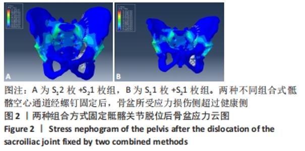
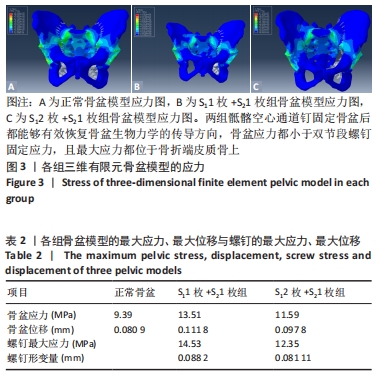
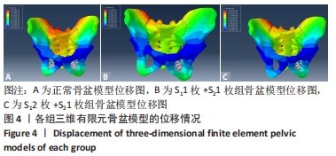
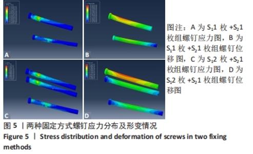
|