Chinese Journal of Tissue Engineering Research ›› 2022, Vol. 26 ›› Issue (33): 5265-5270.doi: 10.12307/2022.712
Previous Articles Next Articles
Establishment and validation of finite element model of cervical facet joint in children
He Yujie1, Li Pei2, Xue Mingming3, Li Zhijun1, Li Xiaohe1, Wang Xing1, Gao Mingjie1, Wu Chao2, Kang Zhijie4, Zhang Shaojie1, Wang Haiyan1
- 1Department of Human Anatomy of Basic Medical College, 2First Clinical Medical College, 3Department of Physiology of Basic Medical College, 4Graduate school, Inner Mongolia Medical University, Hohhot 010010, Inner Mongolia Autonomous Region, China
-
Received:2021-08-25Accepted:2021-11-13Online:2022-11-28Published:2022-03-30 -
Contact:Wang Haiyan, Master, Professor, Master’s supervisor, Department of Human Anatomy of Basic Medical College, Inner Mongolia Medical University, Hohhot 010010, Inner Mongolia Autonomous Region, China Zhang Shaojie, Master, Professor, Department of Human Anatomy of Basic Medical College, Inner Mongolia Medical University, Hohhot 010010, Inner Mongolia Autonomous Region, China -
About author:He Yujie, Master, Lecturer, Department of Human Anatomy of Basic Medical College, Inner Mongolia Medical University, Hohhot 010010, Inner Mongolia Autonomous Region, China Li Pei, First Clinical Medical College, Inner Mongolia Medical University, Hohhot 010010, Inner Mongolia Autonomous Region, China He Yujie and Li Pei contributed equally to this article. -
Supported by:National Natural Science Foundation of China, No. 81860383 (to LZJ); Natural Science Foundation of Inner Mongolia Autonomous Region, No. 2021MS08086 (to WHY); Natural Science Foundation of Inner Mongolia Autonomous Region, No. 2019MS08017 (to ZSJ); Undergraduate Science and Technology Innovation "Cultivation of Talents" Project of Inner Mongolia Medical University in 2020, No. YCPY20200015 (to WHY), No. YCPY20200018 (to HYJ); College Student Enlightenment Project of the First Clinical Medical University of Inner Mongolia Medical University in 2020, No. FYQMJH2020033 (to WHY); “Trinity” College Student Innovation and Entrepreneurship Cultivation Project of Inner Mongolia Medical University in 2020, No. SWYT2020025, SWYT2021007 (to WHY), No. SWYT2020021, SWYT2021016 (to HYJ); School-Level Scientific Research Project "Achievement Transformation" Project of Inner Mongolia Medical University in 2020, No. YKD2020CGZH009 (to WHY); Inner Mongolia Autonomous Region-Level University Student Innovation and Entrepreneurship Training Program, No. 201910132019 (to WHY); Science and Technology Development Program of Inner Mongolia Autonomous Region, No. 2019GG115 (to LZJ)
CLC Number:
Cite this article
He Yujie, Li Pei, Xue Mingming, Li Zhijun, Li Xiaohe, Wang Xing, Gao Mingjie, Wu Chao, Kang Zhijie, Zhang Shaojie, Wang Haiyan. Establishment and validation of finite element model of cervical facet joint in children[J]. Chinese Journal of Tissue Engineering Research, 2022, 26(33): 5265-5270.
share this article
Add to citation manager EndNote|Reference Manager|ProCite|BibTeX|RefWorks
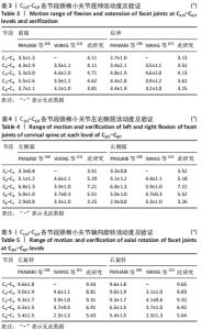
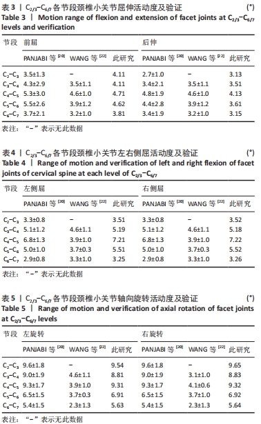
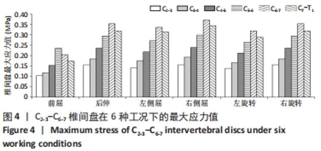
2.1 4岁儿童全颈椎模型几何验证 基于CT影像学资料,依据建模对象的实际形态,使用相关建模软件,成功建立了包含C0-T1、椎间盘、关节突关节、韧带等结构的全颈椎模型,且根据以往文献赋予该实验模型各结构相应的材料属性,共计182 519个单元,327 790个节点,见表2,模仿颈部牵引受力运动状态。 2.2 关节突关节活动度及验证 屈伸、侧屈和轴向旋转状态下的C2/3-C6/7各椎间小关节活动度均在PANJABI等[20]所测参考值范围内;左、右侧屈状态下的C4/5和C5/6节段以及轴向旋转状态下的所有节段小关节活动度均大于WANG等[22]所测参考值范围,见表3-5。在前屈、后伸、左侧屈和右侧屈状态下,小关节活动度最大值均位于C4/5(4.71°,4.13°,7.21°,7.22°),而在轴向旋转状态下,活动度最大值则位于C2/3(9.54°,9.65°)。"
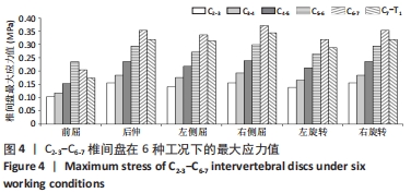
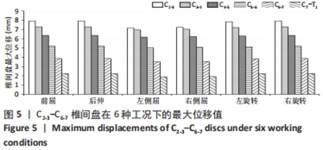
2.3 椎间盘最大应力、位移值 椎间盘最大应力在右侧屈状态下明显大于其余运动状态,除前屈状态最大应力值位于C5-6(0.235 MPa),其余状态椎间盘最大应力值均位于C6-7。C2-3节段椎间盘在6种工况下的最大应力均小于其余节段,与其最大位移值呈反比关系,见图4,5。"
"
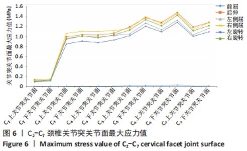
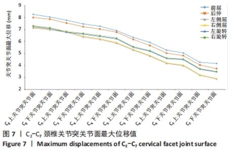
2.4 关节突关节面最大应力、位移值 6种运动状态下,同一椎骨的下关节突关节面最大应力均大于上关节突关节面,随椎序的增加呈递增趋势,峰值位于后伸位C6下关节突关节面(1.481 MPa),谷值位于前屈位C2上关节突关节面(0.005 MPa);关节突关节面的最大位移:前屈>后伸>旋转>侧屈,下关节突关节面最大位移均小于上关节突关节面,随椎序的增加逐渐递减,最大位移出现在前屈位C2(8.266 mm),见图6,7。"
"
| [1] O’LEARY SA, PASCHOS NK, LINK JM, et al. Facet Joints of the Spine: Structure-Function Relationships, Problems and Treatments, and the Potential for Regeneration. Annu Rev Biomed Eng. 2018;20:145-170. [2] 付林,马剑雄,马信龙,等.关节突关节的生物力学研究进展[J].中华骨科杂志,2015,35(9):970-974. [3] WANG QA, GUO C, SUN MJ, et al. Three-dimensional spiral CT observation of the facet joints of the lower cervical spine and its clinical significance. Eur Spine J. 2021;30(6):1536-1541. [4] YIN J, LIU Z, LI C, et al. Effect of facet-joint degeneration on the in vivo motion of the lower lumbar spine. J Orthop Surg Res. 2020;15(1):340. [5] LITTLE JW, GRIEVE T, CANTU J, et al. Reliability of Human Lumbar Facet Joint Degeneration Severity Assessed by Magnetic Resonance Imaging. J Manipulative Physiol Ther. 2020;43(1):43-49. [6] LEE SH, SON DW, LEE JS, et al. Relationship Between Endplate Defects, Modic Change, Facet Joint Degeneration, and Disc Degeneration of Cervical Spine. Neurospine. 2020;17(2):443-452. [7] WINKELSTEIN BA, NIGHTINGALE RW, RICHARDSON WJ, et al. The cervical facet capsule and its role in whiplash injury: a biomechanical investigation. Spine (Phila Pa 1976). 2000;25(10):1238-1246. [8] BOGDUK N. On cervical zygapophysial joint pain after whiplash. Spine (Phila Pa 1976). 2011;36(25 Suppl):S194-199. [9] 林福,付坤飞,全仁夫.有限元法在颈椎生物力学研究中的应用进展[J].中医正骨,2021,33(6):60-69. [10] DOGRU SC, ARSLAN YZ. Effect of Model Parameters on the Biomechanical Behavior of the Finite Element Cervical Spine Model. Appl Bionics Biomech. 2021;2021:5593037. [11] HERRON MR, PARK J, DAILEY AT, et al. Febio finite element models of the human cervical spine. J Biomech. 2020;113:110077. [12] 崔世海,封笑丹,李海岩,等. 3岁儿童颈部屈伸运动生物力学响应研究[J].天津科技大学学报,2020,35(1):67-71. [13] 李琨,李志军,张少杰,等.有限元动态仿真建立4岁儿童“枕-寰-枢”关节模型[J].中国组织工程研究,2021,25(24):3773-3778. [14] 吕文乐,阮世捷,李海岩,等.6岁儿童全颈有限元模型的构建及验证[J].医用生物力学,2016,31(2):95-101. [15] 冯会梅,刘路,张少杰,等.有限元动态仿真建立8岁儿童颈椎关节突关节模型[J].中国组织工程研究,2019,23(32):5181-5187. [16] EVILL G, ESWARAN SK, FARAHMAND F, et al. The influence of boundary conditions and loading mode on high-resolution finite element-computed trabecular tissue properties. Bone. 2009;44(4):573-578. [17] CAI XY, SANG D, YUCHI CX, et al. Using finite element analysis to determine effects of the motion loading method on facet joint forces after cervical disc degeneration. Comput Biol Med. 2020;116:103519. [18] WANG Z, ZHAO H, LIU JM, et al. Resection or degeneration of uncovertebral joints altered the segmental kinematics and load-sharing pattern of subaxial cervical spine: A biomechanical investigation using a C2-T1 finite element model. J Biomech. 2016;49(13):2854-2862. [19] KALLEMEYN N, GANDHI A, KODE S, et al. Validation of a C2-C7 cervical spine finite element model using specimen-specific flexibility data. Med Eng Phys. 2010;32(5):482-489. [20] PANJABI MM, CRISCO JJ, VASAVADA A, et al. Mechanical properties of the human cervical spine as shown by three-dimensional load-displacement curves. Spine (Phila Pa 1976). 2001;26(24):2692-2700. [21] ZHANG QH, TEO EC, NG HW, et al. Finite element analysis of moment-rotation relationships for human cervical spine. J Biomech. 2006;39(1):189-193. [22] WANG H, ZHOU C, YU Y, et al. Quantifying the ranges of relative motions of the intervertebral discs and facet joints in the normal cervical spine. J Biomech. 2020;112:110023. [23] MENGONI M. Biomechanical modelling of the facet joints: a review of methods and validation processes in finite element analysis. Biomech Model Mechanobiol. 2021;20(2):389-401. [24] MANCHIKANTI L, KOSANOVIC R, CASH KA, et al. Assessment of Prevalence of Cervical Facet Joint Pain with Diagnostic Cervical Medial Branch Blocks: Analysis Based on Chronic Pain Model. Pain Physician. 2020;23(6):531-540. [25] PENG B, YANG L, LI Y, et al. Cervical Proprioception Impairment in Neck Pain-Pathophysiology, Clinical Evaluation, and Management: A Narrative Review. Pain Ther. 2021;10(1):143-164. [26] PURUSHOTHAMAN Y, CHOI H, YOGANANDAN N, et al. A Comparison Study of Four Cervical Disk Arthroplasty Devices Using Finite Element Models. Asian Spine J. 2021;15(3):283-293. [27] XIAO LX, LIU CS, ZHONG SZ, et al. Effect of a Traction Exercise Neck Brace on Cervical Spondylopathy Radiculopathy: A Clinical Study and Finite Element Analysis. Evid Based Complement Alternat Med. 2021;2021:8825150. [28] LIU J, WANG R, WANG H, et al. Biomechanical Comparison of a New Memory Compression Alloy Plate versus Traditional Titanium Plate for Anterior Cervical Discectomy and Fusion: A Finite Element Analysis. Biomed Res Int. 2020;2020:5769293. [29] 王宇,雷建银,辛浩,等.椎间盘退变颈椎(C2-C7)在正常承载与推拿下的有限元分析[J].中国组织工程研究,2020,24(27):4278-4284. [30] YOGANANDAN N, CHOI H, PURUSHOTHAMAN Y, et al. Effects of different severities of disc degeneration on the range of motion of cervical spine. J Craniovertebr Junction Spine. 2020;11(4):269-275. [31] SCHMIDT H, HEUER F, CLAES L, et al. The relation between the instantaneous center of rotation and facet joint forces - A finite element analysis. Clin Biomech (Bristol, Avon). 2008;23(3):270-278. [32] 和雨洁,张少杰,李志军,等. 4-6岁儿童下颈椎关节突关节的三维数字化形态特征[J].中国组织工程研究,2019,23(28):4558-4563. [33] 赵卫东,徐波,张美超,等.颈椎三维运动对关节突关节压力的作用[J].中国组织工程研究与临床康复,2010,14(22):3996-3999. [34] MCCLURE P, SIEGLER S, NOBILINI R. Three-dimensional flexibility characteristics of the human cervical spine in vivo. Spine (Phila Pa 1976). 1998;23(2):216-223. |
| [1] | Wei Guoqiang, Li Yunfeng, Wang Yi, Niu Xiaofen, Che Lifang, Wang Haiyan, Li Zhijun, Shi Guopeng, Bai Ling, Mo Kai, Zhang Chenchen, Xu Yangyang, Li Xiaohe. Biomechanical analysis of non-uniform material femur under different loads [J]. Chinese Journal of Tissue Engineering Research, 2022, 26(9): 1318-1322. |
| [2] | Lü Qianyi, Chen Xinyi, Zheng Huie, He Haolong, Li Qilong, Chen Chutao, Tian Haomei. Stress and displacement of normal lumbar vertebra and posterior structure with different elbow pressing methods [J]. Chinese Journal of Tissue Engineering Research, 2022, 26(9): 1346-1350. |
| [3] | Zhang Yufang, Lü Meng, Mei Zhao. Construction and verification of a full spine biomechanical model of adolescent scoliosis [J]. Chinese Journal of Tissue Engineering Research, 2022, 26(9): 1351-1356. |
| [4] | Zhang Jichao, Dong Yuefu, Mou Zhifang, Zhang Zhen, Li Bingyan, Xu Xiangjun, Li Jiayi, Ren Meng, Dong Wanpeng. Finite element analysis of biomechanical changes in the osteoarthritis knee joint in different gait flexion angles [J]. Chinese Journal of Tissue Engineering Research, 2022, 26(9): 1357-1361. |
| [5] | Bai Zixing, Cao Xuhan, Sun Chengyi, Yang Yanjun, Chen Si, Wen Jianmin, Lin Xinxiao, Sun Weidong. Construction and biomechanical analysis of ankle joint finite element model in gait cycle [J]. Chinese Journal of Tissue Engineering Research, 2022, 26(9): 1362-1366. |
| [6] | Liu Feng, Feng Yi. Finite element analysis of different Kirschner wire tension bands on transverse patella fractures during gait cycle [J]. Chinese Journal of Tissue Engineering Research, 2022, 26(9): 1367-1371. |
| [7] | Li Jian, Bao Zhengqi, Zhou Pinghui, Zhu Ruizhi, Li Zhixiang, Wang Jinzi. Effects of posterior single open-door laminoplasty and anterior cervical corpectomy fusion on cervical sagittal balance parameters in the treatment of multilevel cervical spondylotic myelopathy [J]. Chinese Journal of Tissue Engineering Research, 2022, 26(6): 949-953. |
| [8] | Yi Xinrong, Jia Fuquan, He Xin, Zhang Shaojie, Ren Xiaoyan, Li Zhijun. Establishment of cervical bone age equation for male adolescents aged 8-16 years old in Hohhot based on thin-slice CT [J]. Chinese Journal of Tissue Engineering Research, 2022, 26(6): 954-958. |
| [9] | Wen Mingtao, Liang Xuezhen, Li Jiacheng, Xu Bo, Li Gang. Mechanical stability of Sanders II type calcaneal fractures fixed by two internal fixation methods [J]. Chinese Journal of Tissue Engineering Research, 2022, 26(6): 838-842. |
| [10] | Wang Hailong, Li Long, Maihemuti·Yakufu, Chen Hongtao, Liu Xu, Yilihamu·Tuoheti. Finite element analysis of stress distribution of acetabular prosthesis in the Lewinnek safety zone [J]. Chinese Journal of Tissue Engineering Research, 2022, 26(6): 843-847. |
| [11] | Huang Hao, Hong Song, Wa Qingde. Finite element analysis of the effect of femoral component rotation on patellofemoral joint contact pressure in total knee arthroplasty [J]. Chinese Journal of Tissue Engineering Research, 2022, 26(6): 848-852. |
| [12] | Zheng Yongze, Zheng Liqin, He Xingpeng, Chen Xinmin, Li Musheng, Li Pengfei, Lin Ziling. Extended finite element modeling analysis of femoral neck fracture based on ABAQUS software [J]. Chinese Journal of Tissue Engineering Research, 2022, 26(6): 853-857. |
| [13] | Li Shuo, Su Peng, Zhang Li, Wu Qiulong, Hu Xiangyu, Lai Yuliang. Positive effect of supracondylar femoral osteotomy on the correction of knee varus based on three-dimensional reconstruction and finite element analysis [J]. Chinese Journal of Tissue Engineering Research, 2022, 26(6): 858-863. |
| [14] | Wei Bing, Chang Shan. Finite element analysis of different angles of nail placement in sagittal plane of spinal fracture [J]. Chinese Journal of Tissue Engineering Research, 2022, 26(6): 864-869. |
| [15] | Liu Yuhang, Zhou Jianqiang, Xu Xuebin, Qu Xingyue, Li Ziyu, Li Kun, Wang Xing, Li Zhijun, Li Xiaohe, Zhang Shaojie. Establishment and validation of finite element model of lower cervical spine in 6-year-old children [J]. Chinese Journal of Tissue Engineering Research, 2022, 26(6): 870-874. |
| Viewed | ||||||
|
Full text |
|
|||||
|
Abstract |
|
|||||