Chinese Journal of Tissue Engineering Research ›› 2022, Vol. 26 ›› Issue (14): 2138-2143.doi: 10.12307/2022.473
Previous Articles Next Articles
Co-transplantation of acellular allogeneic dermis and autologous split-thickness skin for repairing diabetic foot wound
Gao Lei, Qin Xinyuan, Li Tianbo, Wang Shuo, Yu Zeyang, Wang Jiangning
- Department of Orthopaedic Surgery, Beijing Shijitan Hospital, Capital Medical University, Beijing 100038, China
-
Received:2021-07-02Revised:2021-07-05Accepted:2021-08-11Online:2022-05-18Published:2021-12-21 -
Contact:Wang Jiangning, MD, Chief physician, Department of Orthopaedic Surgery, Beijing Shijitan Hospital, Capital Medical University, Beijing 100038, China -
About author:Gao Lei, MD, Attending physician, Department of Orthopaedic Surgery, Beijing Shijitan Hospital, Capital Medical University, Beijing 100038, China -
Supported by:the Capital Clinical Application Research and Achievement Popularization, No. Z171100001017070 (to WJN)
CLC Number:
Cite this article
Gao Lei, Qin Xinyuan, Li Tianbo, Wang Shuo, Yu Zeyang, Wang Jiangning. Co-transplantation of acellular allogeneic dermis and autologous split-thickness skin for repairing diabetic foot wound[J]. Chinese Journal of Tissue Engineering Research, 2022, 26(14): 2138-2143.
share this article
Add to citation manager EndNote|Reference Manager|ProCite|BibTeX|RefWorks
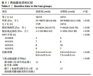
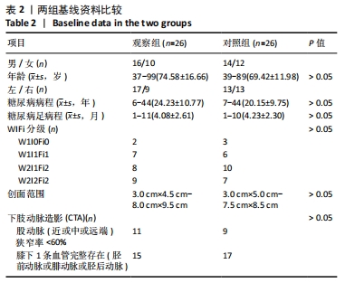
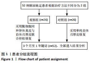
2.1 参与者数量分析 纳入患者52例,分为2组,经过3个月至1年随访,全部进入结果分析。 2.2 两组基线资料比较 两组患者性别、年龄、糖尿病足侧别、糖尿病及糖尿病足病程、WIFi分级、糖尿病足创面的病变范围及下肢动脉造影检查等一般资料进行比较,差异均无显著性意义(P > 0.05),具有可比性,见表2。"
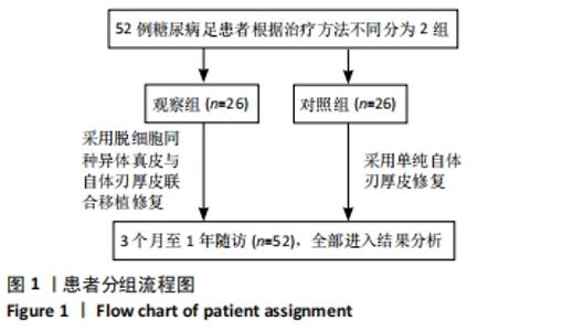
两组患者分组流程图,见图1。"

2.3 两组患者皮片成活率及再手术率 观察组皮片成活率显著高于对照组皮片成活率(P < 0.05);观察组再手术率显著低于对照组再手术率(P < 0.05),见表3。"
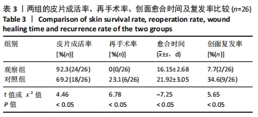

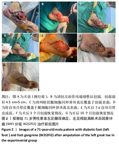
2.4 两组患者创面愈合情况及时间 最终两组患者创面均愈合,观察组愈合时间较对照组显著缩短(t=-7.25,P < 0.05)。创面愈合的评估标准:创面周围组织无红肿热痛的感染迹象,未见脓性渗出;植皮区域颜色红润,边缘区域干燥无颜色发黑结痂现象,见表3。 2.5 随访复发率 两组患者术后均获1年随访,随访期间观察组2例患者创面复发再次入院治疗,对照组9例患者创面复发再次入院治疗,观察组创面复发率低于对照组(P < 0.05),见表3。 2.6 典型病例介绍 观察组1例男性患者,71岁,左足糖尿病足、左足拇趾离断术后跖骨坏疽(WIFi分级W2I2Fi2),治疗前后图片,见图2。"

2.7 不良反应及生物相容性 对照组术后8例患者发生创面植皮修复区域感染,其中6例患者植皮区域组织坏死,植皮未成活,经二次手术修复,2例患者经过换药治疗后愈合;观察组术后2例发生感染,创面修复失败,经过换药治疗后愈合。两组患者均未发生局部及全身与脱细胞同种异体真皮移植相关的不良反应(移植排斥反应)。"

| [1] LECHLEITNER M, ABRAHAMIAN H, FRANCESCONI C, et al. Diabetische Neuropathie und diabetischer Fuß (Update 2019) [Diabetic neuropathy and diabetic foot syndrome (Update 2019)]. Wien Klin Wochenschr. 2019;131(Suppl 1):141-150. [2] YAZDANPANAH L, SHAHBAZIAN H, NAZARI I, et al. Incidence and Risk Factors of Diabetic Foot Ulcer: A Population-Based Diabetic Foot Cohort (ADFC Study)-Two-Year Follow-Up Study. Int J Endocrinol. 2018; 2018:7631659. [3] 王江宁, 高磊.糖尿病足慢性创面治疗的新进展[J].中国修复重建外科杂志,2018,32(7):832-837 [4] ZELEN CM, ORGILL DP, SERENA TE, et al. An aseptically processed, acellular, reticular, allogenic human dermis improves healing in diabetic foot ulcers: A prospective, randomised, controlled, multicentre follow-up trial. Int Wound J. 2018;15(5):731-739. [5] SARKOZYOVA N, DRAGUNOVA J, BUKOVCAN P, et al. Preparation and processing of human allogenic dermal matrix for utilization in reconstructive surgical procedures. Bratisl Lek Listy. 2020;121(6):386-394. [6] 郭海雷,凌翔伟,刘政军,等.刃厚头皮与异体脱细胞真皮基质复合移植修复特大面积烧伤患者手部深度创面[J].中华烧伤杂志, 2019,35(12):876-878. [7] WEAVER ML, HICKS CW, CANNER JK, et al. The Society for Vascular Surgery Wound, Ischemia, and foot Infection (WIfI) classification system predicts wound healing better than direct angiosome perfusion in diabetic foot wounds. J Vasc Surg. 2018;68(5):1473-1481. [8] 柯昌能,刘坡,陈杰明,等.脱细胞同种异体真皮与自体刃厚皮复合移植烧伤功能部位修复创面[J].中国组织工程研究,2015, 19(29):4652-4656. [9] BORDIANU A, BOBIRCĂ F, PĂTRAŞCU T. Skin Grafting in the Treatment of Diabetic Foot Soft Tissue Defects. Chirurgia (Bucur). 2018;113(5):644-650. [10] 薛春利,胡志成,杨祖贤,等.异体脱细胞真皮基质治疗糖尿病足溃疡临床效果荟萃分析[J].中华烧伤杂志,2016,32(12):725-729. [11] SANNIEC K, NGUYEN T, VAN ASTEN S,et al. Split-Thickness Skin Grafts to the Foot and Ankle of Diabetic Patients. J Am Podiatr Med Assoc. 2017;107(5):365-368. [12] ZELEN CM, ORGILL DP, SERENA T, et al. A prospective, randomised, controlled, multicentre clinical trial examining healing rates, safety and cost to closure of an acellular reticular allogenic human dermis versus standard of care in the treatment of chronic diabetic foot ulcers. Int Wound J. 2017;14(2):307-315. [13] CAMPITIELLO F, MANCONE M, DELLA CORTE A,et al. To evaluate the efficacy of an acellular flowable matrix in comparison with a wet dressing for the treatment of patients with diabetic foot ulcers: a randomized clinical trial. Updates Surg. 2017;69(4):523-529. [14] WIDJAJA W, TAN J, MAITZ PKM. Efficacy of dermal substitute on deep dermal to full thickness burn injury: a systematic review. ANZ J Surg. 2017;87(6):446-452. [15] PIRAYESH A, HOEKSEMA H, RICHTERS C, et al. Glyaderm(®) dermal substitute: clinical application and long-term results in 55 patients. Burns. 2015;41(1):132-144. [16] BOHAC M, VARGA I, POLAK S, et al. Delayed post mastectomy breast reconstructions with allogeneic acellular dermal matrix prepared by a new decellularizationmethod. Cell Tissue Bank. 2018;19(1):61-68. [17] SPECHT M, KELM S, MIRASTSCHIJSKI U. Eignung biologischer azellulärer dermaler Matrices als Hautersatz [Suitability of biological acellular dermal matrices as a skin replacement]. Handchir Mikrochir Plast Chir. 2020;52(6):533-544. [18] GOODARZI P, FALAHZADEH K, NEMATIZADEH M, et al. Tissue Engineered Skin Substitutes. Adv Exp Med Biol. 2018;1107:143-188. [19] NITA M, PLISZCZYŃSKI J, KOWALEWSKI C, et al. New Treatment of Wound Healing With Allogenic Acellular Human Skin Graft: Preclinical Assessment and In Vitro Study. Transplant Proc. 2020;52(7):2204-2207. [20] KUO S, KIM HM, WANG Z, et al. Comparison of two decellularized dermal equivalents. J Tissue Eng Regen Med. 2018;12(4):983-990. [21] HUANG W, CHEN Y, WANG N, et al. The Efficacy and Safety of Acellular Matrix Therapy for Diabetic Foot Ulcers: A Meta-Analysis of Randomized Clinical Trials. J Diabetes Res. 2020;2020:6245758. [22] DASGUPTA A, ORGILL D, GALIANO RD,et al. A Novel Reticular Dermal Graft Leverages Architectural and Biological Properties to Support Wound Repair. Plast Reconstr Surg Global Open. 2016;4(10):e1065. [23] CHO H, BLATCHLEY MR, DUH EJ, et al. Acellular and cellular approaches to improve diabetic wound healing. Advanced Drug Delivery Reviews. 2019;146:267-288. [24] AHMEDBEYLI C, IPCI SD, CAKAR G, et al. Laterally positioned flap along with acellular dermal matrix graft in the management of maxillary localized recessions. Clin Oral Investig. 2019;23(2):595-601. [25] KUO KC, LIN RZ, TIEN HW, et al. Bioengineering vascularized tissue constructs using an injectable cell-laden enzymatically crosslinked collagen hydrogel derived from dermal extracellular matrix. Acta Biomater. 2015;27:151-166. [26] WOOD BC, MANTILLA-RIVAS E, GOLDRICH A, et al. Correction of Microstomia Reconstruction with the Use of Acellular Dermal Matrix for Buccal Reconstruction. J Craniofac Surg. 2019;30(3):736-738. [27] YANG Z, WU X, CHEN C, et al. Comparison of type I tympanoplasty with acellular dermal allograft and cartilage perichondrium. Acta Oto-Laryngol. 139:10:833-836. [28] HU Z, ZHU J, CAO X, et al. Composite Skin Grafting with Human Acellular Dermal Matrix Scaffold for Treatment of Diabetic Foot Ulcers: A Randomized Controlled Trial. J Am Coll Surg. 2016;222(6):1171-1179. [29] CAZZELL S, VAYSER D, PHAM H, et al. A randomized clinical trial of a human acellular dermal matrix demonstrated superior healing rates for chronic diabetic foot ulcers over conventional care and an active acellular dermal matrix comparator. Wound Repair Regen. 2017;25(3): 483-497. [30] ZELEN CM, ORGILL DP, SERENA TE, et al. Human Reticular Acellular Dermal Matrix in the Healing of Chronic Diabetic Foot Ulcerations that Failed Standard Conservative Treatment: A Retrospective Crossover Study. Wounds. 2017;29(2):39-45. [31] LEE M, JUN D, CHOI H, et al. Clinical Efficacy of Acellular Dermal Matrix Paste in Treating Diabetic Foot Ulcers. Wounds. 2020;32(1):50-56. [32] CAZZELL S. A Randomized Controlled Trial Comparing a Human Acellular Dermal Matrix Versus Conventional Care for the Treatment of Venous Leg Ulcers. Wounds. 2019;31(3):68-74. [33] LUTHRINGER M, MUKHERJEE T, ARGUELLO-ANGARITA M, et al. Human-derived Acellular Dermal Matrix Grafts for Treatment of Diabetic Foot Ulcers: A Systematic Review and Meta-analysis. Wounds. 2020;32(2):57-65. [34] KIM YH, HWANG KT, KIM KH,et al. Application of acellular human dermis and skin grafts for lower extremity reconstruction. J Wound Care. 2019;28(Sup4):S12-S17. [35] ÁLVARO-AFONSO FJ, GARCÍA-ÁLVAREZ Y, LÁZARO-MARTÍNEZ JL,et al. Advances in Dermoepidermal Skin Substitutes for Diabetic Foot Ulcers. Curr Vasc Pharmacol. 2020;18(2):182-192. [36] TCHERO H, HERLIN C, BEKARA F, et al. Failure rates of artificial dermis products in treatment of diabetic foot ulcer: A systematic review and network meta-analysis. Wound Repair Regen. 2017;25(4):691-696. [37] WALTERS J, CAZZELL S, PHAM H,et al. Healing Rates in a Multicenter Assessment of a Sterile, Room Temperature, Acellular Dermal Matrix Versus Conventional Care Wound Management and an Active Comparator in the Treatment of Full-Thickness Diabetic Foot Ulcers. Eplasty. 2016;16:e10. [38] TOGNETTI L, PIANIGIANI E, IERARDI F, et al. The use of human acellular dermal matrices in advanced wound healing and surgical procedures: State of the art. Dermatol Ther. 2021;34(4):e14987. [39] JEON H, KIM J, YEO H, et al. Treatment of diabetic foot ulcer using matriderm in comparison with a skin graft. Arch Plast Surg. 2013;40(4): 403-408. [40] TCHANQUE-FOSSUO CN, DAHLE SE, LEV-TOV H, et al. Cellular versus acellular matrix devices in the treatment of diabetic foot ulcers: Interim results of a comparative efficacy randomized controlled trial. J Tissue Eng Regen Med. 2019;13(8):1430-1437. |
| [1] | Zhao Zixi, Xu Jun, Ding Min, Li Xiwen, Zhang Jinghang, Wang Penghua. Changes of type I and III collagen and matrix metalloproteinase 2 and 9 on the wound of diabetic foot ulcer with external application of medical collagen dressing [J]. Chinese Journal of Tissue Engineering Research, 2022, 26(10): 1544-1550. |
| [2] | Dong Hongfei, Huang Qilin, Yang Xiong, Li Shuai, Gu Rui, Sun Hongyu, Tang Lijun. Dermal extracellular matrix hydrogel in promoting acute skin wound healing in rats [J]. Chinese Journal of Tissue Engineering Research, 2022, 26(10): 1555-1560. |
| [3] | Wang Xianyao, Guan Yalin, Liu Zhongshan. Strategies for improving the therapeutic efficacy of mesenchymal stem cells in the treatment of nonhealing wounds [J]. Chinese Journal of Tissue Engineering Research, 2021, 25(7): 1081-1087. |
| [4] | Gan Lili, Xiong Na, Liu Yanfei. Hydrogel as drug scaffold in skin wound repair: challenges of clinical application possibilities [J]. Chinese Journal of Tissue Engineering Research, 2021, 25(22): 3578-3583. |
| [5] | Li Wenhui, Liu Guobin. Knowledge mapping analysis on the international research of diabetic foot: a visual analysis based on CiteSpace [J]. Chinese Journal of Tissue Engineering Research, 2021, 25(20): 3178-3184. |
| [6] | Wang Qi, Yang Xiaoshuang, Wang Dali. Effects of hydrogel three-dimensional culture on the characteristics of human amniotic mesenchymal stem cells and paracrine effect [J]. Chinese Journal of Tissue Engineering Research, 2020, 24(22): 3460-3466. |
| [7] | Society for Prevention and Control of Tissue Inflammation and Injury of Chinese Preventive Medicine Association, Society of Parenteral and Enteral Nutrition of Chinese Medical Association, Diabetic Foot Group, Committee of Peripheral Vascular Disease, China Society of Integrated Traditional Chinese and Western Medicine. Medical nutrition treatment guideline for diabetic foot [J]. Chinese Journal of Tissue Engineering Research, 2019, 23(35): 5682-5689. |
| [8] | Zhang Liming, Cai Jieyun, Pan Fuqiang, Wang Jing. Application and prospect of adipose-derived stem cells in wound inflammation [J]. Chinese Journal of Tissue Engineering Research, 2019, 23(25): 4089-4093. |
| [9] | Huang Yongqing, Shen Jie, Yuan Sijie, Pan Daoyan. Value of erythrocyte distribution width, fibrinogen and D-dimer in predicting the occurrence and development risk of diabetic foot [J]. Chinese Journal of Tissue Engineering Research, 2019, 23(23): 3716-3721. |
| [10] | Yang Qi, Zhang Yonghong, Lu Yanjun. Transdermal continuous oxygen therapy repairs diabetic foot ulcer: a systematic review [J]. Chinese Journal of Tissue Engineering Research, 2019, 23(23): 3767-3772. |
| [11] | Li Tao, Zhang Chunling, Long Yi, Di Tietao, Chen Lu, Zhao Wei, Long Li, Huang Qiang, Tang Lisha, Luo Kaizhong. Inhibitory effects of nanosilver sponge with Danhuang powder on Pseudomonas aeruginosa [J]. Chinese Journal of Tissue Engineering Research, 2019, 23(22): 3495-3499. |
| [12] | Huang Ruina, Huang Ruijia, Niu Caili, Qiu Wenbo, Wu Xiaowan, Wang Xiaojun, Wang Haijiao, Yang Chaojie. Effect of silver dressing on diabetic foot ulcer: a Meta-analysis [J]. Chinese Journal of Tissue Engineering Research, 2019, 23(2): 323-328. |
| [13] | Niu Caili, Huang Ruina, Xu Ziqi, Lu Yongmei, Huang Yongming, Zeng Xiuyun. Efficacy and safety of platelet-rich plasma in the treatment of diabetic foot ulcer: a meta-analysis [J]. Chinese Journal of Tissue Engineering Research, 2019, 23(14): 2285-2291. |
| [14] | Gao Wei, Lin Zhenxun, Zhen Puxiang, Chen Yan, Kuang Xiaocong, Hua Qikai. Macrophages promote the healing of severe diabetic foot wounds after tibial transverse transport [J]. Chinese Journal of Tissue Engineering Research, 2018, 22(36): 5811-5815. |
| [15] | Wang Bing-yang, Niu Guang-ming, Du Hua, Weng Li-xin. Different wound dressings in the treatment of diabetic foot ulcers [J]. Chinese Journal of Tissue Engineering Research, 2016, 20(34): 5155-5162. |
| Viewed | ||||||
|
Full text |
|
|||||
|
Abstract |
|
|||||