[1] WOLBANK S, PETERBAUER A, FAHRNER M, et al. Dose-dependent immunomodulatory effect of human stem cells from amniotic membrane: a comparison with human mesenchymal stem cells from adipose tissue. Tissue Eng. 2007;13(6):1173-1183.
[2] BAILO M, SONCINI M, VERTUA E, et al. Engraftment potential of human amnion and chorion cells derived from term placenta.Transplantation.2004;78(10):1439-1448.
[3] HARA J, TOTTORI J, ANDERS M, et al. Trehalose effectiveness as a cryoprotectant in 2D and 3D cell cultures of human embryonic kidney cells.Artif Cells Nanomed Biotechnol.2017;45(3): 609-616.
[4] YU HS, LEE EJ, SEO SJ, et al. Feasibility of silica-hybridized collagen hydrogels as three-dimensional cell matrices for hard tissue engineering.J Biomater Appl.2015;30(3): 338-350.
[5] MIKOS AG, SARAKINOS G, LEITE SM, et al. Laminated three-dimensional biodegradable foams for use in tissue engineering. Biomaterials.1993;14(5):323-330.
[6] ISHAUG-RILEY SL, CRANE-KRUGER GM, YASZEMSKI MJ, et al.Three-dimensional culture of rat calvarial osteoblasts in porous biodegradable polymers.Biomaterials. 1998;19(15): 1405-1412.
[7] HOLY CE, YAKUBOVICH R. Processing cell-seeded polyester scaffolds for histology.J Biomed Mater Res. 2000; 50(2):276-279.
[8] ZANONI M, PICCININI F, ARIENTI C, et al. 3D tumor spheroid models for in vitro therapeutic screening: a systematic approach to enhance the biological relevance of data obtained. Sci Rep. 2016; 6:19103.
[9] FROEHLICH K, HAEGER JD, HEGER J, et al. Generation of Multicellular Breast Cancer Tumor Spheroids: Comparison of Different Protocols.J Mammary Gland Biol. 2016;21(3-4): 89-98.
[10] KNIGHT E, PRZYBORSKI S. Advances in 3D cell culture technologies enabling tissue-like structures to be created in vitro.J Anat.2015;227(6):746-756.
[11] THOMAS CH, COLLIER JH, SFEIR CS, et al.Engineering gene expression and protein synthesis by modulation of nuclear shape.Proc Natl Acad Sci USA.2002;99(4): 1972-1977.
[12] VERGANI L,GRATTAROLA M, NICOLINI C. Modifications of chromatin structure and gene expression following induced alterations of cellular shape.Int J Biochem Cell B. 2004;36(8): 1447-1461.
[13] JANMEY PA, MILLER RT. Mechanisms of mechanical signaling in development and disease. J Cell Biol. 2011;124 (Pt 1):9-18.
[14] SHEKARAN A, LAM A, SIM E, et al. Biodegradable ECM-coated PCL microcarriers support scalable human early MSC expansion and in vivo bone formation.Cytotherapy. 2016;18(10):1332-1344.
[15] MAGUIRE T, NOVIK E, SCHLOSS R, et al. Alginate-PLL microencapsulation: effect on the differentiation of embryonic stem cells into hepatocytes.Biotechnol Bioeng.2006;93(3): 581-591.
[16] OCCHETTA P, CENTOLA M, TONNARELLI B, et al. High-Throughput Microfluidic Platform for 3D Cultures of Mesenchymal Stem Cells, Towards Engineering Developmental Processes.Sci Rep.2015; 5:10288.
[17] ANTONI D, BURCKEL H, JOSSET E, et al. Three-dimensional cell culture: a breakthrough in vivo. Int J Mol Sci. 2015;16(3):5517-5527.
[18] LAM J, LOWRY WE, CARMICHAEL ST, et al. Delivery of iPS-NPCs to the Stroke Cavity within a Hyaluronic Acid Matrix Promotes the Differentiation of Transplanted Cells. Adv Funct Mater.2014; 24(44):7053-7062.
[19] LIANG X, DING Y, ZHANG Y, et al. Paracrine mechanisms of mesenchymal stem cell-based therapy: current status and perspectives.Cell Transplant.2014;23(9):1045-1059.
[20] SONG M, HEO J, CHUN JY, et al. The paracrine effects of mesenchymal stem cells stimulate the regeneration capacity of endogenous stem cells in the repair of a bladder-outlet-obstruction-induced overactive bladder.Stem Cells Dev.2014; 23(6):654-663.
[21] 岳凤文.IL-10-hAMSCs对巨噬细胞表型转化及共培养上清液对成纤维细胞的影响研究[D].遵义:遵义医学院,2018.
[22] LEE EJ, PARK SJ, KANG SK, et al. Spherical bullet formation via E-cadherin promotes therapeutic potency of mesenchymal stem cells derived from human umbilical cord blood for myocardial infarction.Mol Ther. 2012;20(7): 1424-1433.
[23] RETTINGER CL, FOURCAUDOT AB, HONG SJ, et al.In vitro characterization of scaffold-free three-dimensional mesenchymal stem cell aggregates. Cell Tissue Res. 2014; 358(2):395-405.
[24] HO SS, MURPHY KC, BINDER BY, et al. Increased Survival and Function of Mesenchymal Stem Cell Spheroids Entrapped in Instructive Alginate Hydrogels. Stem Cell Transl Med.2016;5(6):773-781.
[25] AGGARWAL S, PITTENGER MF. Human mesenchymal stem cells modulate allogeneic immune cell responses.Blood. 2005; 105(4):1815-1822.
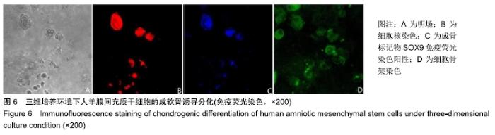
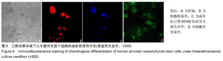
|