Chinese Journal of Tissue Engineering Research ›› 2021, Vol. 25 ›› Issue (28): 4516-4522.doi: 10.12307/2021.067
Previous Articles Next Articles
Influence of plasma spraying and electrochemical deposition of hydroxyapatite coating morphology on bone marrow mesenchymal stem cells
Sun Yang1, Luo Mingran1, Zheng Li1, Hu Weifan1, Yuan Feng2
- 1Graduate School of Xuzhou Medical University, Xuzhou 221000, Jiangsu Province, China; 2Department of Orthopedics, Affiliated Hospital of Xuzhou Medical University, Xuzhou 221000, Jiangsu Province, China
-
Received:2020-10-15Revised:2020-10-17Accepted:2020-11-13Online:2021-10-08Published:2021-05-21 -
Contact:Yuan Feng, Professor, Department of Orthopedics, Affiliated Hospital of Xuzhou Medical University, Xuzhou 221000, Jiangsu Province, China -
About author:Sun Yang, Master candidate, Physician, Graduate School of Xuzhou Medical University, Xuzhou 221000, Jiangsu Province, China -
Supported by:the Natural Science Foundation of Jiangsu Province, No. BE2016647 (to YF)
CLC Number:
Cite this article
Sun Yang, Luo Mingran, Zheng Li, Hu Weifan, Yuan Feng. Influence of plasma spraying and electrochemical deposition of hydroxyapatite coating morphology on bone marrow mesenchymal stem cells[J]. Chinese Journal of Tissue Engineering Research, 2021, 25(28): 4516-4522.
share this article
Add to citation manager EndNote|Reference Manager|ProCite|BibTeX|RefWorks
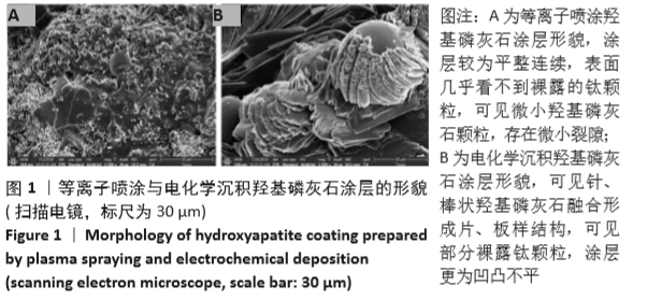
2.1 羟基磷灰石涂层钛支架的表征 如图1所示,两种涂层的表面形貌差异很大,电化学沉积组涂层可见由羟基磷灰石针状结构聚集并组合成片状、板状结构,表面仍可见小部分未被涂层覆盖的钛颗粒,涂层较为粗糙;等离子喷涂组涂层表面与其截然不同,熔融的羟基磷灰石均匀地喷涂在支架表面,涂层较为平整,表面可见细小的颗粒状、球状羟基磷灰石堆积,但涂层中可见细小裂纹。"


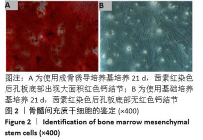
2.2 骨髓间充质干细胞的鉴定 如图2所示,将第3代骨髓间充质干细胞采用成骨诱导培养基培养21 d后,细胞呈复层样生长,十分密集,细胞已逐渐从骨髓间充质干细胞的梭形变为多边形,形成地板装样形态,茜素红染色可见 6孔板底出现大面积红色钙结节,部分钙结节中心为红棕色;对照组骨髓间充质干细胞使用基础培养基培养21 d后,茜素红染色并无红色钙结节,细胞形态依旧为梭形。"
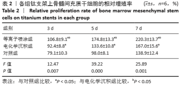
2.3 骨髓间充质干细胞与支架共培养后的增殖 如表2所示,随着培养时间的延长,3组细胞增殖A值增加,等离子喷涂组、电化学沉积组培养各时间点的A值高于对照组(P < 0.05),并且等离子喷涂组培养各时间点的A值高于电化学沉积组(P < 0.05)。"

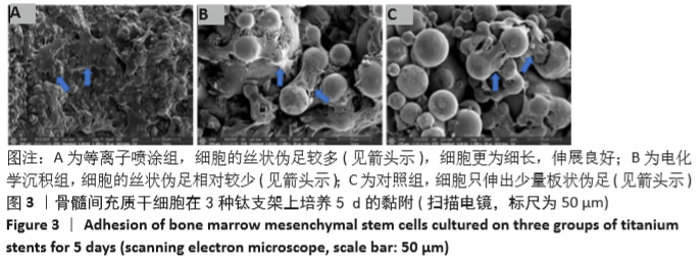
2.4 骨髓间充质干细胞与支架共培养后的黏附及形态 共培养5 d后的扫描电镜显示,3组支架上均可见骨髓间充质干细胞黏附,其中等离子喷涂组细胞铺展面积最大,细胞出现多个丝状伪足向周围延伸且伸展充分;电化学沉积组的细胞多黏附在片状、针棒状羟基磷灰石涂层上,细胞丝状伪足伸出较少且延展不充分;对照组支架上的细胞形态多是黏附并包裹钛颗粒,细胞铺展面积最小,仅有少部分细胞出现明显丝状伪足,见图3。"


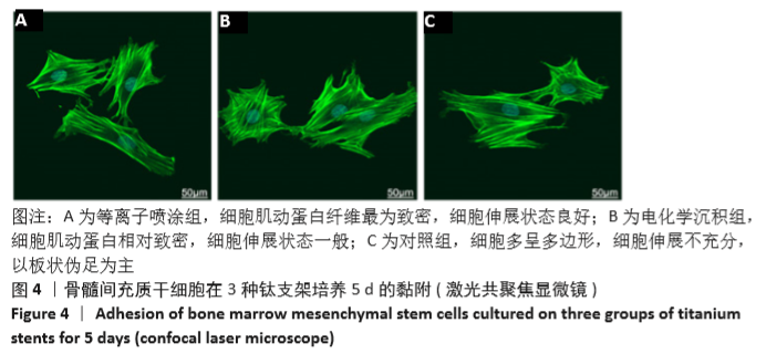
激光共聚焦显微镜下可见,等离子喷涂组骨髓间充质干细胞肌动蛋白纤维最为致密,细胞更为细长,细胞伸展状态良好,拥有紧密的丝状延展;电化学沉积组的细胞肌动蛋白相对致密,少部分细胞呈细长状,细胞伸展状态一般;对照组支架上的骨髓间充质干细胞肌动蛋白相对稀疏,细胞多呈多边形,细胞伸展不充分,以板状伪足为主,见图4。"

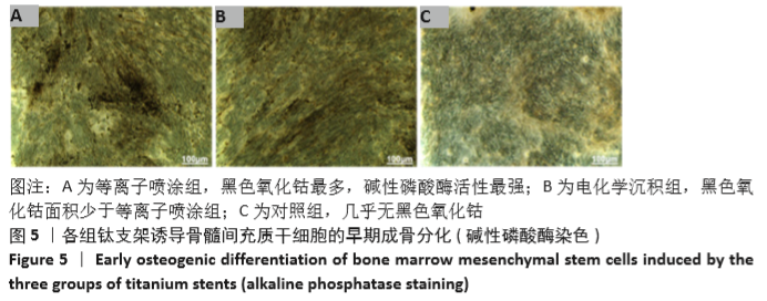
2.5 骨髓间充质干细胞与支架共培养后的成骨分化 培养第14天的碱性磷酸酶染色显示,等离子喷涂组细胞中显现大面积黑色氧化钴,显示出高碱性磷酸酶活性;电化学沉积组中黑色氧化钴面积偏少,与对照组几乎无黑色氧化钴相比碱性磷酸酶活性有所增强,但逊于等离子喷涂组,见图5。"
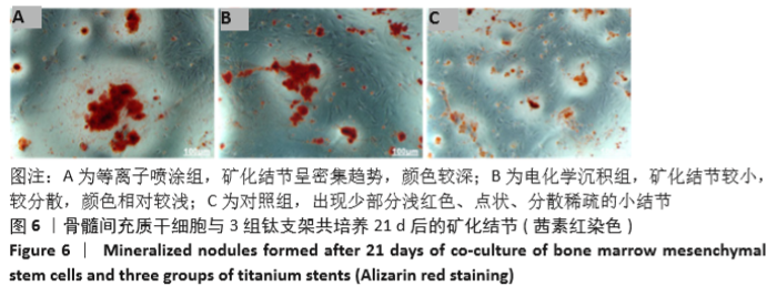
共培养21 d后的茜素红染色显示,等离子喷涂组的矿化结节呈密集趋势,已开始聚集出现团块状结节,结节颜色呈红色,中心区域已呈红褐色;电化学沉积组中的矿化结节较小,较分散,颜色相对偏浅;对照组中出现少部分浅红色、点状、分散稀疏的小结节,见图6。"

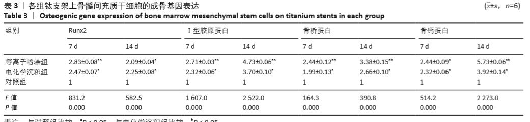
为方便统一计算、统计分析,将对照组中数据进行标准化处理为1。相同时间点下,等离子喷涂组、电化学沉积组的骨桥蛋白、Ⅰ型胶原蛋白、骨钙蛋白、Runx2 mRNA表达均高于对照组(P < 0.05)。除7 d的骨钙蛋白mRNA与14 d的Runx2 mRNA表达无差异外,等离子喷涂组相同时间下的骨桥蛋白、Ⅰ型胶原蛋白、骨钙蛋白、Runx2 mRNA表达均高于电化学沉积组(P < 0.05),见表3。"
| [1] AHMADI SM, HEDAYATI R, LI Y, et al. Fatigue performance of additively manufactured meta-biomaterials: the effects of topology and material type. Acta Biomater. 2018;65:292-304. [2] GADIA A, SHAH K, NENE A. Emergence of Three-Dimensional Printing Technology and Its Utility in Spine Surgery. Asian Spine J. 2018;12(2): 365-371. [3] TRAUNER KB. The Emerging Role of 3D Printing in Arthroplasty and Orthopedics. J Arthroplasty. 2018;33(8):2352-2354. [4] PATI F, SONG T H, RIJAL G, et al. Ornamenting 3D printed scaffolds with cell-laid extracellular matrix for bone tissue regeneration. Biomaterials. 2015;37:230-241. [5] LIU A, XUE GH, SUN M, et al. 3D printing surgical implants at the clinic: A experimental study on anterior cruciate ligament reconstruction. Sci Rep. 2016;6:21704. [6] SILVIA S, SEIJI Y, FRANCESCO B, et al. A critical review of multifunctional titanium surfaces: New frontiers for improving osseointegration and host response, avoiding bacteria contamination. Acta Biomater. 2019;83:37-54. [7] LEUKERS B, GÜLKAN H, IRSEN SH, et al. Hydroxyapatite scaffolds for bone tissue engineering made by 3D printing.J Mater Sci Mater Med. 2005;16(12):1121-1124. [8] RINCÓN-LÓPEZ JA, HERMANN-MUÑOZ JA, GIRALDO-BETANCUR AL, et al. Synthesis, Characterization and In Vitro Study of Synthetic and Bovine-Derived Hydroxyapatite Ceramics: A Comparison. Materials (Basel). 2018;11(3):333. [9] ZHU J, SUN HH, WO J, et al. Duration of electrochemical deposition affects the morphology of hydroxyapatite coatings on 3D-printed titanium scaffold as well as the functions of adhered MC3T3-E1 cells. J Orthop Sci. 2020;25(4):708-714. [10] VILARDELL AM, CINCA N, GARCIA-GIRALT N, et al. In-vitro comparison of hydroxyapatite coatings obtained by cold spray and conventional thermal spray technologies. Mater Sci Eng C Mater Biol Appl. 2020; 107:110306. [11] XUEREB M, CAMILLERI J, ATTARD N. Systematic Review of Current Dental Implant Coating Materials and Novel Coating Techniques. Int J Prosthodont. 2015;28(1):51-59. [12] 李成龙,李文戈,赵远涛,等.降低等离子喷涂涂层孔隙率的研究进展[J].机械工程材料,2020,44(5):60-65. [13] 杨学兵,张林伟.水蒸气处理对钛合金表面羟基磷灰石涂层结构的影响[J].表面技术,2020,49(9):167-174,205. [14] FATHYUNES L, KHALIL-ALLAFI J. Effect of employing ultrasonic waves during pulse electrochemical deposition on the characteristics and biocompatibility of calcium phosphate coatings. Ultrason Sonochem. 2018;42:293-302. [15] 王天云,张青,朱良均,等.利用电化学法结合丝素膜调控羟基磷灰石沉积及其形貌[J].浙江大学学报(农业与生命科学版),2018, 44(2):209-214. [16] ALBAYRAK O, EL-ATWANI O, ALTINTAS S. Hydroxyapatite coating on titanium substrate by electrophoretic deposition method: Effects of titanium dioxide inner layer on adhesion strength and hydroxyapatite decomposition. Surf Coat Technol. 2008;202(11):2482-2487. [17] KINNAIRD T, STABILE E, BURNETT MS, et al. Marrow-Derived Stromal Cells Express Genes Encoding a Broad Spectrum of Arteriogenic Cytokines and Promote In Vitro and In Vivo Arteriogenesis Through Paracrine Mechanisms. Circ Res. 2004;94(5):678-685. [18] AMNA T. Valorization of Bone Waste of Saudi Arabia by Synthesizing Hydroxyapatite. Appl Biochem Biotechnol. 2018;186:779-788. [19] LIN K, WU C, CHANG J, et al. Advances in synthesis of calcium phosphate crystals with controlled size and shape. Acta Biomater. 2014;10(10):4071-102. [20] EPPLE M. Review of potential health risks associated with nanoscopic calcium phosphate. Acta Biomater. 2018;77:1-14. [21] SHUMIN P, YUAN H, PING H, et al. Fabrication of Two Distinct Hydroxyapatite Coatings and Their Effects on MC3T3-E1 Cell Behavior. Colloids Surf B Biointerfaces. 2018;171:40-48. [22] KUBOKI Y, JIN Q, TAKITA H. Geometry of carriers controlling phenotypic expression in BMP-induced osteogenesis and chondrogenesis. J Bone Joint Surg Am. 2001;83-A Suppl 1(Pt 2):S105-115. [23] HAYAKAWA T, KAWASHITA M, TAKAOAKA GH. Coating of hydroxyapatite films on titanium substrates by electrodeposition under pulse current. J Ceram Soc Jpn. 2008;116(1349):68-73. [24] HULBERT SF, YOUNG FA, MATHEWS RS, et al. Potential of ceramic materials as permanently implantable skeletal prostheses. J Biomed Mater Res. 1970;4(3):433-456. [25] MASAHIRO N, MASAHIRO Y, MASATO W, et al. Activation of Osteoblastic Function on Titanium Surface with Titanium-Doped Hydroxyapatite Nanoparticle Coating: An In Vitro Study. Int J Oral Maxillofac Implants. 2017;32(4):779-791. [26] WANG J, WANG M, CHEN F, et al. Nano-Hydroxyapatite Coating Promotes Porous Calcium Phosphate Ceramic-Induced Osteogenesis Via BMP/Smad Signaling Pathway. Int J Nanomedicine. 2019;14: 7987-8000. [27] HAMIDABADI HG, SHAFAROUDI MM, SEIFI M, et al. Repair of Critical-Sized Rat Calvarial Defects With Three-Dimensional Hydroxyapatite-Gelatin Scaffolds and Bone Marrow Stromal Stem Cells. Med Arch. 2018;72(2):88-93. [28] ROYCROFT A, MAYOR R. Michael Abercrombie: contact inhibition of locomotion and more. Int J Dev Biol. 2018;62(1-2-3):5-13. [29] SIPAHI R, ZUPANC GKH. Stochastic cellular automata model of neurosphere growth: Roles of proliferative potential, contact inhibition, cell death, and phagocytosis. J Theor Biol. 2018;445:151-165. [30] CUI JG, CHEN G, PERRY AS, et al. Transient Cell-to-Cell Signaling before Mitosis in Cultures of human Bone Marrow-Derived Mesenchymal Stem/Stromal Cells. Stem Cells Dev. 2019;28(2):120-128. [31] PAPAGEORGIOU P, VALLMAJO-MARTIN Q, KISIELOW M, et al. Expanded skeletal stem and progenitor cells promote and participate in induced bone regeneration at subcritical BMP-2 dose. Biomaterials. 2019;217: 119278. [32] LI X, ZOU Q, LI W, et al. Intracellular Interaction of Hydroxyapatite-Based Nanocrystals with Uniform Shape and Traceable Fluorescence. Inorg Chem. 2018;57(21):13739-13748. [33] PEI YF, LIU L, LIU TL, et al. Joint Association Analysis Identified 18 New Loci for Bone Mineral Density. J Bone Miner Res. 2019;34(6): 1086-1094. [34] LIN Z, HE H, WANG M, et al. MicroRNA-130a controls bone marrow mesenchymal stem cell differentiation towards the osteoblastic and adipogenic fate. Cell Prolif. 2019;52:e12688. |
| [1] | Du Xiupeng, Yang Zhaohui. Effect of degree of initial deformity of impacted femoral neck fractures under 65 years of age on femoral neck shortening [J]. Chinese Journal of Tissue Engineering Research, 2021, 25(9): 1410-1416. |
| [2] | Zhang Shangpu, Ju Xiaodong, Song Hengyi, Dong Zhi, Wang Chen, Sun Guodong. Arthroscopic suture bridge technique with suture anchor in the treatment of acromioclavicular dislocation [J]. Chinese Journal of Tissue Engineering Research, 2021, 25(9): 1417-1422. |
| [3] | Liang Yan, Zhao Yongfei, Xu Shuai, Zhu Zhenqi, Wang Kaifeng, Liu Haiying, Mao Keya. Imaging evaluation of short-segment fixation and fusion for degenerative lumbar scoliosis assisted by highly selective nerve root block [J]. Chinese Journal of Tissue Engineering Research, 2021, 25(9): 1423-1427. |
| [4] | Zhou Jihui, Li Xinzhi, Zhou You, Huang Wei, Chen Wenyao. Multiple problems in the selection of implants for patellar fracture [J]. Chinese Journal of Tissue Engineering Research, 2021, 25(9): 1440-1445. |
| [5] | Wang Debin, Bi Zhenggang. Related problems in anatomy mechanics, injury characteristics, fixed repair and three-dimensional technology application for olecranon fracture-dislocations [J]. Chinese Journal of Tissue Engineering Research, 2021, 25(9): 1446-1451. |
| [6] | Chen Junming, Yue Chen, He Peilin, Zhang Juntao, Sun Moyuan, Liu Youwen. Hip arthroplasty versus proximal femoral nail antirotation for intertrochanteric fractures in older adults: a meta-analysis [J]. Chinese Journal of Tissue Engineering Research, 2021, 25(9): 1452-1457. |
| [7] | Chen Jinping, Li Kui, Chen Qian, Guo Haoran, Zhang Yingbo, Wei Peng. Meta-analysis of the efficacy and safety of tranexamic acid in open spinal surgery [J]. Chinese Journal of Tissue Engineering Research, 2021, 25(9): 1458-1464. |
| [8] | Hu Kai, Qiao Xiaohong, Zhang Yonghong, Wang Dong, Qin Sihe. Treatment of displaced intra-articular calcaneal fractures with cannulated screws and plates: a meta-analysis of 15 randomized controlled trials [J]. Chinese Journal of Tissue Engineering Research, 2021, 25(9): 1465-1470. |
| [9] | Huang Dengcheng, Wang Zhike, Cao Xuewei. Comparison of the short-term efficacy of extracorporeal shock wave therapy for middle-aged and elderly knee osteoarthritis: a meta-analysis [J]. Chinese Journal of Tissue Engineering Research, 2021, 25(9): 1471-1476. |
| [10] | Xu Feng, Kang Hui, Wei Tanjun, Xi Jintao. Biomechanical analysis of different fixation methods of pedicle screws for thoracolumbar fracture [J]. Chinese Journal of Tissue Engineering Research, 2021, 25(9): 1313-1317. |
| [11] | Jiang Yong, Luo Yi, Ding Yongli, Zhou Yong, Min Li, Tang Fan, Zhang Wenli, Duan Hong, Tu Chongqi. Von Mises stress on the influence of pelvic stability by precise sacral resection and clinical validation [J]. Chinese Journal of Tissue Engineering Research, 2021, 25(9): 1318-1323. |
| [12] | Zhang Tongtong, Wang Zhonghua, Wen Jie, Song Yuxin, Liu Lin. Application of three-dimensional printing model in surgical resection and reconstruction of cervical tumor [J]. Chinese Journal of Tissue Engineering Research, 2021, 25(9): 1335-1339. |
| [13] | Zhang Yu, Tian Shaoqi, Zeng Guobo, Hu Chuan. Risk factors for myocardial infarction following primary total joint arthroplasty [J]. Chinese Journal of Tissue Engineering Research, 2021, 25(9): 1340-1345. |
| [14] | Wei Wei, Li Jian, Huang Linhai, Lan Mindong, Lu Xianwei, Huang Shaodong. Factors affecting fall fear in the first movement of elderly patients after total knee or hip arthroplasty [J]. Chinese Journal of Tissue Engineering Research, 2021, 25(9): 1351-1355. |
| [15] | Wang Jinjun, Deng Zengfa, Liu Kang, He Zhiyong, Yu Xinping, Liang Jianji, Li Chen, Guo Zhouyang. Hemostatic effect and safety of intravenous drip of tranexamic acid combined with topical application of cocktail containing tranexamic acid in total knee arthroplasty [J]. Chinese Journal of Tissue Engineering Research, 2021, 25(9): 1356-1361. |
| Viewed | ||||||
|
Full text |
|
|||||
|
Abstract |
|
|||||