[1] Barry F, Murphy M. Mesenchymal stem cells in joint disease and repair.Nat Rev Rheumatol. 2013;9(10):584-594.
[2] Qi Y, Feng G, Yan W. Mesenchymal stem cell-based treatment for cartilage defects in osteoarthritis. Mol Biol Rep. 2012;39(5):5683-5689.
[3] Heydarkhan-Hagvall S, Schenke-Layland K, Yang JQ, et al. Human adipose stem cells: a potential cell source for cardiovascular tissue engineering. Cells Tissues Organs. 2008;187(4):263-274.
[4] Hynes K, Menicanin D, Han J, et al. Mesenchymal stem cells from iPS cells facilitate periodontal regeneration. J Dent Res. 2013;92(9):833-839.
[5] Nekanti U, Rao VB, Bahirvani AG, et al. Long-term expansion and pluripotent marker array analysis of Wharton's jelly-derived mesenchymal stem cells. Stem Cells Dev. 2010;19(1):117-130.
[6] Wexler SA, Donaldson C, Denning-Kendall P, et al. Adult bone marrow is a rich source of human mesenchymal 'stem' cells but umbilical cord and mobilized adult blood are not. Br J Haematol. 2003;121(2):368-374.
[7] Secco M, Zucconi E, Vieira NM, et al. Multipotent stem cells from umbilical cord: cord is richer than blood. Stem Cells. 2008;26(1):146-150.
[8] Chen MY, Lie PC, Li ZL, et al. Endothelial differentiation of Wharton's jelly-derived mesenchymal stem cells in comparison with bone marrow-derived mesenchymal stem cells. Exp Hematol. 2009;37(5):629-640.
[9] Wu R, Tang Y, Zang W, et al. MicroRNA-128 regulates the differentiation of rat bone mesenchymal stem cells into neuron-like cells by Wnt signaling. Mol Cell Biochem. 2014;387(1-2):151-158.
[10] Ohgushi H. Osteogenically differentiated mesenchymal stem cells and ceramics for bone tissue engineering. Expert Opin Biol Ther. 2014;14(2):197-208.
[11] Panfilov IA, de Jong R, Takashima S, et al. Clinical study using adipose-derived mesenchymal-like stem cells in acute myocardial infarction and heart failure. Methods Mol Biol. 2013;1036:207-212.
[12] Bhonde RR, Sheshadri P, Sharma S, et al. Making surrogate β-cells from mesenchymal stromal cells: perspectives and future endeavors. Int J Biochem Cell Biol. 2014;46:90-102.
[13] 田新,符仁义,陈艳,等脐带间充质干细胞分离及向成骨与脂肪细胞的分化[J].四川大学学报,2008,39(1):26-29.
[14] Zhang X, Nan Y, Wang H, et al. Model microgravity enhances endothelium differentiation of mesenchymal stem cells. Naturwissenschaften. 2013;100(2):125-133.
[15] Guan XM, Cheng M, Li H, et al. Biological properties of bone marrow-derived early and late endothelial progenitor cells in different culture media. Mol Med Rep. 2013;8(6):1722-1728.
[16] Lai WH, Ho JC, Chan YC, et al. Attenuation of hind-limb ischemia in mice with endothelial-like cells derived from different sources of human stem cells. PLoS One. 2013;8(3): e57876.
[17] Portalska KJ, Chamberlain MD, Lo C, et al. Collagen modules for in situ delivery of mesenchymal stromal cell-derived endothelial cells for improved angiogenesis. J Tissue Eng Regen Med. 2013 Apr 17. [Epub ahead of print]
[18] Vishnubalaji R, Manikandan M, Al-Nbaheen M, et al. In vitro differentiation of human skin-derived multipotent stromal cells into putative endothelial-like cells. BMC Dev Biol. 2012;12:7.
[19] Marchal JA, Picón M, Perán M, et al. Purification and long-term expansion of multipotent endothelial-like cells with potential cardiovascular regeneration. Stem Cells Dev. 2012; 21(4):562-574.
[20] Yan D, Wang X, Li D, et al. Macrophages overexpressing VEGF, transdifferentiate into endothelial-like cells in vitro and in vivo. Biotechnol Lett. 2011;33(9):1751-1758.
[21] Marchand M, Anderson EK, Phadnis SM, et al. Concurrent generation of functional smooth muscle and endothelial cells via a vascular progenitor. Stem Cells Transl Med. 2014;3(1): 91-97.
[22] Choi M, Lee HS, Naidansaren P, et al. Proangiogenic features of Wharton's jelly-derived mesenchymal stromal/stem cells and their ability to form functional vessels. Int J Biochem Cell Biol. 2013;45(3):560-570.
[23] 刘红,俞小芳,滕杰,等.低氧预处理对大鼠骨髓间充质干细胞迁移能力的影响[J].中华医学杂志,2012,92(10):703-719.
[24] 朱斌,王云雅,张志勇,等.大鼠内皮祖细胞的分离、培养与定向诱导分化[J].现代中西医结合杂志,2009,18(6):608-611.
[25] 邵琴,王长谦,范华骅,等.脐血、外周血内皮祖细胞分化为内皮细胞的实验研究[J].心脏杂志,2006,18(1):18-22.
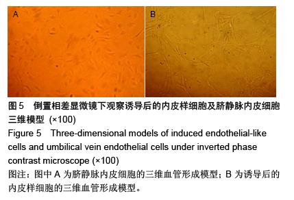
.jpg)