中国组织工程研究 ›› 2013, Vol. 17 ›› Issue (48): 8455-8460.doi: 10.3969/j.issn.2095-4344.2013.48.025

• 骨与关节临床实践 clinical practice of the bone and joint • 上一篇    

不同病因致股骨头缺血性坏死的影像对比

张德洲,易雪冰,钟  鉴   

  1. 四川省骨科医院放射科,四川省成都市  610041
  • 出版日期:2013-11-26 发布日期:2013-11-26
  • 作者简介:张德洲,男,1966年生,四川省西充县人,1990年泸州医学院毕业,副主任医师,主要从事医学影像学研究。 fangsheke3361@sina.com

Imaging comparison for avascular necrosis of the femoral head induced by different etiologies

Zhang De-zhou, Yi Xue-bing, Zhong Jian   

  1. Department of Radiology, Sichuan Orthopaedic Hospital, Chengdu  610041, Sichuan Province, China
  • Online:2013-11-26 Published:2013-11-26
  • About author:Zhang De-zhou, Associate chief physician, Department of Radiology, Sichuan Orthopaedic Hospital, Chengdu 610041, Sichuan Province, China fangsheke3361@sina.com

摘要:

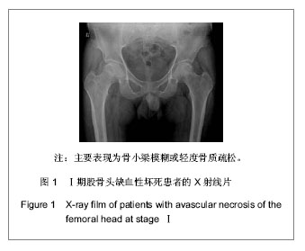
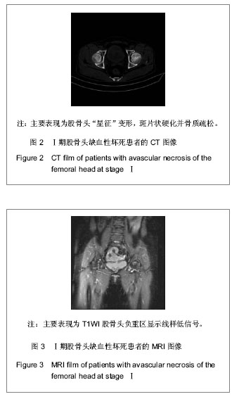
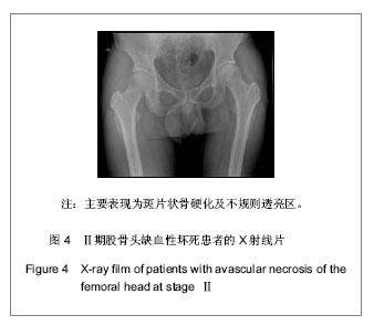
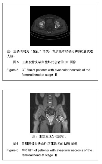
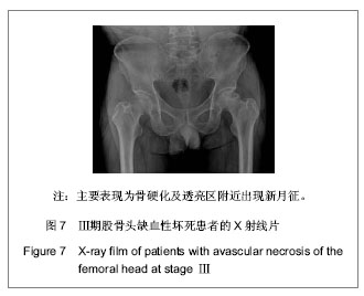
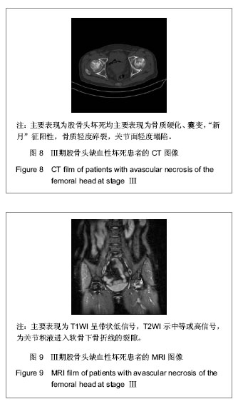
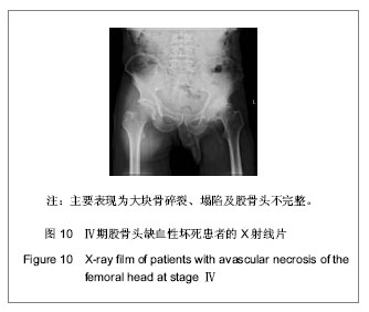
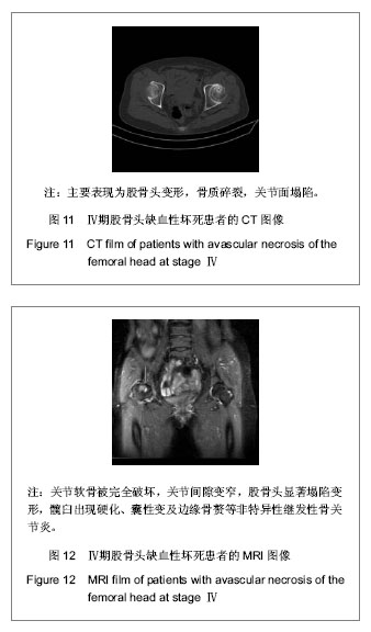
背景:股骨头缺血性坏死的临床诊断主要依靠影像学检查,对不同病因所致股骨头缺血性坏死的影像学对比研究目前报道甚少。
目的:对不同病因股骨头缺血性坏死的影像学表现进行比较研究,观察不同病因股骨头缺血性坏死的影像学表现有无差别。
方法:收集经临床及影像学检查证实的股骨头缺血性坏死54例共计60个髋关节影像资料,根据病因将其分为特发性、酒精性、激素性、股骨颈骨折性4类,将这4类股骨头缺血坏死的X射线、CT、MRI表现进行对比分析。
结果与结论:60个股骨头缺血性坏死髋关节中特发性12个、酒精性21个、激素性15个、股骨颈骨折性12个。X射线、CT主要表现依次为:股骨头“星征”变形或 “星征”消失;“新月”征阳性,骨质轻度碎裂,关节面轻度塌陷;股骨头变形,骨质碎裂,关节面塌陷,髋关节退性行改变,并以CT更可靠。股骨头缺血性坏死分期MRI均表现为:Ⅰ期T1WI股骨头负重区显示线样低信号、T2WI 加权呈高信号为主要改变;Ⅱ期T1WI为新月形边界清楚的不均匀信号,T2WI加权呈中等稍高信号,周围不均匀稍低信号环绕,呈典型的双线征;Ⅲ期股骨头变形,软骨下骨折、塌陷、新月体形成。T1WI呈带状低信号, T2WI示中等或高信号;Ⅳ、Ⅴ期关节软骨被完全破坏,关节间隙变窄,股骨头显著塌陷变形,髋臼出现硬化、囊性变及边缘骨赘等非特异性继发性骨关节炎。提示不同病因相同分期股骨头缺血性坏死的X射线、CT、MRI表现特点及规律是相同的。

关键词: 骨关节植入物, 骨与关节临床实践, 股骨头缺血性坏死, X射线, CT, MRI, 特发, 酒精, 激素, 股骨颈骨折

Abstract:

BACKGROUND: Imaging examination is the main strategy for clinical diagnosis of avascular necrosis of the femoral head. However, comparative studies of imaging findings between different causes of avascular necrosis of the femoral head are rarely reported.
OBJECTIVE: To compare imaging findings between different causes of avascular necrosis of femoral head, and to determine whether the imaging findings of different causes of avascular necrosis are different.
METHODS: A total of 54 cases of avascular necrosis of the femoral head (60 hips) were confirmed by clinical and imaging findings, and divided into idiopathic, alcoholic, hormonal and femoral neck fractural groups according to the causes. X-ray, CT, MRI findings of these four kinds of avascular necroses were analyzed.
RESULTS AND CONCLUSION: Sixty hips of avascular necrosis of the femoral head included 12 idiopathic hips, 21 alcoholic hips, 15 hormonal hips, and 12 femoral neck fractural hips. X-ray and CT main performances were as follows: “star sign” deformation or disappearance of the femoral head; positive “crescent” sign, mild 
bone fracture, mild collapse of the articular surface; femoral head deformation, bone fracture, articular surface collapse, hip degeneration. MRI of four categories of avascular necroses appeared as phase Ⅰ: line-like low signal of weight-bearing area of femoral head on T1WI, high signal on T2WI as the main change. Phase Ⅱ: clear boundary crescent-shaped uneven signal on T1WI, T2WI displayed moderately higher, uneven slightly lower signal around, showing a typical two-line sign. Phase Ⅲ: femoral head deformation, subchondral fracture, collapse, crescent formation, zonal low signal on T1WI, medium or high signal on T2WI. Phase Ⅳ, Ⅴ: complete destruction of articular cartilage, joint space narrowing, significant collapse and deformation of the femoral head, non-specific secondary osteoarthritis of the acetabulum, such as sclerosis, cystic degeneration and marginal osteophyte. The X-ray, CT, MRI performances of four types of avascular necroses at the same period were basically the same.

Key words: femur head necrosis, femoral neck fractures, diagnosis, differential, X-ray, magnetic resonance imaging

中图分类号: