中国组织工程研究 ›› 2023, Vol. 27 ›› Issue (17): 2669-2674.doi: 10.12307/2023.186

• 组织构建细胞学实验 cytology experiments in tissue construction • 上一篇    

少突胶质前体细胞延长神经胶质母细胞瘤大鼠高剂量放疗后的生存期

高  月1,蔺建文1,李  迪1,蓝晓艳1,李  深2,储成艳1   

  1. 1大连医科大学附属大连市中心医院,辽宁省大连市  116033;2首都医科大学附属北京世纪坛医院,北京市  100038
  • 收稿日期:2022-02-18 接受日期:2022-05-28 出版日期:2023-06-18 发布日期:2022-10-24
  • 通讯作者: 储成艳,博士,主治医师,大连医科大学附属大连市中心医院,辽宁省大连市 116033
  • 作者简介:高月,女,1990年生,辽宁省朝阳市人,汉族,2016年大连医科大学毕业,硕士,主要从事神经损伤修复方面的研究工作。
  • 基金资助:
    2021年度中央引导地方科技发展资金计划(自由探索类基础研究),项目负责人:储成艳;2020年大连市医学科学研究计划项目(2011005),项目负责人:储成艳;大连市医学重点专科“登峰计划”(大连市中心医院神经医学中心专项资金项目,2022zz215),项目负责人:储成艳

Oligodendrocyte progenitor cells prolong the survival of glioblastoma-bearing rats after high-dose radiotherapy

Gao Yue1, Lin Jianwen1, Li Di1, Lan Xiaoyan1, Li Shen2, Chu Chengyan1   

  1. 1Dalian Municipal Central Hospital, Dalian Medical University, Dalian 116033, Liaoning Province, China; 2Beijing Shijitan Hospital, Capital Medical University, Beijing 100038, China
  • Received:2022-02-18 Accepted:2022-05-28 Online:2023-06-18 Published:2022-10-24
  • Contact: Chu Chengyan, MD, Attending physician, Dalian Municipal Central Hospital, Dalian Medical University, Dalian 116033, Liaoning Province, China
  • About author:Gao Yue, Master, Dalian Municipal Central Hospital, Dalian Medical University, Dalian 116033, Liaoning Province, China
  • Supported by:
    The Fund Project of Central Government for Guiding Development of the Local Science and Technology (Free Exploration for Basic Research) in 2021 (to CCY); Dalian Medical Science Research Program Project in 2020, No. 2011005 (to CCY); Dalian “Peak Climbing Plan” for Medical Key Specialty, 2022zz215 (to CCY)

摘要:

文题释义:
放疗性脑损伤:放疗是治疗神经胶质瘤的一线治疗手段。然而,辐射可造成少突胶质细胞丢失等健康脑组织的损伤,进而引起一系列不良反应,目前尚缺乏有效的修复手段。
少突胶质前体细胞:作为重要的祖细胞库,少突胶质前体细胞可快速分化为成熟的少突胶质细胞,替代损伤、死亡的少突胶质细胞以维持中枢神经系统髓鞘结构及功能的稳定。
背景:胶质母细胞瘤是成人常见的恶性脑肿瘤,目前尚缺乏有效的治疗方法,临床预后不良。
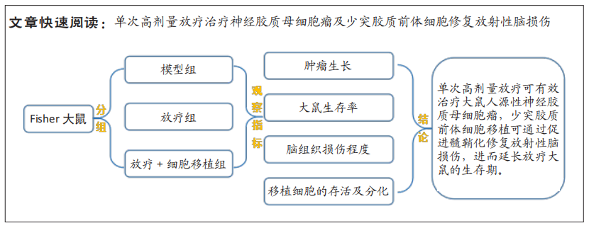
目的:观察单次高剂量放疗对神经胶质母细胞瘤的疗效,以及少突胶质前体细胞修复放射性脑损伤的可行性。
方法:通过质粒转染构建可表达荧光素酶(Luc)的人源性U-87 MG肿瘤细胞系。15只Fisher 344大鼠制备Luc-U-87 MG神经胶质母细胞瘤模型,随机分为肿瘤模型组(n=3)、放疗组(n=6)和放疗+细胞移植组(n=6),后两组放疗方式为单次80 Gy辐射;放疗后5周,放疗+细胞移植组大鼠联合少突胶质前体细胞移植治疗。利用活体成像的方法监测肿瘤细胞的生长;MRI观察放疗引起的脑组织影像学变化;采用Kaplan-Meier生存分析明确细胞移植对放疗大鼠生存率的影响;对移植细胞的存活及分化情况进行组织学观察。
结果与结论:①体外生物成像示转染后的U-87 MG细胞与Luc底物产生反应发出生物信号,信号强度与转染细胞的数量呈线性正相关;②动物活体成像结果显示,肿瘤模型大鼠的颅内胶质母细胞瘤信号持续上升直至接种第5周(全部死亡);放疗大鼠在治疗后2周,肿瘤信号开始衰减,4周后未见肿瘤发光信号;③放疗后5周,T2WI上可见放疗大鼠坏死的肿瘤组织;10周后未见肿瘤显影,但出现脑组织损伤信号,少突胶质前体细胞移植大鼠脑组织未见类似信号;15周后,放疗组和放疗+细胞移植组大鼠在T2WI上均可见高、低混杂的组织损伤信号,但放疗+细胞移植组的损伤信号程度明显低于单纯放疗组;④生存分析结果显示模型组、放疗组及放疗+细胞移植组大鼠的中位生存期分别为30 d,114.5 d和232.5 d,各组大鼠的中位生存期之间差异均有显著性意义(P < 0.01);⑤组织学观察到存活的移植细胞,呈多极样,部分细胞表达髓鞘碱性蛋白,且放疗+细胞移植组的髓鞘碱性蛋白表达明显高于放疗组(P < 0.01);⑥上述结果表明,单次高剂量放疗可有效治疗大鼠人源性神经胶质母细胞瘤,少突胶质前体细胞移植可通过修复放射性脑损伤延长放疗大鼠的生存期,促进髓鞘化是其修复损伤脑组织的机制之一。
https://orcid.org/0000-0003-2916-2882(高月)
中国组织工程研究杂志出版内容重点:组织构建;骨细胞;软骨细胞;细胞培养;成纤维细胞;血管内皮细胞;骨质疏松;组织工程

Abstract: BACKGROUND: Glioblastoma is a common malignant brain tumor for adults with poor prognosis and remains lack of efficient treatments. 
OBJECTIVE: To investigate the efficacy of single high-dose radiotherapy on glioblastoma and the feasibility of oligodendrocyte progenitor cells in the repair of radiation-induced brain injury.
METHODS: Plasmid transfection was used to develop human U-87 MG cell line expressing luciferase (Luc). Fifteen Fisher 344 rats bearing Luc-U-87 MG glioblastoma were randomly divided into tumor model group (n=3), radiotherapy group (n=6), and oligodendrocyte progenitor cell transplantation group (n=6). Single high-dose radiotherapy (80 Gy) was given in the latter two groups and cell transplantation was performed in the last group 5 weeks after radiotherapy. In vivo imaging was employed to monitor the tumor growth. MRI was performed to observe the cerebral structure changes induced by radiation. Kaplan-Meier analysis was used to determine the effect of cell transplantation on the survival rate of irradiated rats. Survival and differentiation of transplanted cells were histologically observed. 
RESULTS AND CONCLUSION: In vitro imaging results showed that the transfected U-87 MG cells reacted with the Luc substrate to produce bioluminescence signals. Signal intensity had a positive linear correlation with the number of transfected cells. Intracranial glioblastoma signals in the tumor model rats continued to increase until their death at 5 weeks after tumor inoculation. Two weeks after radiotherapy, the bioluminescence signal started to decline in the radiotherapy group and was not detected at 4 weeks. Necrotic tumor tissue was observed on T2WI at 5 weeks after radiotherapy but not at 10 weeks, at which timepoint the abnormal signal indicative of brain injury appeared. However, there were no similar signals in the cell transplantation group. Fifteen weeks later, T2WI showed hypo- and hyperintensity signals in the brain parenchyma for all the rats which received irradiation, whereas the injury signal in the radiotherapy group was stronger than that in the cell transplantation group. The survival analysis results revealed the median survival time of the tumor model group, radiotherapy group, and cell transplantation group were 30, 114.5, and 232.5 days, respectively. There were significant differences in the median survival time among groups (P < 0.01). Histological findings showed the transplanted cells survived with multipolarity and some of them expressed myelin basic protein. Moreover, the expression of myelin basic protein in the cell transplantation group was significantly higher than that in the radiotherapy group (P < 0.01). All these findings indicate that single high-dose radiotherapy can effective treat human-derived glioblastoma in rats. Oligodendrocyte progenitor cells transplanted are capable of repairing radiation-induced brain injury, thereby prolonging the survival of irradiated rats. Remyelination is one of the mechanisms accounting for brain tissue repair.  

Key words: glioblastoma, radiotherapy, radiation-induced injury, oligodendrocyte progenitor cell, remyelination, survival time, MRI, rat

中图分类号: