Chinese Journal of Tissue Engineering Research ›› 2015, Vol. 19 ›› Issue (12): 1852-1857.doi: 10.3969/j.issn.2095-4344.2015.12.010
Previous Articles Next Articles
Nano-hydroxyapatite film as a support to improve the proliferation of human umbilical vein endothelial cells
Cheng Guang-cun, Yan Zhong-ya, Li Chun-sheng, Yan Yu, Wei Xiao-yong
- Department of Cardiothoracic Surgery, Anhui Provincial Hospital Affiliated to Anhui Medical University, Hefei 230001, Anhui Province, China
-
Revised:2015-03-01Online:2015-03-19Published:2015-03-19 -
Contact:Yan Zhong-ya, Professor, Chief physician, Department of Cardiothoracic Surgery, Anhui Provincial Hospital Affiliated to Anhui Medical University, Hefei 230001, Anhui Province, China -
About author:Cheng Guang-cun, Master, Associate chief physician, Associate professor, Department of Cardiothoracic Surgery, Anhui Provincial Hospital Affiliated to Anhui Medical University, Hefei 230001, Anhui Province, China -
Supported by:the Natural Science Foundation of Anhui Province, No. 1308085MH143
CLC Number:
Cite this article
Cheng Guang-cun, Yan Zhong-ya, Li Chun-sheng, Yan Yu, Wei Xiao-yong. Nano-hydroxyapatite film as a support to improve the proliferation of human umbilical vein endothelial cells[J]. Chinese Journal of Tissue Engineering Research, 2015, 19(12): 1852-1857.
share this article

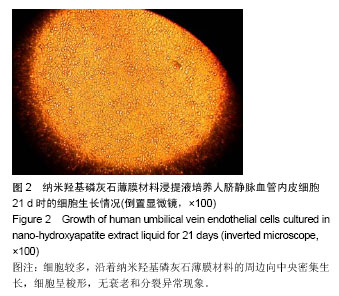
2.1 倒置显微镜观察 细胞传代接种后24 h可见部分类圆形人脐静脉血管内皮细胞贴壁生长;3 d时,细胞贴壁增多,呈铺路石样,散在分布;五六天时,细胞进一步增多,细胞间相互接触,排列紊乱;7-9 d时,细胞基本铺满瓶底,呈梭形或多边形。 2.2 人脐静脉血管内皮细胞在纳米羟磷灰石薄膜材料浸提液中的生长情况 倒置显微镜下观察纳米羟基磷灰石薄膜材料浸提液培养人脐静脉血管内皮细胞的贴附形态,早期(1 d内)可见纳米羟基磷灰石薄膜材料周边少量人脐静脉血管内皮细胞生长良好,随着培养时间的延长,3 d后细胞生长明显增多,沿着纳米羟基磷灰石薄膜材料的周边向中央密集生长,细胞呈梭形,无衰老和分裂异常现象(图2)。"
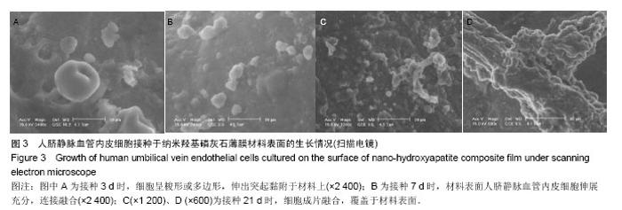
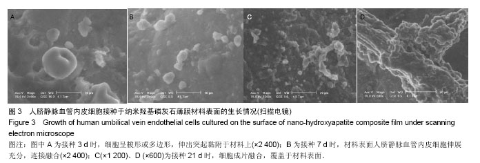
2.3 扫描电镜观察人脐静脉血管内皮细胞接种于纳米羟基磷灰石薄膜材料的生长情况 共培养1 d时,可见纳米羟基磷灰石薄膜材料表面有人脐静脉血管内皮细胞附着;3 d时,细胞呈梭形或多边形,伸出突起黏附于材料上(图3A);7 d时,可见纳米羟基磷灰石薄膜材料表面人脐静脉血管内皮细胞伸展充分,连接融合(图3B);21 d时,可见人脐静脉血管内皮细胞成片融合,覆盖于纳米羟基磷灰石薄膜材料表面(图3C,D)。"
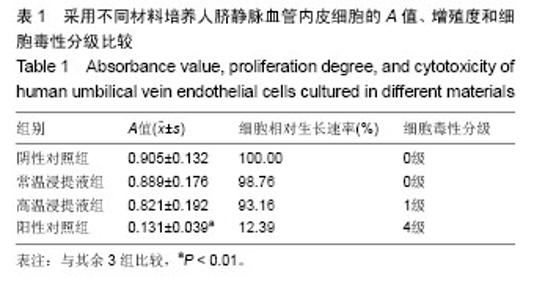
2.4 MTT法测定人脐静脉血管内皮细胞与纳米羟基磷灰石薄膜材料共培养的增殖情况 72 h后,与阴性对照组比较,常温与高温浸提液组对人脐静脉血管内皮细胞的增殖均无显著影响(P > 0.05);常温与高温浸提液组人脐静脉血管内皮细胞增殖优于阳性对照组(P < 0.01),见表1。"

| [1] 范瑞新,吴若彬,肖学钧,等.二尖瓣主动脉瓣三尖瓣同时置换治疗重症风湿性瓣膜病[J].中华胸心血管外科杂志, 2003,19(4): 208-210. [2] 赵铁夫,王盛宇,周其文,等.心脏瓣膜种类选择对老年患者生活质量影响的临床对照研究[J].中国胸心血管外科临床杂志, 2013,20(4):430-434. [3] Conrotto F, D'Ascenzo F,Salizzoni S,et al. A gender based analysis of predictors of all cause death after transcatheter aortic valve implantation.Am J Cardiol.2014;114(8): 1269-1274. [4] Chopard R,Perrotti A,Durst C,et al. Six-year outcomes after non-resective mitral valve repair with artificial chordae using removable clips.J Heart Valve Dis.2014;23(3): 364-369. [5] Noble S,Frangos E,Samaras N,et al.Transcatheter aortic valve implantation in nonagenarians: effective and safe. Eur J Intern Med.2013;24(8):750-755. [6] Baraki H,Al Ahmad A,Jeng-Singh S,et al.Pacemaker dependency after isolated aortic valve replacement: do conductance disorders recover over time? Interact Cardiovasc Thorac Surg.2013;16(4):476-481. [7] Sfeir PM,Abchee AB,Ghazzal Z,et al.Endovascular transcatheter aortic valve implantation: an evolving standard.J Cardiothorac Vasc Anesth.2013;27(4):765-778. [8] Said SM,Burkhart HM,Dearani JA.Surgical management of congenital (non-Ebstein) tricuspid valve regurgitation. Semin Thorac Cardiovasc Surg Pediatr Card Surg Annu. 2012;15(1): 46-60. [9] Coats L,Tsang V,Khambadkone S,et al.The potential impact of percutaneous pulmonary valve stent implantation on right ventricular outflow tract re-intervention.Eur J Cardiothorac Surg.2005;27(4):536-543. [10] Sun W,Martin C,Pham T.Computational modeling of cardiac valve function and intervention. Annu Rev Biomed Eng. 2014;16(1):53-76. [11] Zahn R,Gerckens U,Linke A,et al.Predictors of one-year mortality after transcatheter aortic valve implantation for severe symptomatic aortic stenosis.Am J Cardiol.2013; 112(2):272-9. [12] Helder MR,Schaff HV,Dearani JA,et al.Management of mitral regurgitation in Marfan syndrome: Outcomes of valve repair versus replacement and comparison with myxomatous mitral valve disease.J Thorac Cardiovasc Surg.2014;148(3): 1020-1024. [13] Claiborne TE,Sheriff J,Kuetting M,et al.In vitro evaluation of a novel hemodynamically optimized trileaflet polymeric prosthetic heart valve.J Biomech Eng.2013;135(2):21021. [14] Jackson MJ,Robinson GM,Ali N,et al.Surface engineering of artificial heart valve disks using nanostructured thin films deposited by chemical vapour deposition and sol-gel methods.J Med Eng Technol.2006;30(5):323-329. [15] Ma L,Sines G.Fatigue behavior of a pyrolytic carbon. J Biomed Mater Res.2000; 51(1):61-68. [16] Sneckenberger DS,Stinebring DR,Deutsch S,et al. Mitral heart valve cavitation in an artificial heart environment.J Heart Valve Dis.1996;5(2):216-227. [17] Fenglan X,Yubao L,Xiaoming Y,et al.Preparation and in vivo investigation of artificial cornea made of nano-hydroxyapatite/ poly(vinyl alcohol) hydrogel composite.J Mater Sci Mater Med. 2007;18(4):635-640. [18] Ito M,Kotani Y,Hojo Y,et al.Evaluation of hydroxyapatite ceramic vertebral spacers with different porosities and their binding capability to the vertebral body: an experimental study in sheep.J Neurosurg Spine.2007;6(5):431-437. [19] Siriphannon P,Monvisade P,Jinawath S,et al.Preparation and characterization of hydroxyapatite/poly (ethylene glutarate) biomaterials.J Biomed Mater Res A. 2007;81(2):381-91. [20] 王晓伟,叶福林,徐志云,等.人脐静脉血管内皮细胞构建组织工程心脏瓣膜及其生理功能[J].江苏医药,2007,33(2):156-158. [21] 王晓伟,徐志云,张宝仁,等.人脐静脉内皮细胞用于构建组织工程心脏瓣膜的实验研究[J].第二军医大学学报,2003,24(12): 1287-120. [22] Weymann A,Schmack B,Okada T,et al.Reendothelialization of human heart valve neoscaffolds using umbilical cord-derived endothelial cells.Circ J.2013;77(1): 207-216. [23] Sha JM,Yan ZY,Cheng GC,et al.In-vitro seeding of human umbilical cord vein endothelial cells on hydroxyapatite for mechanical heart valve applications.J Heart Valve Dis.2010; 19(4):506-512. [24] Martínez GJ,Seco M,Jaijee SK,et al.Introduction of an interdisciplinary heart team-based transcatheter aortic valve implantation programme: short and mid-term outcomes.Intern Med J.2014;44(9):876-883. [25] 郝和平.医疗器械生物学评价标准实施指南[M].北京:中国标准出版社,2002: 100-10. [26] Orme NM,Wright TC,Harmon GE,et al.Imaging Pandora's Box: incidental findings in elderly patients evaluated for transcatheter aortic valve replacement.Mayo Clin Proc.2014; 89(6):747-753. [27] Sadat-Shojai M,Khorasani MT,Jamshidi A.3-Dimensional cell-laden nano-hydroxyapatite/protein hydrogels for bone regeneration applications.Mater Sci Eng C Mater Biol Appl. 2015;49:835-843. [28] de Bruyn JR,Goiko M,Mozaffari M,et al.Dynamic light scattering study of inhibition of nucleation and growth of hydroxyapatite crystals by osteopontin. PLoS One.2013; 8(2):e56764. [29] Moroi A,Ueki K,Okabe K,et al.Comparison between unsintered hydroxyapatite/poly-L-lactic acid mesh and titanium mesh in bone regeneration of rabbit mandible. Implant Dent.2013;22(3):255-262. [30] Smolen D,Chudoba T,Malka I,et al.Highly biocompatible, nanocrystalline hydroxyapatite synthesized in a solvothermal process driven by high energy density microwave radiation.Int J Nanomedicine.2013;8:653-668. |
| [1] | Pu Rui, Chen Ziyang, Yuan Lingyan. Characteristics and effects of exosomes from different cell sources in cardioprotection [J]. Chinese Journal of Tissue Engineering Research, 2021, 25(在线): 1-. |
| [2] | Jiang Xin, Qiao Liangwei, Sun Dong, Li Ming, Fang Jun, Qu Qingshan. Expression of long chain non-coding RNA PGM5-AS1 in serum of renal transplant patients and its regulation of human glomerular endothelial cells [J]. Chinese Journal of Tissue Engineering Research, 2021, 25(5): 741-745. |
| [3] | Jiang Tao, Ma Lei, Li Zhiqiang, Shou Xi, Duan Mingjun, Wu Shuo, Ma Chuang, Wei Qin. Platelet-derived growth factor BB induces bone marrow mesenchymal stem cells to differentiate into vascular endothelial cells [J]. Chinese Journal of Tissue Engineering Research, 2021, 25(25): 3937-3942. |
| [4] | Chen Siqi, Xian Debin, Xu Rongsheng, Qin Zhongjie, Zhang Lei, Xia Delin. Effects of bone marrow mesenchymal stem cells and human umbilical vein endothelial cells combined with hydroxyapatite-tricalcium phosphate scaffolds on early angiogenesis in skull defect repair in rats [J]. Chinese Journal of Tissue Engineering Research, 2021, 25(22): 3458-3465. |
| [5] | Mo Jianling, He Shaoru, Feng Bowen, Jian Minqiao, Zhang Xiaohui, Liu Caisheng, Liang Yijing, Liu Yumei, Chen Liang, Zhou Haiyu, Liu Yanhui. Forming prevascularized cell sheets and the expression of angiogenesis-related factors [J]. Chinese Journal of Tissue Engineering Research, 2021, 25(22): 3479-3486. |
| [6] | Cao Yang, Zhang Junping, Peng Li, Ding Yi, Li Guanghui. Isolation and culture of rabbit aortic endothelial cells and biological characteristics [J]. Chinese Journal of Tissue Engineering Research, 2021, 25(19): 3000-3003. |
| [7] | Wang Huili, Chen Zhijiang, Wu Bingyi. Glia maturation factor-gamma inhibits the proliferation of human colorectal cancer LoVo cells and affects the cytoskeletal motion of human umbilical vein endothelial cells [J]. Chinese Journal of Tissue Engineering Research, 2021, 25(13): 2055-2059. |
| [8] | Liu Tao, Zhang Nini, Huang Guilin . Relationship between extracellular vesicles and radiation-induced tissue injury [J]. Chinese Journal of Tissue Engineering Research, 2021, 25(13): 2121-2126. |
| [9] | Fan Haixia, Tan Qingkun, Wang Hong, Cheng Huanzhi, Liu Xue, Ching-chang Ko, Geng Haixia. Rabbit skull defects repaired by the hydroxyapatite/geltin scaffold combined with bone marrow mesenchymal stem cells and umbilical vein endothelial cells [J]. Chinese Journal of Tissue Engineering Research, 2021, 25(10): 1495-1499. |
| [10] | Gu Jingjing, Zhou Rui, Yang Tingting, Yang Xiaoping, Xu Fei, Zheng Bo. Supporting effect of human skeletal muscle-derived myoendothelial cells on hematopoietic stem/progenitor cells in vitro [J]. Chinese Journal of Tissue Engineering Research, 2021, 25(1): 50-55. |
| [11] | Zhang Jian, Chen Miao, Li Weixin, Ye Yichao, Xu Huiyou, Ma Ke, Chen Xuyi, Sun Hongtao, Zhang Sai. Collagen/heparin sulfate scaffolds loaded with brain-derived neurotrophic factor promote neurological and locomotor function recovery in rats after traumatic brain injury [J]. Chinese Journal of Tissue Engineering Research, 2020, 24(34): 5538-5544. |
| [12] | Chen Jia, Yang Yiqiang, Hu Chen, Chen Qi, Zhao Tian, Yong Min, Ma Dongyang, Ren Liling. Fabrication of prevascularized osteogenic differentiated cell sheet based on human bone marrow mesenchymal stem cells and human umbilical vein endothelial cells [J]. Chinese Journal of Tissue Engineering Research, 2020, 24(31): 4934-4940. |
| [13] | Cao Baichuan, Zeng Gaofeng, Gao Yunbing, Deng Guiying, Cen Zhongxi, Zhang Chuanyang, Guo Yande, Zong Shaohui. Heat-shock endothelial cells induce differentiation of bone marrow mesenchymal stem cells into vascular endothelial cells [J]. Chinese Journal of Tissue Engineering Research, 2020, 24(19): 3023-3028. |
| [14] | Cao Yang, Hu Ping, Tian Min, Wei Fang, Gu Qing, Lü Hongbin. Effects of 6-phosphofructokinase-2/fructose-2,6-bisphosphatase 3 on tubule formation of human umbilical vein endothelial cells [J]. Chinese Journal of Tissue Engineering Research, 2020, 24(14): 2217-2222. |
| [15] | Zhao Jianfeng, Geng Yu, Chen Qianbo, Yang Jinghui, Li Yan. Changes in insulin resistance and inflammatory factors in cataract patients with glaucoma after phacoemulsification and trabeculectomy: a self-controlled trial [J]. Chinese Journal of Tissue Engineering Research, 2020, 24(11): 1750-1755. |
| Viewed | ||||||
|
Full text |
|
|||||
|
Abstract |
|
|||||