Chinese Journal of Tissue Engineering Research ›› 2014, Vol. 18 ›› Issue (50): 8080-8087.doi: 10.3969/j.issn.2095-4344.2014.50.008
Previous Articles Next Articles
Damage of chemotherapy agents to bone marrow mesenchymal stem cells
Wang Jin-huan, Wang Zhen-ling, Wang Xiao-fang, Zhao Zhi-gang
- Department of Hemooncology, Tianjin University Cancer Institute & Hospital, Tianjin 300060, China
-
Received:2014-11-24Online:2014-12-03Published:2014-12-03 -
Contact:Zhao Zhi-gang, Associate chief physician, Department of Hemooncology, Tianjin University Cancer Institute & Hospital, Tianjin 300060, China -
About author:Wang Jin-huan, Studying for master’s degree, Department of Hemooncology, Tianjin University Cancer Institute & Hospital, Tianjin 300060, China -
Supported by:the National Natural Science Foundation of China, No. 81172833
CLC Number:
Cite this article
Wang Jin-huan, Wang Zhen-ling, Wang Xiao-fang, Zhao Zhi-gang. Damage of chemotherapy agents to bone marrow mesenchymal stem cells[J]. Chinese Journal of Tissue Engineering Research, 2014, 18(50): 8080-8087.
share this article
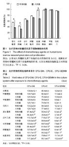
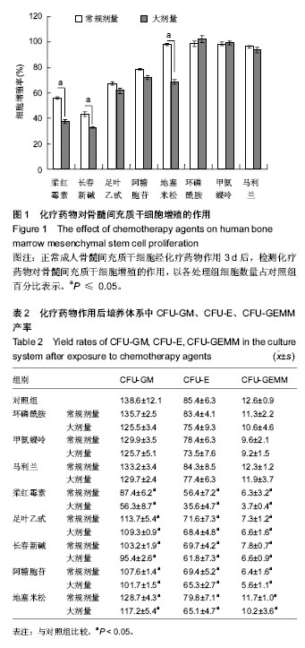
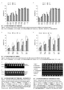
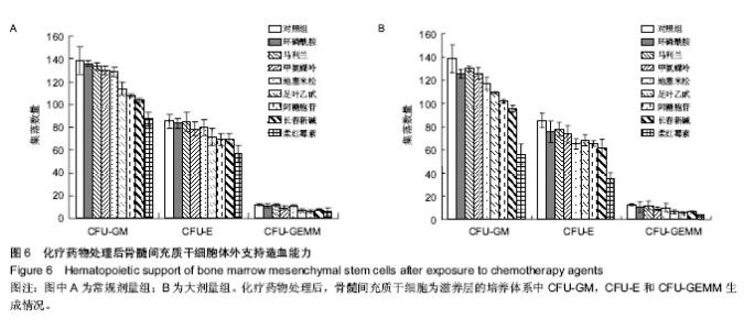
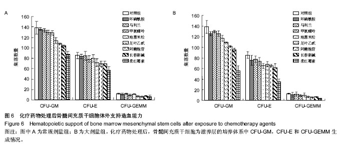
2.1 化疗药物对骨髓间充质干细胞增殖的影响 为了检测化疗药物对骨髓间充质干细胞的影响,不同剂量(常规剂量组,大剂量组)、不同类型化疗药物(环磷酰胺、阿糖胞苷、甲氨蝶呤、足叶乙甙、地塞米松、长春新碱、柔红霉素、马利兰)加入骨髓间充质干细胞中,处理3 d后检测骨髓间充质干细胞的增殖情况。发现任何浓度的环磷酰胺、甲氨蝶呤、马利兰和常规剂量的地塞米松不影响骨髓间充质干细胞的增殖;而常规剂量和大剂量长春新碱、足叶乙甙、柔红霉素、阿糖胞苷和大剂量地塞米松均不同程度抑制骨髓间充质干细胞的增殖,见图1。 2.2 化疗药物作用后骨髓间充质干细胞的恢复 如图2所示,柔红霉素、足叶乙甙和阿糖胞苷持续性抑制骨髓间充质干细胞的增殖。当柔红霉素、足叶乙甙和阿糖胞苷撤除后,继续培养9 d,骨髓间充质干细胞仍处于增殖受抑状态。而长春新碱、地塞米松对骨髓间充质干细胞的增殖抑制作用随着药物的撤除有所恢复,但是不能恢复到正常。环磷酰胺、甲氨蝶呤和马利兰作用后骨髓间充质干细胞的早期(3 d)和晚期(12 d)的增殖能力并不发生改变。 2.3 化疗药物诱导骨髓间充质干细胞凋亡 5种检测的化疗药物(阿糖胞苷、柔红霉素、长春新碱、足叶乙甙、地塞米松)均可以诱导骨髓间充质干细胞凋亡。不同类型、不同剂量的化疗药物诱导骨髓间充质干细胞凋亡的程度存在差异。大剂量组化疗药物对骨髓间充质干细胞的凋亡作用要明显强于常规剂量组(P < 0.05),不同类型化疗药物诱导骨髓间充质干细胞凋亡的作用存在差异,其中大剂量组柔红霉素诱导细胞凋亡的能力最强,而地塞米松诱导凋亡的作用最弱;此外,不同类型、不同剂量化疗药物对骨髓间充质干细胞的凋亡率与处理时间成正相关。随着处理时间的延长,细胞凋亡率上升,见图3。 2.4 化疗药物处理后骨髓间充质干细胞的分化能力 化疗药物处理后和未处理骨髓间充质干细胞都能在体外向脂肪和成骨诱导分化。成脂肪诱导1周后看见大部分细胞胞体变大,胞浆出现大量脂肪滴,油红O染色证实为脂肪滴,RT-PCR证实诱导后细胞表达脂肪形成特异性基因脂蛋白脂酶。成骨诱导3周后细胞呈现多层生长的结节状,Von kossa 染色证实为钙盐沉积,RT-PCR证实上述被诱导的细胞表达骨形成特异性基因Osteocalcin(图4)。化疗药物处理前后的骨髓间充质干细胞具有相似的体外分化能力。 2.5 造血相关因子的表达 应用RT-PCR方法检测化疗药物处理前后骨髓间充质干细胞中造血相关因子的表达,结果显示干细胞因子(SCF),单核细胞集落刺激因子(M-CSF),白细胞介素6(IL-6)和粒细胞集落刺激因子(G-CSF)在化疗药物处理前后的骨髓间充质干细胞中稳定表达,但是没有检测到粒、单核细胞集落刺激因子,白细胞介素3(IL-3)和血小板生长因子(TPO)的表达(图5)。2.6 体外支持造血能力 为了明确化疗药物是否影响骨髓间充质干细胞支持造血的能力,将不同类型、不同剂量处理后的骨髓间充质干细胞照射后,将骨髓单个核细胞接种其中,在长期骨髓培养基中培养4周后检测CFU生成情况。各种化疗药物均不同程度影响骨髓间充质干细胞支持造血的能力,见图6。"
"

"
| [1] Arthur A, Zannettino A, Panagopoulos R,et al.EphB/ephrin-B interactions mediate human MSC attachment, migration and osteochondral differentiation.Bone. 2011;48(3):533-542. [2] Bian L, Zhai DY, Tous E,et al.Enhanced MSC chondrogenesis following delivery of TGF-β3 from alginate microspheres within hyaluronic acid hydrogels in vitro and in vivo. Biomaterials. 2011;32(27):6425-6434. [3] Du Z, Wang L, Zhao Y,et al.Sympathetic denervation-induced MSC mobilization in distraction osteogenesis associates with inhibition of MSC migration and osteogenesis by norepinephrine/adrb3.PLoS One. 2014;9(8):e105976. [4] Jamnig A, Lepperdinger G.From tendon to nerve: an MSC for all seasons.Can J Physiol Pharmacol. 2012;90(3):295-306. [5] Lv F, Lu M, Cheung KM,et al.Intrinsic properties of mesemchymal stem cells from human bone marrow, umbilical cord and umbilical cord blood comparing the different sources of MSC.Curr Stem Cell Res Ther. 2012;7(6):389-399. [6] Bessout R, Sémont A, Demarquay C,et al.Mesenchymal stem cell therapy induces glucocorticoid synthesis in colonic mucosa and suppresses radiation-activated T cells: new insights into MSC immunomodulation.Mucosal Immunol. 2014; 7(3):656-669. [7] Prata Kde L, Orellana MD, De Santis GC,et al.Effects of high-dose chemotherapy on bone marrow multipotent mesenchymal stromal cells isolated from lymphoma patients. Exp Hematol. 2010;38(4):292-300. [8] Stute N, Fehse B, Schröder J,et al.Human mesenchymal stem cells are not of donor origin in patients with severe aplastic anemia who underwent sex-mismatched allogeneic bone marrow transplant.J Hematother Stem Cell Res. 2002; 11(6):977-984. [9] Cilloni D, Carlo-Stella C, Falzetti F,et al.Limited engraftment capacity of bone marrow-derived mesenchymal cells following T-cell-depleted hematopoietic stem cell transplantation.Blood. 2000;96(10):3637-3643. [10] Wu KH, Wu HP, Chan CK,et al.The role of mesenchymal stem cells in hematopoietic stem cell transplantation: from bench to bedsides.Cell Transplant. 2013;22(4):723-729. [11] Epstein RB, Sarpel SC.Autologous bone marrow infusion following high dose chemotherapy of the canine transmissible venereal tumor (TVT).Exp Hematol. 1980;8(6):683-689. [12] Majumdar MK, Thiede MA, Haynesworth SE,et al.Human marrow-derived mesenchymal stem cells (MSCs) express hematopoietic cytokines and support long-term hematopoiesis when differentiated toward stromal and osteogenic lineages.J Hematother Stem Cell Res. 2000; 9(6):841-848. [13] Carrancio S, Romo C, Ramos T,et al.Effects of MSC coadministration and route of delivery on cord blood hematopoietic stem cell engraftment.Cell Transplant. 2013; 22(7):1171-1183. [14] Zhang C, Zhang X, Chen XH.Granulocyte-colony stimulating factor-mobilized mesenchymal stem cells: A new resource for rapid engraftment in hematopoietic stem cell transplantation. Med Hypotheses. 2011;76(2):241-243. [15] Yeh SP, Lo WJ, Lin CL,et al.Anti-leukemic therapies induce cytogenetic changes of human bone marrow-derived mesenchymal stem cells.Ann Hematol. 2012;91(2):163-172. [16] Li J, Law HK, Liu YL,et al.Effect of cisplatin, topotecan, daunorubicin and hydroxyurea on human mesenchymal stem cells.Zhongguo Shi Yan Xue Ye Xue Za Zhi. 2010;18(4): 991-996. [17] Domenech J, Cartron G, Clement N,et al.Persistent decrease in proliferative potential of marrow CD34(+)cells exposed to early-acting growth factors after autologous bone marrow transplantation.Bone Marrow Transplant. 2002;29(7):557-562. [18] Koç ON, Day J, Nieder M,et al.Allogeneic mesenchymal stem cell infusion for treatment of metachromatic leukodystrophy (MLD) and Hurler syndrome (MPS-IH).Bone Marrow Transplant. 2002;30(4):215-222. [19] Li J, Law HK, Lau YL,et al.Differential damage and recovery of human mesenchymal stem cells after exposure to chemotherapeutic agents.Br J Haematol. 2004;127(3): 326-334. [20] Ben-Ishay Z, Barak V.Bone marrow stromal dysfunction in mice administered cytosine arabinoside.Eur J Haematol. 2001;66(4):230-237. [21] Kushwaha P, Khedgikar V, Gautam J,et al.A novel therapeutic approach with Caviunin-based isoflavonoid that en routes bone marrow cells to bone formation via BMP2/Wnt-β-catenin signaling.Cell Death Differ. 2014;5:e1422. [22] Cartron G, Herault O, Benboubker L,et al.Quantitative and qualitative analysis of the human primitive progenitor cell compartment after autologous stem cell transplantation.J Hematother Stem Cell Res. 2002;11(2):359-368. [23] Banfi A, Bianchi G, Galotto M,et al.Bone marrow stromal damage after chemo/radiotherapy: occurrence, consequences and possibilities of treatment.Leuk Lymphoma. 2001;42(5):863-870. |
| [1] | Pu Rui, Chen Ziyang, Yuan Lingyan. Characteristics and effects of exosomes from different cell sources in cardioprotection [J]. Chinese Journal of Tissue Engineering Research, 2021, 25(在线): 1-. |
| [2] | Lin Qingfan, Xie Yixin, Chen Wanqing, Ye Zhenzhong, Chen Youfang. Human placenta-derived mesenchymal stem cell conditioned medium can upregulate BeWo cell viability and zonula occludens expression under hypoxia [J]. Chinese Journal of Tissue Engineering Research, 2021, 25(在线): 4970-4975. |
| [3] | Hou Jingying, Yu Menglei, Guo Tianzhu, Long Huibao, Wu Hao. Hypoxia preconditioning promotes bone marrow mesenchymal stem cells survival and vascularization through the activation of HIF-1α/MALAT1/VEGFA pathway [J]. Chinese Journal of Tissue Engineering Research, 2021, 25(7): 985-990. |
| [4] | Shi Yangyang, Qin Yingfei, Wu Fuling, He Xiao, Zhang Xuejing. Pretreatment of placental mesenchymal stem cells to prevent bronchiolitis in mice [J]. Chinese Journal of Tissue Engineering Research, 2021, 25(7): 991-995. |
| [5] | Liang Xueqi, Guo Lijiao, Chen Hejie, Wu Jie, Sun Yaqi, Xing Zhikun, Zou Hailiang, Chen Xueling, Wu Xiangwei. Alveolar echinococcosis protoscolices inhibits the differentiation of bone marrow mesenchymal stem cells into fibroblasts [J]. Chinese Journal of Tissue Engineering Research, 2021, 25(7): 996-1001. |
| [6] | Fan Quanbao, Luo Huina, Wang Bingyun, Chen Shengfeng, Cui Lianxu, Jiang Wenkang, Zhao Mingming, Wang Jingjing, Luo Dongzhang, Chen Zhisheng, Bai Yinshan, Liu Canying, Zhang Hui. Biological characteristics of canine adipose-derived mesenchymal stem cells cultured in hypoxia [J]. Chinese Journal of Tissue Engineering Research, 2021, 25(7): 1002-1007. |
| [7] | Geng Yao, Yin Zhiliang, Li Xingping, Xiao Dongqin, Hou Weiguang. Role of hsa-miRNA-223-3p in regulating osteogenic differentiation of human bone marrow mesenchymal stem cells [J]. Chinese Journal of Tissue Engineering Research, 2021, 25(7): 1008-1013. |
| [8] | Lun Zhigang, Jin Jing, Wang Tianyan, Li Aimin. Effect of peroxiredoxin 6 on proliferation and differentiation of bone marrow mesenchymal stem cells into neural lineage in vitro [J]. Chinese Journal of Tissue Engineering Research, 2021, 25(7): 1014-1018. |
| [9] | Zhu Xuefen, Huang Cheng, Ding Jian, Dai Yongping, Liu Yuanbing, Le Lixiang, Wang Liangliang, Yang Jiandong. Mechanism of bone marrow mesenchymal stem cells differentiation into functional neurons induced by glial cell line derived neurotrophic factor [J]. Chinese Journal of Tissue Engineering Research, 2021, 25(7): 1019-1025. |
| [10] | Duan Liyun, Cao Xiaocang. Human placenta mesenchymal stem cells-derived extracellular vesicles regulate collagen deposition in intestinal mucosa of mice with colitis [J]. Chinese Journal of Tissue Engineering Research, 2021, 25(7): 1026-1031. |
| [11] | Pei Lili, Sun Guicai, Wang Di. Salvianolic acid B inhibits oxidative damage of bone marrow mesenchymal stem cells and promotes differentiation into cardiomyocytes [J]. Chinese Journal of Tissue Engineering Research, 2021, 25(7): 1032-1036. |
| [12] | Wang Xianyao, Guan Yalin, Liu Zhongshan. Strategies for improving the therapeutic efficacy of mesenchymal stem cells in the treatment of nonhealing wounds [J]. Chinese Journal of Tissue Engineering Research, 2021, 25(7): 1081-1087. |
| [13] | Wang Shiqi, Zhang Jinsheng. Effects of Chinese medicine on proliferation, differentiation and aging of bone marrow mesenchymal stem cells regulating ischemia-hypoxia microenvironment [J]. Chinese Journal of Tissue Engineering Research, 2021, 25(7): 1129-1134. |
| [14] | Kong Desheng, He Jingjing, Feng Baofeng, Guo Ruiyun, Asiamah Ernest Amponsah, Lü Fei, Zhang Shuhan, Zhang Xiaolin, Ma Jun, Cui Huixian. Efficacy of mesenchymal stem cells in the spinal cord injury of large animal models: a meta-analysis [J]. Chinese Journal of Tissue Engineering Research, 2021, 25(7): 1142-1148. |
| [15] | Chen Junyi, Wang Ning, Peng Chengfei, Zhu Lunjing, Duan Jiangtao, Wang Ye, Bei Chaoyong. Decalcified bone matrix and lentivirus-mediated silencing of P75 neurotrophin receptor transfected bone marrow mesenchymal stem cells to construct tissue-engineered bone [J]. Chinese Journal of Tissue Engineering Research, 2021, 25(4): 510-515. |
| Viewed | ||||||
|
Full text |
|
|||||
|
Abstract |
|
|||||