Chinese Journal of Tissue Engineering Research ›› 2014, Vol. 18 ›› Issue (25): 3998-4003.doi: 10.3969/j.issn.2095-4344.2014.25.011
Previous Articles Next Articles
Fibrin gel enhances osteogenic differentiation of rat mesenchymal stem cells
Mai Xia, Li Wei, Wang Ying-hui, Zha La Ga Hu, Chen Xiao-yi
- Research Room of Cell Biology and Medical Genetics, Department of Clinical Medicine, Logistics College of Chinese People’s Armed Police Forces, Tianjin 300039, China
-
Received:2014-05-28Online:2014-06-18Published:2014-06-18 -
Contact:Chen Xiao-yi, Master, Professor, Master’s supervisor, Research Room of Cell Biology and Medical Genetics, Department of Clinical Medicine, Logistics College of Chinese People’s Armed Police Forces, Tianjin 300039, China -
About author:Mai Xia, Studying for master’s degree, Experimentalist, Research Room of Cell Biology and Medical Genetics, Department of Clinical Medicine, Logistics College of Chinese People’s Armed Police Forces, Tianjin 300039, China -
Supported by:the Key Program of Chinese People’s Armed Police Forces, No. WKH2009Z04; the Program of Logistics College of Chinese People’s Armed Police Forces, No. WHM201209
CLC Number:
Cite this article
Mai Xia, Li Wei, Wang Ying-hui, Zha La Ga Hu, Chen Xiao-yi. Fibrin gel enhances osteogenic differentiation of rat mesenchymal stem cells[J]. Chinese Journal of Tissue Engineering Research, 2014, 18(25): 3998-4003.
share this article
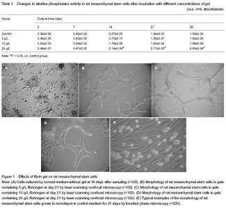
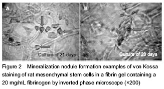
Fibrin gel effects on morphology of rat bone marrow mesenchymal stem cells Microscopy image of a typical cell colony was seen at 7 days by inverted phase microscope. Laser scanning confocal microscopy results demonstrated that the majority of passage 3 cells stained by calcein dye in the gels were viable in all gel formulations and at all incubation time. Different cell morphologies were observed depending on the formulation of the fibrin gel. In gels containing 5 g/L of fibrinogen concentration cells started to elongate as early as day 3 and formed an interconnected network within the gels up to day 21. On the other hand, in gels containing 20 g/L of fibrinogen concentration cells tended to remain mostly round or ‘‘star-shaped’’ up to day 21 and formed some agglomeration, but not an interconnected network. Cells in no gels showed a spindle-shape or cuboidal morphology up to day 21, but no interconnected network was found during prolonged incubation time (Figure 1). Fibrin gel affected the proliferation of rat mesenchymal stem cells Alkaline phosphatase activity was relatively stable for all the formulations at 3 and 7 days. The alkaline phosphatase activity in cell-seeded fibrin gels containing a high (10, 20 g/L) fibrinogen concentration became significant at 14 days, and continued to increase up to 28 days, but that remained still lower in no gels and containing a low (5 g/L) fibrinogen concentration cultures of rat mesenchymal stem cells. In contrast to the proliferation results, alkaline phosphatase activity was higher in gels containing a high fibrinogen concentration (Table 1). Von Kossa staining Von Kossa staining in a formulation containing a high (20 g/L) fibrinogen concentration showed the presence of mineralization nodules after 21 and 28 days of incubation. The nodules appeared in areas with cells, and mostly in the areas with holes in the fibrin gels. No nodules were observed on top of the gel, which was covered by a monolayer of cells (Figure 2)."

"
| [1] Caplan AI. Mesenchymal stem cells. J Orthop Res. 1991; 9(5):641-650. [2] Pittenger MF, Mackay AM, Beck SC, et al. Multilineage potential of adult human mesenchymal stem cells. Science. 1999;284(5411):143-147. [3] Bianco P, Robey PG, Simmons PJ. Mesenchymal stem cells: revisiting history, concepts, and assays. Cell Stem Cell. 2008;2(4):313-319. [4] Jaiswal RK, Jaiswal N, Bruder SP, et al. Adult human mesenchymal stem cell differentiation to the osteogenic or adipogenic lineage is regulated by mitogen-activated protein kinase. J Biol Chem. 2000;275(13):9645-9652. [5] Wang X, Wang Y, Gou W, et al. Role of mesenchymal stem cells in bone regeneration and fracture repair: a review. Int Orthop. 2013;37(12):2491-2498. [6] Cao L, Liu G, Gan Y, et al. The use of autologous enriched bone marrow MSCs to enhance osteoporotic bone defect repair in long-term estrogen deficient goats. Biomaterials. 2012;33(20):5076-5084. [7] Guan M, Yao W, Liu R, et al. Directing mesenchymal stem cells to bone to augment bone formation and increase bone mass. Nat Med. 2012;18(3):456-462. [8] Pino AM, Rosen CJ, Rodríguez JP. In osteoporosis, differentiation of mesenchymal stem cells (MSCs) improves bone marrow adipogenesis. Biol Res. 2012; 45(3):279-287. [9] Dohan Ehrenfest DM, Doglioli P, de Peppo GM, et al. Choukroun's platelet-rich fibrin (PRF) stimulates in vitro proliferation and differentiation of human oral bone mesenchymal stem cell in a dose-dependent way. Arch Oral Biol. 2010;55(3):185-194. [10] Wong C, Inman E, Spaethe R, et al. Fibrin-based biomaterials to deliver human growth factors. Thromb Haemost. 2003;89(3):573-582. [11] Gorodetsky R, Clark RA, An J, et al. Fibrin microbeads (FMB) as biodegradable carriers for culturing cells and for accelerating wound healing. J Invest Dermatol. 1999; 112(6): 866-872. [12] Horch RE, Bannasch H, Kopp J, et al. Single-cell suspensions of cultured human keratinocytes in fibrin-glue reconstitute the epidermis. Cell Transplant. 1998;7(3): 309-317. [13] Catelas I, Dwyer JF, Helgerson S. Controlled release of bioactive transforming growth factor beta-1 from fibrin gels in vitro. Tissue Eng Part C Methods. 2008;14(2):119-128. [14] Mosesson MW, Siebenlist KR, Meh DA. The structure and biological features of fibrinogen and fibrin. Ann N Y Acad Sci. 2001;936:11-30. [15] Marktl W, Rudas B. The effect of factor XIII on wound granulation in the rat. Thromb Diath Haemorrh. 1974;32 (2-3):578-581. [16] Lin B, Cai ZG, Yu GY, et al. Osteoblastoma of the maxilla and mandible: a report of 2 cases and literature review. Chin J Dent Res. 2012;15(2):153-158. [17] Nöth U, Steinert AF, Tuan RS. Technology insight: adult mesenchymal stem cells for osteoarthritis therapy. Nat Clin Pract Rheumatol. 2008;4(7):371-380. [18] Yu Y, Shao B, Shuai Y, et al. Role of bone marrow-derived mesenchymal stem cells in treating colitis through Fas/FasL-mediated immune regulation. Xi Bao Yu Fen Zi Mian Yi Xue Za Zhi. 2013;29(10):1028-1031. [19] Ishii M, Shibata R, Numaguchi Y, et al. Enhanced angiogenesis by transplantation of mesenchymal stem cell sheet created by a novel magnetic tissue engineering method. Arterioscler Thromb Vasc Biol. 2011;31(10): 2210-2215. [20] Li X, van Blitterswijk CA, Feng Q, et al. The effect of calcium phosphate microstructure on bone-related cells in vitro. Biomaterials. 2008;29(23):3306-3316. [21] Sun N, Yang L, Li Y, et al. Effect of advanced oxidation protein products on the proliferation and osteogenic differentiation of rat mesenchymal stem cells. Int J Mol Med. 2013;32(2):485-491. [22] Tang X, Sheng L, Xie F, et al. Differentiation of bone marrow-derived mesenchymal stem cells into chondrocytes using chondrocyte extract. Mol Med Rep. 2012;6(4): 745-749. [23] Scotti C, Pozzi A, Mangiavini L, et al. Healing of meniscal tissue by cellular fibrin glue: an in vivo study. Knee Surg Sports Traumatol Arthrosc. 2009;17(6):645-651. [24] Chien CS, Ho HO, Liang YC, et al. Incorporation of exudates of human platelet-rich fibrin gel in biodegradable fibrin scaffolds for tissue engineering of cartilage. J Biomed Mater Res B Appl Biomater. 2012;100(4):948-955. [25] Sha'ban M, Yoon SJ, Ko YK, et al. Fibrin promotes proliferation and matrix production of intervertebral disc cells cultured in three-dimensional poly(lactic-co-glycolic acid) scaffold. J Biomater Sci Polym Ed. 2008;19(9):1219- 1237. [26] Even-Ram S. Fibrin gel model for assessment of cellular contractility. Methods Mol Biol. 2009;522:251-259. [27] de Jonge N, Kanters FM, Baaijens FP, et al. Strain-induced collagen organization at the micro-level in fibrin-based engineered tissue constructs. Ann Biomed Eng. 2013;41(4): 763-774. [28] Gerard C, Forest MA, Beauregard G, et al. Fibrin gel improves the survival of transplanted myoblasts. Cell Transplant. 2012;21(1):127-137. [29] Lee F, Kurisawa M. Formation and stability of interpenetrating polymer network hydrogels consisting of fibrin and hyaluronic acid for tissue engineering. Acta Biomater. 2013;9(2):5143-5152. [30] Allan P, Uitte de Willige S, Abou-Saleh RH, et al. Evidence that fibrinogen γ' directly interferes with protofibril growth: implications for fibrin structure and clot stiffness. J Thromb Haemost. 2012;10(6):1072-1080. [31] Beck GR Jr. Inorganic phosphate as a signaling molecule in osteoblast differentiation. J Cell Biochem. 2003;90(2):234- 243. [32] Simão AM, Beloti MM, Cezarino RM, et al. Membrane-bound alkaline phosphatase from ectopic mineralization and rat bone marrow cell culture. Comp Biochem Physiol A Mol Integr Physiol. 2007;146(4): 679-687. [33] Kim A, Benning MM, OkLee S, et al. Divergence of chemical function in the alkaline phosphatase superfamily: structure and mechanism of the P-C bond cleaving enzyme phosphonoacetate hydrolase. Biochemistry. 2011;50(17): 3481-3494. [34] Tobe BT, Hou J, Crain AM, et al. Phosphoproteomic analysis: an emerging role in deciphering cellular signaling in human embryonic stem cells and their differentiated derivatives. Stem Cell Rev. 2012;8(1):16-31. [35] Brill LM, Xiong W, Lee KB, et al. Phosphoproteomic analysis of human embryonic stem cells. Cell Stem Cell. 2009;5(2):204-213. [36] Tian XF, Heng BC, Ge Z, et al. Comparison of osteogenesis of human embryonic stem cells within 2D and 3D culture systems. Scand J Clin Lab Invest. 2008;68(1):58-67. [37] Bensaïd W, Triffitt JT, Blanchat C, et al. A biodegradable fibrin scaffold for mesenchymal stem cell transplantation. Biomaterials. 2003;24(14):2497-2502. [38] Ho W, Tawil B, Dunn JC, et al. The behavior of human mesenchymal stem cells in 3D fibrin clots: dependence on fibrinogen concentration and clot structure. Tissue Eng. 2006;12(6):1587-1595. [39] Catelas I, Sese N, Wu BM, et al. Human mesenchymal stem cell proliferation and osteogenic differentiation in fibrin gels in vitro. Tissue Eng. 2006;12(8):2385-2396. [40] Rose LC, Fitzsimmons R, Lee P, et al. Effect of basic fibroblast growth factor in mouse embryonic stem cell culture and osteogenic differentiation. J Tissue Eng Regen Med. 2013;7(5):371-382. [41] Luan J, Cui Y, Zhang Y, et al. Effect of CXCR4 inhibitor AMD3100 on alkaline phosphatase activity and mineralization in osteoblastic MC3T3-E1 cells. Biosci Trends. 2012;6(2):63-69. [42] Mauney JR, Blumberg J, Pirun M, et al. Osteogenic differentiation of human bone marrow stromal cells on partially demineralized bone scaffolds in vitro. Tissue Eng. 2004;10(1-2):81-92. [43] Kitamura S, Ohgushi H, Hirose M, et al. Osteogenic differentiation of human bone marrow-derived mesenchymal cells cultured on alumina ceramics. Artif Organs. 2004;28(1):72-82. [44] Lee SJ, Atala A. Scaffold technologies for controlling cell behavior in tissue engineering. Biomed Mater. 2013;8(1): 010201 |
| [1] | Lin Qingfan, Xie Yixin, Chen Wanqing, Ye Zhenzhong, Chen Youfang. Human placenta-derived mesenchymal stem cell conditioned medium can upregulate BeWo cell viability and zonula occludens expression under hypoxia [J]. Chinese Journal of Tissue Engineering Research, 2021, 25(在线): 4970-4975. |
| [2] | Pu Rui, Chen Ziyang, Yuan Lingyan. Characteristics and effects of exosomes from different cell sources in cardioprotection [J]. Chinese Journal of Tissue Engineering Research, 2021, 25(在线): 1-. |
| [3] | Hou Jingying, Yu Menglei, Guo Tianzhu, Long Huibao, Wu Hao. Hypoxia preconditioning promotes bone marrow mesenchymal stem cells survival and vascularization through the activation of HIF-1α/MALAT1/VEGFA pathway [J]. Chinese Journal of Tissue Engineering Research, 2021, 25(7): 985-990. |
| [4] | Shi Yangyang, Qin Yingfei, Wu Fuling, He Xiao, Zhang Xuejing. Pretreatment of placental mesenchymal stem cells to prevent bronchiolitis in mice [J]. Chinese Journal of Tissue Engineering Research, 2021, 25(7): 991-995. |
| [5] | Liang Xueqi, Guo Lijiao, Chen Hejie, Wu Jie, Sun Yaqi, Xing Zhikun, Zou Hailiang, Chen Xueling, Wu Xiangwei. Alveolar echinococcosis protoscolices inhibits the differentiation of bone marrow mesenchymal stem cells into fibroblasts [J]. Chinese Journal of Tissue Engineering Research, 2021, 25(7): 996-1001. |
| [6] | Fan Quanbao, Luo Huina, Wang Bingyun, Chen Shengfeng, Cui Lianxu, Jiang Wenkang, Zhao Mingming, Wang Jingjing, Luo Dongzhang, Chen Zhisheng, Bai Yinshan, Liu Canying, Zhang Hui. Biological characteristics of canine adipose-derived mesenchymal stem cells cultured in hypoxia [J]. Chinese Journal of Tissue Engineering Research, 2021, 25(7): 1002-1007. |
| [7] | Geng Yao, Yin Zhiliang, Li Xingping, Xiao Dongqin, Hou Weiguang. Role of hsa-miRNA-223-3p in regulating osteogenic differentiation of human bone marrow mesenchymal stem cells [J]. Chinese Journal of Tissue Engineering Research, 2021, 25(7): 1008-1013. |
| [8] | Lun Zhigang, Jin Jing, Wang Tianyan, Li Aimin. Effect of peroxiredoxin 6 on proliferation and differentiation of bone marrow mesenchymal stem cells into neural lineage in vitro [J]. Chinese Journal of Tissue Engineering Research, 2021, 25(7): 1014-1018. |
| [9] | Zhu Xuefen, Huang Cheng, Ding Jian, Dai Yongping, Liu Yuanbing, Le Lixiang, Wang Liangliang, Yang Jiandong. Mechanism of bone marrow mesenchymal stem cells differentiation into functional neurons induced by glial cell line derived neurotrophic factor [J]. Chinese Journal of Tissue Engineering Research, 2021, 25(7): 1019-1025. |
| [10] | Duan Liyun, Cao Xiaocang. Human placenta mesenchymal stem cells-derived extracellular vesicles regulate collagen deposition in intestinal mucosa of mice with colitis [J]. Chinese Journal of Tissue Engineering Research, 2021, 25(7): 1026-1031. |
| [11] | Pei Lili, Sun Guicai, Wang Di. Salvianolic acid B inhibits oxidative damage of bone marrow mesenchymal stem cells and promotes differentiation into cardiomyocytes [J]. Chinese Journal of Tissue Engineering Research, 2021, 25(7): 1032-1036. |
| [12] | Wang Xianyao, Guan Yalin, Liu Zhongshan. Strategies for improving the therapeutic efficacy of mesenchymal stem cells in the treatment of nonhealing wounds [J]. Chinese Journal of Tissue Engineering Research, 2021, 25(7): 1081-1087. |
| [13] | Wang Shiqi, Zhang Jinsheng. Effects of Chinese medicine on proliferation, differentiation and aging of bone marrow mesenchymal stem cells regulating ischemia-hypoxia microenvironment [J]. Chinese Journal of Tissue Engineering Research, 2021, 25(7): 1129-1134. |
| [14] | Kong Desheng, He Jingjing, Feng Baofeng, Guo Ruiyun, Asiamah Ernest Amponsah, Lü Fei, Zhang Shuhan, Zhang Xiaolin, Ma Jun, Cui Huixian. Efficacy of mesenchymal stem cells in the spinal cord injury of large animal models: a meta-analysis [J]. Chinese Journal of Tissue Engineering Research, 2021, 25(7): 1142-1148. |
| [15] | Chen Junyi, Wang Ning, Peng Chengfei, Zhu Lunjing, Duan Jiangtao, Wang Ye, Bei Chaoyong. Decalcified bone matrix and lentivirus-mediated silencing of P75 neurotrophin receptor transfected bone marrow mesenchymal stem cells to construct tissue-engineered bone [J]. Chinese Journal of Tissue Engineering Research, 2021, 25(4): 510-515. |
| Viewed | ||||||
|
Full text |
|
|||||
|
Abstract |
|
|||||