Chinese Journal of Tissue Engineering Research ›› 2014, Vol. 18 ›› Issue (13): 2059-2064.doi: 10.3969/j.issn.2095-4344.2014.13.016
Previous Articles Next Articles
Percutaneous annulus fibrosus needle puncture versus muscular annulus fibrosus knife penetration for establishing a rabbit model of intervertebral disc degeneration
广西柳州市工人医院,广西壮族自治区柳州市 545005
- Liuzhou Workers Hospital, Liuzhou 545005, Guangxi Zhuang Autonomous Region, China
-
Received:2014-01-28Online:2014-03-26Published:2014-03-26 -
Contact:Li Bing, Ph.D., Chief physician, Liuzhou Workers Hospital, Liuzhou 545005, Guangxi Zhuang Autonomous Region, China -
About author:He Qing, Studying for master’s degree, Attending physician, Liuzhou Workers Hospital, Liuzhou 545005, Guangxi Zhuang Autonomous Region, China -
Supported by:Natural Science Foundation of Guangxi Zhuang Autonomous Region of China, No. 2010GXNSFA013257
CLC Number:
Cite this article
He Qing, Li Bing, Zhuo Xiang-long, Lai Cao-sheng, Tie Chao-en. Percutaneous annulus fibrosus needle puncture versus muscular annulus fibrosus knife penetration for establishing a rabbit model of intervertebral disc degeneration[J]. Chinese Journal of Tissue Engineering Research, 2014, 18(13): 2059-2064.
share this article
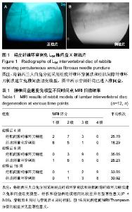
2.1 实验动物数量分析 纳入的新西兰大白兔36只,均进入结果分析,无死亡,无需补充,最终计入结果分析的数量36只。 2.2 实验动物穿刺并发症、成功率及手术时间分析 经肌间隙纤维环刀刺组1兔穿刺后伤口出现红肿积液,予以青霉素注射后,伤口换药后,伤口愈合良好,感染率为5.6%,而经皮纤维环穿刺组无感染,两组均未出现肢体瘫痪等其它并发症,两组所有动物16周后均出现影像学变化,提示两组动物造模成功率为100%。 纤维环穿刺组的穿刺时间(19.64±1.03) min,经肌间隙纤维环刀刺组穿刺时间(30.85±1.11) min,两组相比较差异有显著性意义(P < 0.05)。 2.3 MRI检测数据变化 所有MRI影像学资料由放射科医生进行盲法评估,根据Thompson分级[7],T2信号强度变化分为1-4级。1级:正常;2级:轻度减弱且高信号区域明显缩小;3级:中度减弱;4级:严重减弱。经秩和检验两造模组间比较差异有显著性意义(P < 0.05),穿刺后8周时与穿刺后4周时相同,但16周时两组间MRI Thompsom分级比较差异无显著性意义(P > 0.05),见表1。 2.4 MRI扫描结果 MRI Thompsom分级4,8,16周逐渐增加,退变程度加重,穿刺后4周时针剌组MRI中实验椎处椎间盘的改变不明显(图2A),而经肌间隙刀刺组见实验椎处椎间盘T2信号明显变弱(图2B),经秩和检验两造模组间MRI Thompsom分级比较差异有显著性意义(P < 0.05),穿刺后8周时与穿刺后4周时相同,但16周时两组间MRI Thompsom分级比较差异无显著性意义(P > 0.05,表1)。"

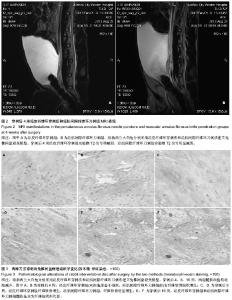
2.5 穿刺后兔椎间盘病理组织学观察结果 正常的髓核组织(L5/6)呈透明胶质样,镜下见细胞均匀分布,形态正常,随着时间的推移,髓核组织逐渐混浊,髓核细胞数量不断减少,少数可见有血管翳的生长。 经皮纤维环穿刺组和经肌间隙纤维化刀刺组造模后4周髓核组织髓核细胞逐渐减少,形态不规则,分布不均匀,纤维软骨组织增生,经肌间隙纤维环刀刺组较经皮纤维环穿刺组更为显著。 经皮纤维环穿刺组和经肌间隙纤维化刀刺组造模后第16周髓核中主要为纤维软骨组织,伴有极少量髓核细胞,髓核几乎被纤维软骨组织替代,纤维环排列不整齐,与髓核界限不清,可知16周时两组实验动物椎间盘均发生了明显的退变(图3)。"

| [1] Key J, Ford L. Experimental intervertebral-disc lesions. J Bone Joint Surg. 1948;30(3):621-630.[2] 郭常安,胡有谷,吴新彦,等.腰椎间盘退变动物模型的建立[J].中华外科杂志,2000,38(7):548- 551.[3] 吴健,唐天驷,王根,等.兔腰椎间盘退变模型的建立及影像学分析[J].苏州大学学报,2007,27(4):552-554.[4] 蒋新华,张伶,陈建宇,等.透视引导下穿刺构建兔椎间盘退变模型的影像和病理变化[J].中国组织工程研究与临床康复,2011, 15(48):8931-8934.[5] Zhou RP, Zhang ZM, Wang L, et al. Establishing a disc degeneration model using computed tomography-guided percutaneous puncture technique in the rabbit. J Surg Res. 2013;181(2):e65-74. [6] 辛龙,韩国灿,赵凤东,等.兔纤维环损伤椎间盘退变模型观察神经长入的实验研究[J].浙江大学学报(医学版),2009,38(5):485-492.[7] 王靖,唐天驷,姚啸生,等.纤维环穿刺诱导椎间盘退变动物模型的实验研究[J].中国脊柱脊髓杂志,2006,16(4):284-287.[8] 谢卯,熊蠡茗,周建国,等.椎间盘退变模型研究进展[J].国际骨科学杂志,2009,30(2):124-126.[9] Stokes IA, Iatridis JC. Mechanical conditions that accelerate intervertebral disc degeneration: overload versus immobilization. Spine (Phila Pa 1976). 2004;29(23): 2724-2732.[10] Masuda K, Aota Y, Muehleman C, et al. A novel rabbit model of mild, reproducible disc degeneration by an anulus needle puncture: correlation between the degree of disc injury and radiological and histological appearances of disc degeneration. Spine (Phila Pa 1976). 2005;30(1):5-14.[11] Hutton WC, Murakami H, Li J, et al. The effect of blocking a nutritional pathway to the intervertebral disc in the dog model. J Spinal Disord Tech. 2004;17(1):53-63.[12] Oda H, Matsuzaki H, Tokuhashi Y, et al. Degeneration of intervertebral discs due to smoking: experimental assessment in a rat-smoking model. J Orthop Sci. 2004;9(2):135-141.[13] Sahlman J, Inkinen R, Hirvonen T, et al. Premature vertebral endplate ossification and mild disc degeneration in mice after inactivation of one allele belonging to the Col2a1 gene for Type II collagen. Spine (Phila Pa 1976). 2001;26(23): 2558-2565.[14] Liu HF, Zhang H, Qiao GX, et al. A novel rabbit disc degeneration model induced by fibronectin fragment. Joint Bone Spine. 2013;80(3):301-306. [15] Moss IL, Zhang Y, Shi P, et al. Retroperitoneal approach to the intervertebral disc for the annular puncture model of intervertebral disc degeneration in the rabbit. Spine J. 2013; 13(3):229-234. [16] Xin L, Han GC, Zhao FD, et al. In vivo study of innervation of degenerative intervertebral discs in rabbit anular-injury model. Zhejiang Da Xue Xue Bao Yi Xue Ban. 2009;38(5):485-492.[17] Han B, Zhu K, Li FC, et al. A simple disc degeneration model induced by percutaneous needle puncture in the rat tail. Spine (Phila Pa 1976). 2008;33(18):1925-1934. [18] Yoon SH, Miyazaki M, Hong SW, et al. A porcine model of intervertebral disc degeneration induced by annular injury characterized with magnetic resonance imaging and histopathological findings. Laboratory investigation. J Neurosurg Spine. 2008;8(5):450-457.[19] 林鸿宽,叶君健.纤维环损伤诱导兔椎间盘退变模型[J].解剖学杂志,2008,31(5):699-702.[20] 胡鹏,刘勇,胡有谷,等.一种羊腰椎间盘退变模型的建立[J].青岛大学医学院学报,2007,43(2):167-168.[21] Holm S, Holm AK, Ekström L, et al. Experimental disc degeneraIion due to endplate injury. J Spinal Disord Tech. 2004;17(1):64-71.[22] Cinotti G, Della Rocca C, Romeo S, et al. Degenerative changes of porcine intervertebral disc induced by vertebral endplate injuries. Spine (Phila Pa 1976). 2005;30(2):174-180.[23] Greg Anderson D, Li X, Tannoury T, et al. A fibronectin fragment stimulates intervertebral disc degeneration in vivo. Spine (Phila Pa 1976). 2003;28(20):2338-2345.[24] Nakamura T, Iribe T, Asou Y, et al. Effects of compressive loading on biomechanical properties of disc and peripheral tissue in a rat tail model. Eur Spine J. 2009;18(11):1595-1603.[25] 熊晓芊,邵增务,裴洪,等.可控轴向压力致兔腰椎间盘退变模型的建立及评价[J].中国病理生理杂志,2008,24(10):2077-2080.[26] 李振宙,侯树勋,刘宁,等.新型山羊腰椎间盘突出模型的建立[J].中华骨科杂志,2007,27(11):838-843.[27] Lotz C. Animal models of intervertebral disc degeneration: lessons learned. Spine. 2004;29(23):2742-2750.[28] 孔杰,王子轩,季爱玉,等.应用微创技术建立恒河猴腰椎间盘早期退变模型[J].中华外科杂志,2008,46(11):835-838.[29] Seol D, Choe H, Ramakrishnan PS, et al. Organ culture stability of the intervertebral disc: rat versus rabbit. J Orthop Res. 2013;31(6):838-846.[30] 吴靖平,陈统一,陈中伟,等.双后肢大鼠椎间盘退变动物模型的建立[J].中华实验外科杂志,2004,21(1):105-107.[31] O’Connell GD, Vresilovic EJ, Elliott DM. Comparison of animals used in disc research to human lumbar disc geometry. Spine. 2007;32(3):328-333.[32] Elliott DM, Sarver JJ. Young investigator award winner: Validation of the mouse and rat disc as mechanical models of the human lumbar disc. Spine. 2004;29(7):713-722.[33] Vo N, Seo HY, Robinson A, et al. Accelerated aging of intervertebral discs in a mouse model of progeria. J Orthop Res. 2010;28(12):1600-1607.[34] Demers CN, Antoniou J, Mwale F. Value and limitations of using the bovine tail as a model for the human lumbar spine. Spine. 2004;29(24):2793-2799.[35] Gruber HE, Gordon B, Williams C, et al. Vertebral endplate and disc changes in the aging sand rat lumbar spine: Cross-sectional analyses of a large male and female population. Spine. 2007;32(23):2529-2536.[36] Cappello R, Bird JL, Pfeiffer D, et al. Notochordal cell produce and assemble extracellular matrix in a distinct manner, whichmay be responsible for the maintenance of healthy nucleus pulposus. Spine. 2006;31(8):873-882.[37] Sowa G, Valada G, Studer R, et al. Characterization of intervertebral disc aging: Longitudinal analysis of a rabbit model by magnetic resonance imaging, histology, and gene expression. Spine. 2008;33(17):1821-1828.[38] 崔运能,李绍林,金大地,等.肌间隙入路腰椎间盘退变模型的构建[J].南方医科大学学报,2012;32(3):404-408. |
| [1] | Wu Xun, Meng Juanhong, Zhang Jianyun, Wang Liang. Concentrated growth factors in the repair of a full-thickness condylar cartilage defect in a rabbit [J]. Chinese Journal of Tissue Engineering Research, 2021, 25(8): 1166-1171. |
| [2] | Song Liming, Su Hailong. Muscle force response characteristics of the slipping leg after an unexpected slip [J]. Chinese Journal of Tissue Engineering Research, 2021, 25(8): 1184-1189. |
| [3] | Li Jiacheng, Liang Xuezhen, Liu Jinbao, Xu Bo, Li Gang. Differential mRNA expression profile and competitive endogenous RNA regulatory network in osteoarthritis [J]. Chinese Journal of Tissue Engineering Research, 2021, 25(8): 1212-1217. |
| [4] | Geng Qiudong, Ge Haiya, Wang Heming, Li Nan. Role and mechanism of Guilu Erxianjiao in treatment of osteoarthritis based on network pharmacology [J]. Chinese Journal of Tissue Engineering Research, 2021, 25(8): 1229-1236. |
| [5] | He Xiangzhong, Chen Haiyun, Liu Jun, Lü Yang, Pan Jianke, Yang Wenbin, He Jingwen, Huang Junhan. Platelet-rich plasma combined with microfracture versus microfracture in the treatment of knee cartilage lesions: a meta-analysis [J]. Chinese Journal of Tissue Engineering Research, 2021, 25(6): 964-969. |
| [6] | Liu Xin, Yan Feihua, Hong Kunhao. Delaying cartilage degeneration by regulating the expression of aquaporins in rats with knee osteoarthritis [J]. Chinese Journal of Tissue Engineering Research, 2021, 25(5): 668-673. |
| [7] | Deng Zhenhan, Huang Yong, Xiao Lulu, Chen Yulin, Zhu Weimin, Lu Wei, Wang Daping. Role and application of bone morphogenetic proteins in articular cartilage regeneration [J]. Chinese Journal of Tissue Engineering Research, 2021, 25(5): 798-806. |
| [8] | Chen Lei, Zheng Rui, Jie Yongsheng, Qi Hui, Sun Lei, Shu Xiong. In vitro evaluation of adipose-derived stromal vascular fraction combined with osteochondral integrated scaffold [J]. Chinese Journal of Tissue Engineering Research, 2021, 25(22): 3487-3492. |
| [9] | Tian Guangzhao, Yang Zhen, Zha Kangkang, Sun Zhiqiang, Li Xu, Sui Xiang, Huang Jingxiang, Guo Quanyi, Liu Shuyun. Regulatory effect of decellularized cartilage matrix on macrophage polarization [J]. Chinese Journal of Tissue Engineering Research, 2021, 25(22): 3545-3550. |
| [10] | Ren Wenbo, Liao Yuanpeng. Visualization analysis of traumatic osteoarthritis research hotspots and content based on CiteSpace [J]. Chinese Journal of Tissue Engineering Research, 2021, 25(21): 3374-3381. |
| [11] | Song Shilei, Chen Yueping, Zhang Xiaoyun, Li Shibin, Lai Yu, Zhou Yi. Potential molecular mechanism of Wuling powder in treating osteoarthritis based on network pharmacology and molecular docking [J]. Chinese Journal of Tissue Engineering Research, 2021, 25(20): 3185-3193. |
| [12] | Wei Jinqiang, Huang Dengcheng, Cao Xuewei, Zhou Jianwei, Sun He, Li Zehui. Analysis of researches on TCM treatments for cartilage diseases in recent 20 years by mapping knowledge domains [J]. Chinese Journal of Tissue Engineering Research, 2021, 25(20): 3202-3209. |
| [13] | Lu Mingfeng, Zhao Lilian, Xing Jisi, He Lilei, Xu Ting, Wang Changbing. Posttraumatic progression of cartilage degeneration following anterior cruciate ligament reconstruction: a second-look arthroscopic analysis [J]. Chinese Journal of Tissue Engineering Research, 2021, 25(2): 222-227. |
| [14] | Zhang Jianhui, Ma Heran, Tan Yi, Wang Zhihui. Knee injury repair using human adipose-derived mesenchymal stem cells-based scaffold-free three-dimensional gel-like construct in pigs [J]. Chinese Journal of Tissue Engineering Research, 2021, 25(19): 2969-2975. |
| [15] | Yang Tengyun, Li Yanlin, Liu Dejian, Wang Guoliang. Application prospects and problems of peripheral blood derived mesenchymal stem cells in cartilage repair of osteoarthritis [J]. Chinese Journal of Tissue Engineering Research, 2021, 25(19): 3071-3076. |
| Viewed | ||||||
|
Full text |
|
|||||
|
Abstract |
|
|||||