Chinese Journal of Tissue Engineering Research ›› 2013, Vol. 17 ›› Issue (7): 1243-1250.doi: 10.3969/j.issn.2095-4344.2013.07.019
Previous Articles Next Articles
Hydrogen peroxide induced oxidative stress in the spleen of metallothionein Ⅰ/Ⅱ knockout mice
He Jing, Wei Si-yu, Liu Feng-yong, Ye Jing, Shen Bing-ling, Yao Xiao-mei
- Department of Pathophysiology, School of Basic Medical Sciences, Tianjin Medical University, Tianjin 300070, China
-
Received:2012-10-15Revised:2012-11-23Online:2013-02-12Published:2013-03-21 -
Contact:Yao Xiao-mei, Ph.D. in neuroscience, Professor, Master’s supervisor, Department of Pathophysiology, School of Basic Medical Sciences, Tianjin Medical University, Tianjin 300070, China jupx@163.com -
About author:He Jing★, Studying for master’s degree, Department of Pathophysiology, School of Basic Medical Sciences, Tianjin Medical University, Tianjin 300070, China hjdelightful@163.com
CLC Number:
Cite this article
He Jing, Wei Si-yu, Liu Feng-yong, Ye Jing, Shen Bing-ling, Yao Xiao-mei. Hydrogen peroxide induced oxidative stress in the spleen of metallothionein Ⅰ/Ⅱ knockout mice[J]. Chinese Journal of Tissue Engineering Research, 2013, 17(7): 1243-1250.
share this article
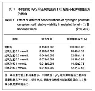
2.1 金属硫蛋白Ⅰ/Ⅱ 敲除鼠脾细胞对H2O2处理呈浓度依赖性 用不同浓度H2O2(0.1,0.2,0.5,1, 2 mmol/L)处理金属硫蛋白Ⅰ/Ⅱ 敲除鼠脾细胞2 h后,MTT测定结果发现,与对照组相比,0.1,0.2,0.5,1,2 mmol/L H2O2组使细胞活力显著下降 (P < 0.01),不同浓度H2O2组间脾细胞活力差异有显著性意义(P < 0.01)。见表1。"


2.2 NAC能够改善H2O2诱导的金属硫蛋白Ⅰ/Ⅱ敲除鼠脾细胞活力 MTT检测表明,与对照组相比,NAC组能显著提高脾细胞活力(P < 0.01),与0.5 mmol/L H2O2相比,NAC+0.5 mmol/L H2O2组能显著改善0.5 mmol/L H2O2诱导的脾细胞活力下降(P < 0.01),与1 mmol/L H2O2相比,NAC+1 mmol/L H2O2组能显著改善 1 mmol/L H2O2诱导的脾细胞活力下降(P < 0.01),但NAC+0.5 mmol/L H2O2组和NAC+1 mmol/L H2O2组与对照组相比,脾细胞活力仍降低,见表2。"


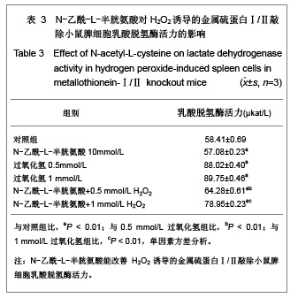
2.3 NAC能减轻H2O2诱导的金属硫蛋白Ⅰ/Ⅱ敲除鼠脾细胞乳酸脱氢酶活性 乳酸脱氢酶是糖酵解过程中一种重要的酶,任何原因引起的细胞损伤均可导致乳酸脱氢酶漏出,使培养液中乳酸脱氢酶活性增加。 处理2 h后乳酸脱氢酶测定结果显示,与对照组相比,NAC组乳酸脱氢酶活力显著降低(P < 0.01),0.5,1 mmol/L H2O2组培养液上清乳酸脱氢酶活力升高(P < 0.01),但增加10 mmol/L NAC处理后,与0.5 mmol/L H2O2相比,NAC+0.5 mmol/L H2O2组培养液上清乳酸脱氢酶活力明显降低(P < 0.01),与1 mmol/L H2O2相比,NAC+1 mmol/L H2O2组培养液上清乳酸脱氢酶活力也明显降低(P < 0.01),但是NAC+0.5 mmol/L H2O2组和NAC+1 mmol/L H2O2组与对照组相比,乳酸脱氢酶活力仍升高,见表3。"

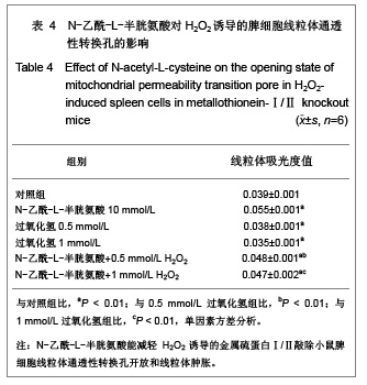
2.4 NAC能减轻H2O2诱导的金属硫蛋白Ⅰ/Ⅱ敲除鼠脾细胞线粒体通透性转换孔开放和线粒体肿胀 线粒体通透性转换孔开放可对甘露醇和蔗糖高通透造成线粒体膨胀,线粒体吸光度值减少。因而,通过检测线粒体吸光度值的变化可反映线粒体通透性转换孔的开放情况[15]。 处理2 h后测定结果显示,与对照组相比,NAC组线粒体吸光度值升高(P < 0.01),0.5、1 mmol/L H2O2 组线粒体吸光度值降低(P < 0.01),但增加10 mmol/L NAC 处理后,与0.5 mmol/L H2O2相比,NAC+0.5 mmol/L H2O2组线粒体吸光度值显著增加(P < 0.01),与 1 mmol/L H2O2相比,NAC+1 mmol/L H2O2组线粒体吸光度值也显著增加(P < 0.01),NAC+0.5 mmol/L H2O2 组和NAC+1 mmol/L H2O2组与对照组相比,线粒体吸光度值也是增高的,说明NAC能明显改善H2O2(0.5, 1 mmol/L)诱导的线粒体通透性转换孔开放和线粒体肿胀。见表4。"
| [1] Thirumoorthy N, Shyam SA, Manisenthil Kumar KT, et al. A review of metallothionein isoforms and their role in pathophysiology. World J Surg Oncol. 2011;9:54.[2] Inoue k, Takano H, Shimada A, et al. Metallothionein as an anti-in?ammatory mediator. Mediators of In?ammation. 2009; 2009:101659.[3] Yao X,Miao W, Li M, et al. Protective effect of albumin on VEGF and brain edema in acute isehemia in rats. Neurosei Lett. 2010;472(3):179-183.[4] Wang M, Wang YM, Li HD, et al. Zhonghua Laonian Xinnaoxueguanbing Zazhi. 2011;13(2):164-167. 王敏,王永明,李海东,等.白蛋白治疗对小鼠脑缺血早期脑组织Toll样受体4和骨髓分化因子88 mRNA表达的影响[J].中华老年心脑血管病杂志,2011,13(2):164-167.[5] Offner H, Vandenbark AA, Hurn PD. Effect of experimental stroke on peripheral immunity: CNS ischemia induces profound immunosuppression. Neuroscience. 2009;158(3): 1098-1111.[6] Kim CH, Vaziri ND, Rodriguez-Iturbe B. Integrin expression and H2O2 production in circulating and splenic leukocytes of obese rats. Obesity (Silver Spring). 2007; 15(9):2209-2216.[7] Liu B, Jian Z, Li Q, et al. Baicalein protects Human melanocytes from H2O2-induced apoptosis via inhibiting mitochondria-dependent caspase activation and the p38 MAPK pathway. Free Radic Biol Med. 2012;53(2):183-193.[8] Li XB, Zhang HC, Guo ZK. Zhongguo Zuzhi Gongcheng Yanjiu. 2012;16(1):22-26. 刘新宾,张红超,郭子宽.骨髓间充质干细胞条件培养液对H2O2损伤大鼠心肌细胞的保护[J].中国组织工程研究, 2012,16(1): 22-26. [9] Gu DS, Li W, Gu WW. Zhongguo Zuzhi Gongcheng Yanjiu. 2011;15(44):8281-8284. 顾东曙,刘闻,顾为望.静脉输注过氧化氢溶液上调小鼠脾脏和外周血CD4+Foxo3+调节性T细胞[J].中国组织工程研究与临床康复,2011,15(44):8281-8284. [10] Yang P, Peairs JJ, Tano R, et al. Oxidant-mediated Akt activation in human RPE cells. Invest Ophthalmol Vis Sci. 2006;47(10):4598-606.[11] Yoshino F, Yoshida A, Okada E, et al. Dental resin curing blue light induced oxidative stress with reactive oxygen. J Photochem Photobiol B. 2012;114:73-78.[12] Nakamura R, Lnoue K, Fujitani Y, et al. In vitro study of the effect of nanoparticle-rch diesel exhaust particles on IL-18 production in splenocytes. J Toxicol Sci. 2011;36(6):823-827.[13] Wu Y, Wang D, Wang X, et al. Caspase 3 is activated through caspase 8 instead of caspase 9 during H2O2-induced apoptosis in HeLa cells. Cell Physiol Biochem. 2011;27(5): 539-546. [14] Ru Y, Yin L, Sun H, et al. A micropreparation of mitochondria from cells using magnetic beads with immounoaf?nity. Anal Biochem. 2012;421(1):219-226.[15] Wang DM, Dong J, Qi ZT, et al. Xian Tiyu Xueyuan Xuebao. 2011;28(4):471-475. 王冬梅,董军,漆正堂,等.动对骨骼肌线粒体通透性转换孔及细胞凋亡的影响[J].西安体育学院学报,2011,28(4):471-475.[16] Petronilli V, Penzo D, Scorrano L, et al. The mitochondrial permeability transition, release of cytochrome c and cell death. J Biol Chem. 2001;267(15):12030-12034. [17] Ajmo CT Jr, Vernon DO, Collier L, et al. The spleen contributes to stroke-induced neurodegeneration. J Neurosci Res. 2008;86(10):2227-2234.[18] Izci Y. Splenectomy may be a prophylactic treatment for cerebral ischemia? Med Hypotheses. 2010;75(4):347-349. [19] Yu X, Guo J, Fang H, et al. Basal metallothionein-Ⅰ/Ⅱ protects against NMDA-mediated oxidative injury in cortical neuron/astrocyte cultures. Toxicology. 2011;282(1-2):16-22.[20] Ghanizadeh A. Gold implants and increased expression of metallothionein-Ⅰ/Ⅱ as a novel hypothesized therapeutic approach for autism. Toxicology. 2011; 283(1):63-64.[21] Hamdi Y, Kaddour H, Vaudry D, et al. The octadecaneuropeptide ODN protects astrocytes against hydrogen peroxide-induced apoptosis via a PKA/MAPK- dependent mechanism. PLoS One. 2012;7(8):e42498. [22] Baumeister P, Huebner T, Reiter M, et al. Reduction of oxidative DNA fragmentation by ascorbic acid, zinc and N-acetylcysteine in nasal mucosa tissue cultures. Anticancer Res. 2009;29(11):4571-4574.[23] Khouri R, Novais F, Santana G, et al. DETC induces Leishmania parasite killing in human in vitro and murine in vivo models: a promising therapeutic alternative in Leishmaniasis. PLoS One. 2010;5(12):e14394.[24] Xie M, Roy R. Increased levels of hydrogen peroxide induce a HIF-1-dependent modi?cation of lipid metabolism in AMPK compromised C. elegans Dauer Larvae. Cell Metab. 2012; 16(3):322-335.[25] Wu JP, Chen HC, Li MH. Bioaccumulation and toxicodynamics of cadmiumt of reshwater planarian and the protective effect of N-Acetylcysteine. Arch Environ Contam Toxicol. 2012;63(2):220-229.[26] Yao X, Li M, He J, et al. Effect of early acute high concentrations of iodide exposure on mitochondrial superoxide production in FRTL cells. Free Radic Biol Med. 2012; 52(8):1343-1352.[27] Li M, Li LY, Chen ZP, et al. Tianjin Yiyao. 2010;38(3): 212-215. 李敏,李兰英,陈组培,等.过氧化氢对FRTL细胞线粒体膜电位和超氧化物生成的影响[J].天津医药,2010,38(3):212-215.[28] Kinnally KW, Peixoto PM, Ryu SY, et al. Is mPTP the gate keeper for necrosis, apoptosis, or both? Biochim Biophys Acta. 2011;1813(4):616-622.[29] Drahota Z, Milerova M, Endlicher R, et al. Developmental changes of the sensitivity of cardiac and liver mitochondrial permeability transition pore to calcium load and oxidative stress. Physiol Res. 2012;61(Suppl 1):S165-172.[30] Loor G, Kondapalli J, Lwase H, et al. Mitochondrial oxidant stress triggers cell death in simulated ischemia-reperfusion. Biochim Biophys Acta. 2011;1813(7):1382-1394.[31] The Ministry of Science and Technology of the People’s Republic of China. Guidance Suggestions for the Care and Use of Laboratory Animals. 2006-09-30. |
| [1] |
Dong Chunyang, Zhou Tianen, Mo Mengxue, Lyu Wenquan, Gao Ming, Zhu Ruikai, Gao Zhiwei.
Action mechanism of metformin combined with Eomecon chionantha Hance dressing in treatment of deep second-degree burn wounds#br#
#br#
[J]. Chinese Journal of Tissue Engineering Research, 2026, 30(8): 2001-2013.
|
| [2] | Yang Xuetao, Zhu Menghan, Zhang Chenxi, Sun Yimin, Ye Ling. Applications and limitations of antioxidant nanomaterials in oral cavity [J]. Chinese Journal of Tissue Engineering Research, 2026, 30(8): 2044-2053. |
| [3] | Chen Ju, Zheng Jinchang, Liang Zhen, Huang Chengshuo, Lin Hao, Zeng Li. Effect and mechanism of beta-caryophyllene in mice with osteoarthritis [J]. Chinese Journal of Tissue Engineering Research, 2026, 30(6): 1341-1347. |
| [4] | Wen Guangwei, Zhen Yinghao, Zheng Taikeng, Zhou Shuyi, Mo Guoye, Zhou Tengpeng, Li Haishan, Lai Yiyi. Effects and mechanisms of isoginkgetin on osteoclastogenesis [J]. Chinese Journal of Tissue Engineering Research, 2026, 30(6): 1348-1358. |
| [5] | Li Linzhen, Jiao Hongzhuo, Chen Weinan, Zhang Mingzhe, Wang Jianlong, Zhang Juntao. Effect of icariin-containing serum on lipopolysaccharide-induced inflammatory damage in human chondrocytes [J]. Chinese Journal of Tissue Engineering Research, 2026, 30(6): 1368-1374. |
| [6] | Lyu Xiaofan, Huang Yi, Ding Liucheng . Mitochondrial mechanism and intervention therapy in diabetic cystopathy [J]. Chinese Journal of Tissue Engineering Research, 2026, 30(6): 1508-1515. |
| [7] | Cao Xinyan, Yu Zifu, Leng Xiaoxuan, Gao Shiai, Chen Jinhui, Liu Xihua. Effect of repetitive transcranial magnetic stimulation and transcranial direct current stimulation on motor function and gait in children with cerebral palsy: a network meta-analysis [J]. Chinese Journal of Tissue Engineering Research, 2026, 30(6): 1539-1548. |
| [8] | Guo Ying, Tian Feng, Wang Chunfang. Potential drug targets for the treatment of rheumatoid arthritis: large sample analysis from European databases [J]. Chinese Journal of Tissue Engineering Research, 2026, 30(6): 1549-1557. |
| [9] | Pan Hongfei, Zhuang Zhenbing, Xu Baiyun, Yang Zhangyang, Lin Kairui, Zhan Bingqing, Lan Jinghan, Gao Heng, Zhang Nanbo, Lin Jiayu. Inhibitory effects of different concentrations of auranofin on M1 macrophage function and its therapeutic potential in diabetic wound healing [J]. Chinese Journal of Tissue Engineering Research, 2026, 30(6): 1390-1397. |
| [10] | Peng Zhiwei, Chen Lei, Tong Lei. Luteolin promotes wound healing in diabetic mice: roles and mechanisms [J]. Chinese Journal of Tissue Engineering Research, 2026, 30(6): 1398-1406. |
| [11] | Jia Jinwen, Airefate·Ainiwaer, Zhang Juan. Effects of EP300 on autophagy and apoptosis related to allergic rhinitis in rats [J]. Chinese Journal of Tissue Engineering Research, 2026, 30(6): 1439-1449. |
| [12] | Yang Zhijie, Zhao Rui, Yang Haolin, Li Xiaoyun, Li Yangbo, Huang Jiachun, Lin Yanping, Wan Lei, HuangHongxing. Postmenopausal osteoporosis: predictive values of muscle mass, grip strength, and appendicular skeletal muscle index [J]. Chinese Journal of Tissue Engineering Research, 2026, 30(5): 1073-1080. |
| [13] | Yin Yongcheng, Zhao Xiangrui, Yang Zhijie, Li Zheng, Li Fang, Ning Bin. Effect and mechanism of peroxiredoxin 1 in microglial inflammation after spinal cord injury [J]. Chinese Journal of Tissue Engineering Research, 2026, 30(5): 1106-1113. |
| [14] | Zhang Jiuxuan, Zhang Jinnan, Sui Xiaofan, Pei Xiaxia, Wei Jianhong, Su Qiang, Li Tian. Effects of ammonia poisoning on cognitive behavior and hippocampal synaptic damage in mice [J]. Chinese Journal of Tissue Engineering Research, 2026, 30(5): 1122-1128. |
| [15] | Sun Yajie, Zhao Xinchen, Bo Shuangling. Spatiotemporal expression of bone morphologic protein 7 in mouse kidney development [J]. Chinese Journal of Tissue Engineering Research, 2026, 30(5): 1156-1161. |
| Viewed | ||||||
|
Full text |
|
|||||
|
Abstract |
|
|||||