Chinese Journal of Tissue Engineering Research ›› 2026, Vol. 30 ›› Issue (5): 1106-1113.doi: 10.12307/2026.035
Previous Articles Next Articles
Effect and mechanism of peroxiredoxin 1 in microglial inflammation after spinal cord injury
Yin Yongcheng1, Zhao Xiangrui1, Yang Zhijie2, Li Zheng2, Li Fang2, Ning Bin2
- 1School of Clinical Medicine, Shandong Second Medical University, Weifang 261053, Shandong Province, China; 2Central Hospital Affiliated to Shandong First Medical University, Shandong First Medical University & Shandong Academy of Medical Sciences, Jinan 250013, Shandong Province, China
-
Received:2024-11-08Accepted:2025-01-09Online:2026-02-18Published:2025-06-23 -
Contact:Ning Bin, Chief physician, Central Hospital Affiliated to Shandong First Medical University, Shandong First Medical University & Shandong Academy of Medical Sciences, Jinan 250013, Shandong Province, China -
About author:Yin Yongcheng, MS candidate, School of Clinical Medicine, Shandong Second Medical University, Weifang 261053, Shandong Province, China -
Supported by:the National Natural Science Foundation of China, No. 82071383 (to NB); Natural Science Foundation of Shandong Province, No. ZR2023MH235 (to LF)
CLC Number:
Cite this article
Yin Yongcheng, Zhao Xiangrui, Yang Zhijie, Li Zheng, Li Fang, Ning Bin. Effect and mechanism of peroxiredoxin 1 in microglial inflammation after spinal cord injury[J]. Chinese Journal of Tissue Engineering Research, 2026, 30(5): 1106-1113.
share this article
Add to citation manager EndNote|Reference Manager|ProCite|BibTeX|RefWorks
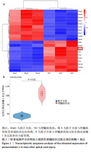
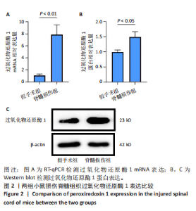
2.1 动物实验结果 2.1.1 实验动物数量分析 12只小鼠全部进入结果分析。 2.1.2 转录组测序分析 利用小鼠假手术组和脊髓损伤组损伤部位脊髓组织的转录组数据筛选差异基因,使用pheatmap和ggplot2包进行可视化分析,结果发现,与假手术组相比,脊髓损伤组过氧化物还原酶1表达水平显著上升(log2FC > 1且P-adj < 0.05),见图1。 2.1.3 过氧化物还原酶1表达 RT-qPCR检测结果显示,与假手术组相比,脊髓损伤组过氧化物还原酶1 mRNA表达高于假手术组(P < 0.01),见图2A;Western blot检测结果显示,脊髓损伤组过氧化物还原酶1蛋白表达高于假手术组(P < 0.05),见图2B,C,这些结果与转录组测序结果一致,进一步证明了脊髓损伤后过氧化物还原酶1表达显著上升。 2.2 体外细胞实验结果 2.2.1 小胶质细胞BV2内过氧化物还原酶1敲除验证 RT-qPCR检测结果显示,敲除组过氧化物还原酶1 mRNA表达低于对照组(P < 0.001),见图3A。Western blot检测结果显示,敲除组过氧化物还原酶1蛋白表达低于对照组(P < 0.01),见图3B,C。结果"

"


"
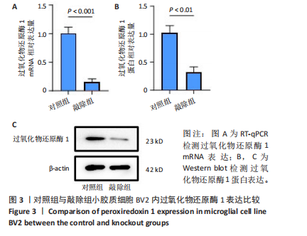
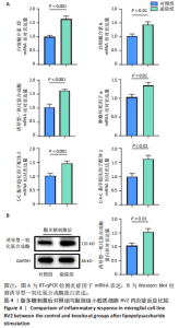
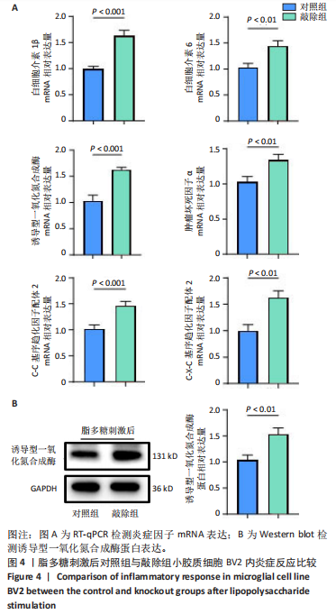
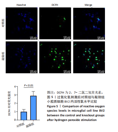
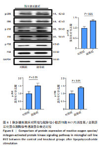
表明过氧化物还原酶1敲除小胶质细胞BV2内模型构建成功。 2.2.2 过氧化物还原酶1敲除加重小胶质细胞BV2内炎症反应 RT-qPCR检测结果显示,敲除组白细胞介素1β、白细胞介素6、肿瘤坏死因子α、C-C基序趋化因子配体2和C-X-C基序趋化因子配体2 mRNA表达均高于对照组(P < 0.01或P < 0.001),见图4A。Western blot检测结果显示,敲除组诱导型一氧化氮合成酶的蛋白表达高于对照组(P < 0.01),见图4B。结果表明,小鼠小胶质细胞系BV2中敲除过氧化物还原酶1基因后炎症反应加重。 2.2.3 敲除过氧化物还原酶1后小胶质细胞BV2内活性氧水平上升 免疫荧光染色结果显示,在过氧化氢刺激下,敲除组活性氧水平高于对照组(P < 0.01),见图5。 2.2.4 敲除过氧化物还原酶1后影响小胶质细胞BV2内活性氧/丝裂原活化蛋白激酶信号通路 最新研究表明,活性氧同丝裂原活化蛋白激酶信号通路密切相关,丝裂原活化蛋白激酶在细胞对各种外界刺激(如细胞因子、氧化应激、热应激和辐射等)的反应中起着关键作用[21-22]。因此,此次研究拟在体外进一步探究过氧化物还原酶1对小胶质细胞BV2内活性氧/丝裂原活化蛋白激酶信号通路的影响,实验结果显示,在脂多糖刺激下,敲除组p-P38、p-JNK和p-ERK蛋白表达均高于对照组(P < 0.01或P < 0.05),见图6,进一步证明过氧化物还原酶1可以通过活性氧/丝裂原活化蛋白激酶信号通路影响小胶质细胞的炎症反应。"

"
"

| [1] WARNER FM, CRAGG JJ, JUTZELER CR, et al. Progression of Neuropathic Pain after Acute Spinal Cord Injury: A Meta-Analysis and Framework for Clinical Trials. J Neurotrauma. 2019;36(9):1461-1468. [2] KATHE C, SKINNIDER MA, HUTSON TH, et al. The neurons that restore walking after paralysis. Nature. 2022;611(7936):540-547. [3] RIVERS CS, FALLAH N, NOONAN VK, et al. Health Conditions: Effect on Function, Health-Related Quality of Life, and Life Satisfaction After Traumatic Spinal Cord Injury. A Prospective Observational Registry Cohort Study. Arch Phys Med Rehabil. 2018;99(3):443-451. [4] BADHIWALA JH, WILSON JR, FEHLINGS MG. Global burden of traumatic brain and spinal cord injury. Lancet Neurol. 2019;18(1):24-25. [5] DIOP M, EPSTEIN D. A Systematic Review of the Impact of Spinal Cord Injury on Costs and Health-Related Quality of Life. Pharmacoecon Open. 2024;8(6):793-808. [6] FREYERMUTH-TRUJILLO X, SEGURA-URIBE JJ, SALGADO-CEBALLOS H, et al. Inflammation: A Target for Treatment in Spinal Cord Injury. Cells. 2022;11(17):2692. [7] AHUJA CS, WILSON JR, NORI S, et al. Traumatic spinal cord injury. Nat Rev Dis Primers. 2017;3(1):17018. [8] 史旭,李瑞语,张兵,等.小胶质细胞极化介导炎症反应在脊髓损伤中的作用[J].中国组织工程研究,2023,27(1):121-129. [9] JEONG SJ, CHO MJ, KO NY, et al. Deficiency of peroxiredoxin 2 exacerbates angiotensin II-induced abdominal aortic aneurysm. Exp Mol Med. 2020;52(9):1587-1601. [10] RHEE SG, KANG SW, JEONG W, et al. Intracellular messenger function of hydrogen peroxide and its regulation by peroxiredoxins. Curr Opin Cell Biol. 2005;17(2):183-189. [11] PARK JG, YOO JY, JEONG SJ, et al. Peroxiredoxin 2 deficiency exacerbates atherosclerosis in apolipoprotein E-deficient mice. Circ Res. 2011; 109(7):739-749. [12] MORIWAKI T, YOSHIMURA A, TAMARI Y, et al. PRDX1 is essential for the viability and maintenance of reactive oxygen species in chicken DT40. Genes Environ. 2021;43(1):35. [13] GUAN X, RUAN Y, CHE X, et al. Dual role of PRDX1 in redox-regulation and tumorigenesis: Past and future. Free Radic Biol Med. 2024;210: 120-129. [14] HAN YH, LIAN XD, LEE SJ, et al. Regulatory effect of peroxiredoxin 1 (PRDX1) on doxorubicin-induced apoptosis in triple negative breast cancer cells. Appl Biol Chem. 2022;65(1):63. [15] LIU CH, KUO SW, HSU LM, et al. Peroxiredoxin 1 induces inflammatory cytokine response and predicts outcome of cardiogenic shock patients necessitating extracorporeal membrane oxygenation: an observational cohort study and translational approach. J Transl Med. 2016;14(1):114. [16] SZELIGA M. Peroxiredoxins in Neurodegenerative Diseases. Antioxidants (Basel). 2020;9(12):1203. [17] KIM S, LEE W, JO H, et al. The antioxidant enzyme Peroxiredoxin-1 controls stroke-associated microglia against acute ischemic stroke. Redox Biol. 2022;54:102347. [18] ZHANG C, SHAO Q, ZHANG Y, et al. Therapeutic application of nicotinamide: As a potential target for inhibiting fibrotic scar formation following spinal cord injury. CNS Neurosci Ther. 2024;30(7):e14826. [19] XUE MT, SHENG WJ, SONG X, et al. Atractylenolide III ameliorates spinal cord injury in rats by modulating microglial/macrophage polarization. CNS Neurosci Ther. 2022;28(7):1059-1071. [20] LI C, ZHAO Z, LUO Y, et al. Macrophage-Disguised Manganese Dioxide Nanoparticles for Neuroprotection by Reducing Oxidative Stress and Modulating Inflammatory Microenvironment in Acute Ischemic Stroke. Adv Sci (Weinh). 2021;8(20):e2101526. [21] LIU J, WEI Y, JIA W, et al. Chenodeoxycholic acid suppresses AML progression through promoting lipid peroxidation via ROS/p38 MAPK/DGAT1 pathway and inhibiting M2 macrophage polarization. Redox Biol. 2022;56:102452. [22] CHEN HR, SUN Y, MITTLER G, et al. MOF-mediated PRDX1 acetylation regulates inflammatory macrophage activation. Cell Rep. 2024;43(9): 114682. [23] FAKHRI S, ABBASZADEH F, MORADI SZ, et al. Effects of Polyphenols on Oxidative Stress, Inflammation, and Interconnected Pathways during Spinal Cord Injury. Oxid Med Cell Longev. 2022;2022:8100195. [24] MAO XY, ZHOU HH, JIN WL. Redox-Related Neuronal Death and Crosstalk as Drug Targets: Focus on Epilepsy. Front Neurosci. 2019;13: 512. [25] HASSANZADEH K, RAHIMMI A. Oxidative stress and neuroinflammation in the story of Parkinson’s disease: Could targeting these pathways write a good ending? J Cell Physiol. 2018;234(1):23-32. [26] SOLLEIRO-VILLAVICENCIO H, RIVAS-ARANCIBIA S. Effect of Chronic Oxidative Stress on Neuroinflammatory Response Mediated by CD4(+)T Cells in Neurodegenerative Diseases. Front Cell Neurosci. 2018;12:114. [27] LIU Z, REN Z, ZHANG J, et al. Role of ROS and Nutritional Antioxidants in Human Diseases. Front Physiol. 2018;9:477. [28] SKINNIDER MA, GAUTIER M, TEO AYY, et al. Single-cell and spatial atlases of spinal cord injury in the Tabulae Paralytica. Nature. 2024; 631(8019):150-163. [29] LI C, WU Z, ZHOU L, et al. Temporal and spatial cellular and molecular pathological alterations with single-cell resolution in the adult spinal cord after injury. Signal Transduct Target Ther. 2022;7(1):65. [30] ROJO AI, MCBEAN G, CINDRIC M, et al. Redox control of microglial function: molecular mechanisms and functional significance. Antioxid Redox Signal. 2014;21(12):1766-1801. [31] JORDÃO MJC, SANKOWSKI R, BRENDECKE SM, et al. Single-cell profiling identifies myeloid cell subsets with distinct fates during neuroinflammation. Science. 2019;363(6425):eaat7554. [32] MENDIOLA AS, RYU JK, BARDEHLE S, et al. Transcriptional profiling and therapeutic targeting of oxidative stress in neuroinflammation. Nat Immunol. 2020;21(5):513-524. [33] YANG GQ, HUANG JC, YUAN JJ, et al. Prdx1 Reduces Intracerebral Hemorrhage-Induced Brain Injury via Targeting Inflammation- and Apoptosis-Related mRNA Stability. Front Neurosci. 2020;14:181. [34] ATTARAN S, SKOKO JJ, HOPKINS BL, et al. Peroxiredoxin-1 Tyr194 phosphorylation regulates LOX-dependent extracellular matrix remodelling in breast cancer. Br J Cancer. 2021;125(8):1146-1157. [35] LU Y, ZHANG XS, ZHOU XM, et al. Peroxiredoxin 1/2 protects brain against H2O2-induced apoptosis after subarachnoid hemorrhage. FASEB J. 2019;33(2):3051-3062. [36] SHICHITA T, HASEGAWA E, KIMURA A, et al. Peroxiredoxin family proteins are key initiators of post-ischemic inflammation in the brain. Nat Med. 2012;18(6):911-917. [37] LIU CH, KUO SW, HSU LM, et al. Peroxiredoxin 1 induces inflammatory cytokine response and predicts outcome of cardiogenic shock patients necessitating extracorporeal membrane oxygenation: an observational cohort study and translational approach. J Transl Med. 2016;14(1):114. [38] KANG JH, LEE HY, KIM NY, et al. Extracellular Prdx1 mediates bacterial infection and inflammatory bone diseases. Life Sci. 2023;333:122140. [39] MORETTON A, KOURTIS S, GAÑEZ ZAPATER A, et al. A metabolic map of the DNA damage response identifies PRDX1 in the control of nuclear ROS scavenging and aspartate availability. Mol Syst Biol. 2023;19(7):e11267. |
| [1] | Zhang Di, Zhao Jun, Ma Guangyue, Sun Hui, Jiang Rong. Mechanism of depression-like behavior in chronic social defeat stress mice based on high-throughput sequencing [J]. Chinese Journal of Tissue Engineering Research, 2026, 30(5): 1139-1146. |
| [2] | Hu Jing, Zhu Ling, Xie Juan, Kong Deying, Liu Doudou. Autophagy regulates early embryonic development in mice via affecting H3K4me3 modification [J]. Chinese Journal of Tissue Engineering Research, 2026, 30(5): 1147-1155. |
| [3] | Sun Yajie, Zhao Xinchen, Bo Shuangling. Spatiotemporal expression of bone morphologic protein 7 in mouse kidney development [J]. Chinese Journal of Tissue Engineering Research, 2026, 30(5): 1156-1161. |
| [4] | Li Haojing, Wang Xin, Song Chenglin, Zhang Shengnan, Chen Yunxin. Therapeutic efficacy of extracorporeal shock wave therapy in the upper trapezius muscle area combined with exercise control training in patients with chronic non-specific neck pain [J]. Chinese Journal of Tissue Engineering Research, 2026, 30(5): 1162-1170. |
| [5] | Liu Yu, Lei Senlin, Zhou Jintao, Liu Hui, Li Xianhui. Mechanisms by which aerobic and resistance exercises improve obesity-related cognitive impairment [J]. Chinese Journal of Tissue Engineering Research, 2026, 30(5): 1171-1183. |
| [6] | Yu Huifen, Mo Licun, Cheng Leping. The position and role of 5-hydroxytryptamine in the repair of tissue injury [J]. Chinese Journal of Tissue Engineering Research, 2026, 30(5): 1196-1206. |
| [7] | Wang Zhengye, Liu Wanlin, Zhao Zhenqun. Advance in the mechanisms underlying miRNAs in steroid-induced osteonecrosis of the femoral head [J]. Chinese Journal of Tissue Engineering Research, 2026, 30(5): 1207-1214. |
| [8] | Bu Yangyang, Ning Xinli, Zhao Chen. Intra-articular injections for the treatment of osteoarthritis of the temporomandibular joint: different drugs with multiple combined treatment options [J]. Chinese Journal of Tissue Engineering Research, 2026, 30(5): 1215-1224. |
| [9] | Wen Fan, Xiang Yang, Zhu Huan, Tuo Yanfang, Li Feng. Exercise improves microvascular function in patients with type 2 diabetes [J]. Chinese Journal of Tissue Engineering Research, 2026, 30(5): 1225-1235. |
| [10] | Liu Xinyue, Li Chunnian, Li Yizhuo, Xu Shifang. Regeneration and repair of oral alveolar bone defects [J]. Chinese Journal of Tissue Engineering Research, 2026, 30(5): 1247-1259. |
| [11] | Leng Xiaoxuan, Zhao Yuxin, Liu Xihua. Effects of different neuromodulatory stimulation modalities on non-motor symptoms in Parkinson’s patients: a network meta-analysis [J]. Chinese Journal of Tissue Engineering Research, 2026, 30(5): 1282-1293. |
| [12] | Wen Xiaolong, Weng Xiquan, Feng Yao, Cao Wenyan, Liu Yuqian, Wang Haitao. Effects of inflammation on serum hepcidin and iron metabolism related parameters in patients with type 2 diabetes mellitus: a meta-analysis [J]. Chinese Journal of Tissue Engineering Research, 2026, 30(5): 1294-1301. |
| [13] | Yang Zeyu, Zhi Liang, Wang Jia, Zhang Jingyi, Zhang Qingfang, Wang Yulong, Long Jianjun. A visualized analysis of research hotspots in high-frequency repetitive transcranial magnetic stimulation from the macroscopic perspective [J]. Chinese Journal of Tissue Engineering Research, 2026, 30(5): 1320-1330. |
| [14] | Yang Zhijie, Zhao Rui, Yang Haolin, Li Xiaoyun, Li Yangbo, Huang Jiachun, Lin Yanping, Wan Lei, HuangHongxing. Postmenopausal osteoporosis: predictive values of muscle mass, grip strength, and appendicular skeletal muscle index [J]. Chinese Journal of Tissue Engineering Research, 2026, 30(5): 1073-1080. |
| [15] | Zhang Jiuxuan, Zhang Jinnan, Sui Xiaofan, Pei Xiaxia, Wei Jianhong, Su Qiang, Li Tian. Effects of ammonia poisoning on cognitive behavior and hippocampal synaptic damage in mice [J]. Chinese Journal of Tissue Engineering Research, 2026, 30(5): 1122-1128. |
| Viewed | ||||||
|
Full text |
|
|||||
|
Abstract |
|
|||||