Chinese Journal of Tissue Engineering Research ›› 2026, Vol. 30 ›› Issue (5): 1114-1121.doi: 10.12307/2026.024
Previous Articles Next Articles
Forskolin promotes C2C12 myoblast differentiation via regulating the ERK and Akt signaling pathways
Huang Liuyan, Zhang Wenxi, Chen Shuwen, Yu Shimei, Dai Zhong, Zuo Changqing
- School of Pharmacy, Guangdong Medical University, Dongguan 523808, Guangdong Province, China
-
Received:2024-12-05Accepted:2025-01-13Online:2026-02-18Published:2025-06-23 -
Contact:Zuo Changqing, PhD, Associate professor, School of Pharmacy, Guangdong Medical University, Dongguan 523808, Guangdong Province, China Co-corresponding author: Dai Zhong, PhD, Associate professor, School of Pharmacy, Guangdong Medical University, Dongguan 523808, Guangdong Province, China -
About author:Huang Liuyan, Master candidate, School of Pharmacy, Guangdong Medical University, Dongguan 523808, Guangdong Province, China -
Supported by:Guangdong Basic and Applied Basic Research Foundation, No. 2023A1515140157 (to ZCQ)
CLC Number:
Cite this article
Huang Liuyan, Zhang Wenxi, Chen Shuwen, Yu Shimei, Dai Zhong, Zuo Changqing. Forskolin promotes C2C12 myoblast differentiation via regulating the ERK and Akt signaling pathways[J]. Chinese Journal of Tissue Engineering Research, 2026, 30(5): 1114-1121.
share this article
Add to citation manager EndNote|Reference Manager|ProCite|BibTeX|RefWorks
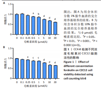
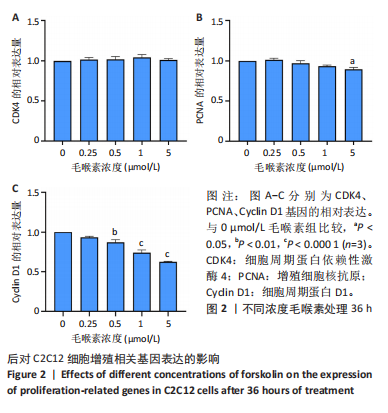
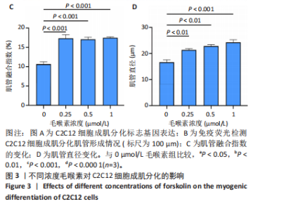
2.1 毛喉素对C2C12细胞活力的影响 首先,使用C2C12成肌细胞研究了不同浓度毛喉素处理的细胞毒性,将C2C12细胞分别与不同浓度的毛喉素(0-20 μmol/L)孵育48 h后进行CCK-8检测。结果显示,无论是在含体积分数1%胎牛血清或是在体积分数10%胎牛血清的培养基中培养,低浓度的毛喉素对C2C12成肌细胞的增殖过程无细胞毒性(图1A),但是当毛喉素浓度增加,细胞活力下降,并在毛喉素浓度> 1 μmol/L时活性下降20%左右(图1B)。这些数据表明,高剂量毛喉素处理会抑制C2C12细胞的活性。 2.2 毛喉素对C2C12细胞增殖相关基因的影响 为了进一步探讨毛喉素是否会影响C2C12细胞的增殖,在毛喉素处理C2C12细胞36 h后进行qRT-PCR检测。结果显示,CDK4表达水平在毛喉素处理的细胞和对照组细胞之间没有差异(图2A);PCNA表达水平在毛喉素处理浓度为5 μmol/L的时候下降(图2B);Cyclin D1表达水平在毛喉素处理浓度> 0.25 μmol/L时开始下降,毛喉素处理浓度为5 μmol/L时CyclinD1表达水平大幅下降(图2C)。以上结果说明高浓度毛喉素对C2C12细胞具有抗增殖作用。 2.3 毛喉素对C2C12细胞成肌分化的影响 2.3.1 qPCR分析 为了研究毛喉素对C2C12细胞成肌分化的影响,用不同浓度的毛喉素(0,0.25,0.5,1 μmol/L)孵育C2C12细胞,并在处理后进行qPCR分析,观察毛喉素对成肌分化标志基因mRNA表达的影响。在成肌分化4 d时,经毛喉素处理的细胞Myh2、Myh4基因表达水平较0 μmol/L毛喉素组增加(图3A),肌肉特异性膜蛋白Myomaker的表达水平也增加,而主要在Ⅰ型慢缩肌纤维中表达的Myh7"


"
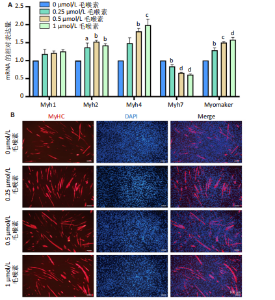
的表达水平降低。因此推测毛喉素除了影响成肌分化过程中成肌细胞的融合,同时也参与了快慢肌纤维的转化。 2.3.2 免疫荧光染色 为了进一步探讨毛喉素对C2C12细胞肌管形成的影响,在C2C12细胞诱导分化第4天时,用抗MyHC抗体进行免疫荧光染色,检测C2C12细胞的分化形态。可以看到0 μmol/L毛喉素组C2C12细胞已经有一定数量分化融合形成的多核肌管,然而,在毛喉素处理的细胞中,多核肌管的数量明显更多,并且肌管的长度和宽度也更长"
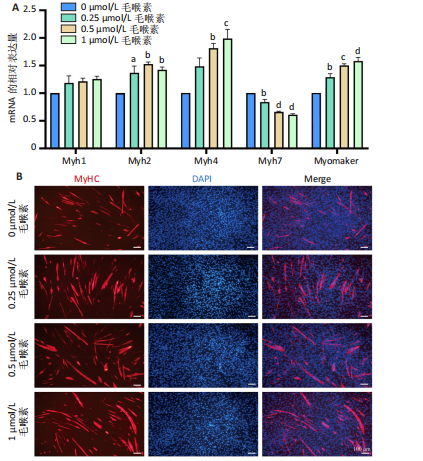
"


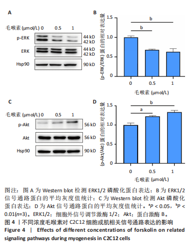
(图3B)。此外,定量分析数据显示:毛喉素处理后的C2C12细胞融合指数上升(图3C),与0 μmol/L毛喉素组相比,毛喉素处理各组肌管直径明显增加(图3D)。总之,与0 μmol/L毛喉素组相比,毛喉素处理各组MyHC阳性肌管的数量明显增多。以上结果表明,毛喉素能有效促进C2C12成肌细胞的分化。 2.4 毛喉素通过调控ERK1/2和Akt信号通路影响C2C12成肌细胞分化 先前研究表明,ERK1/2对哺乳动物骨骼肌生成至关重要 [15]。为了探讨毛喉素促进C2C12细胞分化的分子机制,采用Western blot法检测了毛喉素处理C2C12细胞分化2 d时ERK1/2磷酸化蛋白水平的变化(图4)。与对照组C2C12细胞相比,毛喉素处理2 d的C2C12细胞中磷酸化ERK1/2的蛋白表达显著减少,而总ERK1/2蛋白的水平保持不变(图4A),表明毛喉素的促肌生成作用依赖于ERK1/2信号通路的抑制。此外,在调节肌肉发育的信号通路中,Akt信号通路在肌肉生长中起着核心作用 [16],因此同时检测了Akt信号通路的变化,结果发现,成肌分化2 d时,毛喉素处理组磷酸化Akt的蛋白表达水平升高(图4C),表明毛喉素的促肌生成作用还依赖于Akt信号通路的激活。"
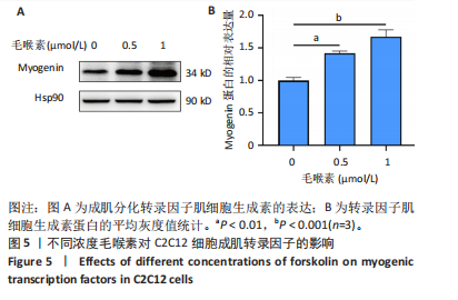
2.5 毛喉素对C2C12细胞成肌转录因子的影响 研究还探索了毛喉素处理是否通过促进肌细胞生成素的表达从而刺激肌源性分化(图5)。为了验证假设,采用Western blot法检测了毛喉素处理后成肌分化2 d肌细胞生成素的蛋白表达,结果表明,与对照组细胞相比,经过毛喉素处理的C2C12细胞中的肌细胞生成素的表达水平在成肌分化2 d时显著增加。因此,结果揭示毛喉素可能通过影响肌细胞生成素的表达从而作用于成肌分化。"

| [1] Progress in heterologous biosynthesis of forskolin. J Ind Microbiol Biotechnol. 2021;48(1-2):kuab009. [2] ROSHNI PT, REKHA PD. Biotechnological interventions for the production of forskolin, an active compound from the medicinal plant, Coleus forskohlii. Physiol Mol Biol Plants. 2024;30(2):213-226. [3] SAPIO L, GALLO M, ILLIANO M, et al. The Natural cAMP Elevating Compound Forskolin in Cancer Therapy: Is It Time? J Cell Physiol. 2017;232(5):922-927. [4] BANGAY G, BRAUNING FZ, ROSATELLA A, et al. Anticancer diterpenes of African natural products: Mechanistic pathways and preclinical developments. Phytomedicine. 2024;129:155634. [5] SALZILLO A, RAGONE A, SPINA A, et al. Forskolin affects proliferation, migration and Paclitaxel-mediated cytotoxicity in non-small-cell lung cancer cell lines via adenylyl cyclase/cAMP axis. Eur J Cell Biol. 2023;102(2):151292. [6] WU J, YUE B. Regulation of myogenic cell proliferation and differentiation during mammalian skeletal myogenesis. Biomed Pharmacother. 2024;174:116563. [7] 吴玲玲, 张小玉, 李晓,等. miR-196b-5p促进成肌细胞增殖分化[J]. 遗传, 2023,45(5):435-446. [8] CHEN JY, PENG SY, CHENG YH, et al. Effect of Forskolin on Body Weight, Glucose Metabolism and Adipocyte Size of Diet-Induced Obesity in Mice. Animals (Basel). 2021;11(3):645. [9] AWALE GM, BARAJAA MA, KAN HM, et al. Regenerative engineering of long bones using the small molecule forskolin. Proc Natl Acad Sci U S A. 2023;120(22):e2219756120. [10] XU C, TABEBORDBAR M, IOVINO S, et al. A zebrafish embryo culture system defines factors that promote vertebrate myogenesis across species. Cell. 2013,155(4):909-921. [11] ZHANG Y, ZHU Y, LI Y, et al. Long-term engraftment of myogenic progenitors from adipose-derived stem cells and muscle regeneration in dystrophic mice. Hum Mol Genet. 2015;24(21):6029-6040. [12] JEONG J, CHOI KH, KIM SH, et al.Combination of cell signaling molecules can facilitate MYOD1-mediated myogenic transdifferentiation of pig fibroblasts. J Anim Sci Biotechnol. 2021;12(1):64. [13] CHEN AE, GINTY DD, FAN CM. Protein kinase A signalling via CREB controls myogenesis induced by Wnt proteins. Nature. 2005; 433(7023):317-322. [14] TAGLIETTI V, KEFI K, RIVERA L, et al. Thyroid-stimulating hormone receptor signaling restores skeletal muscle stem cell regeneration in rats with muscular dystrophy. Sci Transl Med. 2023;15(685):eadd5275. [15] KNIGHT JD, KOTHARY R. The myogenic kinome: protein kinases critical to mammalian skeletal myogenesis. Skelet Muscle. 2011;1:29. [16] HO PT, PARK E, LUONG QXT, et al. Amelioration of Cancer Cachexia by Dalbergia odorifera Extract Through AKT Signaling Pathway Regulation. Nutrients. 2024;16(21):3671. [17] LAURENZA A, SUTKOWSKI EM, SEAMON KB. Forskolin: a specific stimulator of adenylyl cyclase or a diterpene with multiple sites of action? Trends Pharmacol Sci. 1989;10(11):442-447. [18] ZHANG H, LIU Y, LIU J, et al. cAMP-PKA/EPAC signaling and cancer: the interplay in tumor microenvironment. J Hematol Oncol. 2024;17(1):5. [19] LAUTHERBACH N, GONCALVES DAP, SILVEIRA WA, et al. Urocortin 2 promotes hypertrophy and enhances skeletal muscle function through cAMP and insulin/IGF-1 signaling pathways. Mol Metab. 2022; 60:101492. [20] ASLAM M, LADILOV Y. Emerging Role of cAMP/AMPK Signaling. Cells. 2022;11(2):308. [21] TCHAKARSKA G, SOLA B. The double dealing of cyclin D1. Cell Cycle. 2020;19(2):163-178. [22] DIECKMAN LM, FREUDENTHAL BD, WASHINGTON MT. PCNA structure and function: insights from structures of PCNA complexes and post-translationally modified PCNA. Subcell Biochem. 2012;62:281-299. [23] DE LA SERNA IL, ROY K, CARLSON KA, et al. MyoD can induce cell cycle arrest but not muscle differentiation in the presence of dominant negative SWI/SNF chromatin remodeling enzymes. J Biol Chem. 2001; 276(44):41486-41491. [24] DOS SANTOS M, BACKER S, AURADE F, et al. A fast Myosin super enhancer dictates muscle fiber phenotype through competitive interactions with Myosin genes. Nat Commun. 2022;13(1):1039. [25] MO M, ZHANG Z, WANG X, et al. Molecular mechanisms underlying the impact of muscle fiber types on meat quality in livestock and poultry. Front Vet Sci. 2023;10:1284551. [26] MILLAY DP, O’ROURKE JR, SUTHERLAND LB, et al. Myomaker is a membrane activator of myoblast fusion and muscle formation. Nature. 2013;499(7458):301-305. [27] KILANOWSKA A, ZIOLKOWSKA A, STASIAK P, et al. cAMP-Dependent Signaling and Ovarian Cancer. Cells. 2022;11(23):3835. [28] PARK HB, BAEK KH. E3 ligases and deubiquitinating enzymes regulating the MAPK signaling pathway in cancers. Biochim Biophys Acta Rev Cancer. 2022;1877(3):188736. [29] LAKE D, CORREA SA, MULLER J. Negative feedback regulation of the ERK1/2 MAPK pathway. Cell Mol Life Sci. 2016;73(23):4397-4413. [30] 刘波, 黄晓帆, 李冬寒, 等. Forskolin激活cAMP信号通路调控小鼠心肌成纤维细胞结缔组织生长因子的表达[J].华中科技大学学报(医学版),2015,44(1):1-9. [31] CRISTOBAL I, RINCON R, MANSO R, et al. Hyperphosphorylation of PP2A in colorectal cancer and the potential therapeutic value showed by its forskolin-induced dephosphorylation and activation. Biochim Biophys Acta. 2014;1842(9):1823-1829. [32] XIAO D, CALDOW M, KIM HJ, et al. Time-resolved phosphoproteome and proteome analysis reveals kinase signaling on master transcription factors during myogenesis. iScience. 2022;25(6):104489. [33] JIANG X, JI S, CUI S, et al. Apol9a regulates myogenic differentiation via the ERK1/2 pathway in C2C12 cells. Front Pharmacol. 2022;13:942061. [34] CHENG C, ZHANG S, GONG Y, et al. Cordycepin inhibits myogenesis via activating the ERK1/2 MAPK signalling pathway in C2C12 cells. Biomed Pharmacother. 2023,165:115163. [35] FU X, MATSUI T, FUNABA M. Enhancement of vitamin C-induced myogenesis by inhibition of extracellular signal-regulated kinase (ERK) 1/2 pathway. Biochem Biophys Res Commun. 2022;612:57-62. [36] TORTORELLA LL, MILASINCIC DJ, PILCH PF. Critical proliferation-independent window for basic fibroblast growth factor repression of myogenesis via the p42/p44 MAPK signaling pathway.J Biol Chem. 2001;276(17):13709-13717. [37] ENDO T. Postnatal skeletal muscle myogenesis governed by signal transduction networks: MAPKs and PI3K-Akt control multiple steps. Biochem Biophys Res Commun. 2023;682:223-243. [38] EIGLER T, ZARFATI G, AMZALLAG E, et al. ERK1/2 inhibition promotes robust myotube growth via CaMKII activation resulting in myoblast-to-myotube fusion. Dev Cell. 2021;56(24):3349-3363. [39] SCHIAFFINO S, DYAR KA, CICILIOT S, et al. Mechanisms regulating skeletal muscle growth and atrophy. FEBS J. 2013;280(17):4294-4314. [40] NAMKOONG S, KIM CK, CHO YL, et al. Forskolin increases angiogenesis through the coordinated cross-talk of PKA-dependent VEGF expression and Epac-mediated PI3K/Akt/eNOS signaling. Cell Signal. 2009;21(6):906-915. [41] GHORBANI A, JEDDI-TEHRANI M, SAIDPOUR A, et al. PI3K/AKT and Mdm2 activation are associated with inhibitory effect of cAMP increasing agents on DNA damage-induced cell death in human pre-B NALM-6 cells. Arch Biochem Biophys. 2015;566:58-66. [42] CHEN YJ, BASKARAN R, CHANG CF, et al. Decapeptide from Potato Hydrolysate Induces Myogenic Differentiation and Ameliorates High Glucose-Associated Modulations in Protein Synthesis and Mitochondrial Biogenesis in C2C12 Cells. Biomolecules. 2022;12(4):565. [43] QUAN-JUN Y, YAN H, YONG-LONG H, et al. Selumetinib Attenuates Skeletal Muscle Wasting in Murine Cachexia Model through ERK Inhibition and AKT Activation. Mol Cancer Ther. 2017;16(2):334-343. [44] LI H, GUAN K, WANG R, et al. Synergistic effects of MFG-E8 and whey protein on mitigating d-galactose-induced sarcopenia through PI3K/AKT/PGC-1alpha and MAPK/ERK signaling pathways. J Dairy Sci. 2024; 107(1):9-23. |
| [1] | Yang Zhijie, Zhao Rui, Yang Haolin, Li Xiaoyun, Li Yangbo, Huang Jiachun, Lin Yanping, Wan Lei, HuangHongxing. Postmenopausal osteoporosis: predictive values of muscle mass, grip strength, and appendicular skeletal muscle index [J]. Chinese Journal of Tissue Engineering Research, 2026, 30(5): 1073-1080. |
| [2] | Li Kaiying, Wei Xiaoge, Song Fei, Yang Nan, Zhao Zhenning, Wang Yan, Mu Jing, Ma Huisheng. Mechanism of Lijin manipulation regulating scar formation in skeletal muscle injury repair in rabbits [J]. Chinese Journal of Tissue Engineering Research, 2025, 29(8): 1600-1608. |
| [3] | Li Huayuan, Li Chun, Liu Junwei, Wang Ting, Li Long, Wu Yongli. Effect of warm acupuncture on PINK1/Parkin pathway in the skeletal muscle of rats with chronic fatigue syndrome [J]. Chinese Journal of Tissue Engineering Research, 2025, 29(8): 1618-1625. |
| [4] | Wang Xuanqiang, Zhang Wenyang, Li Yang, Kong Weiqian, Li Wei, Wang Le, Li Zhongshan, Bai Shi. Effects of chronic exposure to low-frequency pulsed magnetic fields on contractility and morphology of the quadriceps muscle in healthy adults [J]. Chinese Journal of Tissue Engineering Research, 2025, 29(8): 1634-1642. |
| [5] | Zhao Ruihua, Chen Sixian, Guo Yang, Shi Lei, Wu Chengjie, Wu Mao, Yang Guanglu, Zhang Haoheng, Ma Yong. Wen-Shen-Tong-Du Decoction promoting spinal cord injury repair in mice [J]. Chinese Journal of Tissue Engineering Research, 2025, 29(6): 1118-1126. |
| [6] | Zhang Wenhua, Li Xun, Zhang Weichao, Li Xinying, Ma Guoao, Wang Xiaoqiang . Promoting myogenesis based on the SphK1/S1P/S1PR2 signaling pathway: a new perspective on improving skeletal muscle health through exercise [J]. Chinese Journal of Tissue Engineering Research, 2025, 29(6): 1265-1275. |
| [7] | Xu Tianjie, Fan Jiaxin, Guo Xiaoling, Jia Xiang, Zhao Xingwang, Liu kainan, Wang Qian. Metformin exerts a protective effect on articular cartilage in osteoarthritis rats by inhibiting the PI3K/AKT/mTOR pathway [J]. Chinese Journal of Tissue Engineering Research, 2025, 29(5): 1003-1012. |
| [8] | Wen Zixing, Xu Xin, Zhu Shengqun. Correlations between gastrocnemius morphology parameters and physical activity capacity in elderly females under high-frequency ultrasound [J]. Chinese Journal of Tissue Engineering Research, 2025, 29(5): 1058-1063. |
| [9] | Li Tingyue, Guo Qian, He Wenxi, Wu Jiayuan. Long noncoding RNA TP53TG1 promotes odontogenic and osteogenic differentiation of stem cells from the apical papilla [J]. Chinese Journal of Tissue Engineering Research, 2025, 29(36): 7776-7782. |
| [10] | Cai Zhixing, Xia Qiufang, Chen Lili, Zhu Danyang, Zhu Huiwen, Sun Yanan, Liang Wenyu, Zhao Heqian. Effect of Roujishuncuiyin on the improvement of skeletal muscle insulin resistance in a mouse model of type 2 diabetes mellitus [J]. Chinese Journal of Tissue Engineering Research, 2025, 29(35): 7537-7543. |
| [11] |
Li Yunzhe, Niu Zefan, Wang Zirou, Ai Chongyi, Chen Gang, Wang Xinxing.
Asperosaponin VI promotes osteogenic differentiation of MC3T3-E1 cells under hypoxia environment #br#
#br#
[J]. Chinese Journal of Tissue Engineering Research, 2025, 29(35): 7481-7489.
|
| [12] | Liu Chenchen, Liu Ruize, Bao Mengmeng, Fang Li, Cao Liquan, Wu Jiangbo. Blood flow restriction training intervention in the elderly with sarcopenic obesity [J]. Chinese Journal of Tissue Engineering Research, 2025, 29(32): 6963-6970. |
| [13] | Wang Jiaqian, , Jiang Changjun, Peng Yi, Ma Mi, Li Junhan. Study on the role of aerobic exercise in regulating the CNPY2-mediated AKT/GSK3β pathway for improving non-alcoholic fatty liver [J]. Chinese Journal of Tissue Engineering Research, 2025, 29(30): 6441-6448. |
| [14] | Hu Shujuan, Liu Dang, Ding Yiting, Liu Xuan, Xia Ruohan, Wang Xianwang. Ameliorative effect of walnut oil and peanut oil on atherosclerosis [J]. Chinese Journal of Tissue Engineering Research, 2025, 29(30): 6482-6488. |
| [15] | Zhang Zihan¹, Wang Jiaxin¹, Yang Wenyi², Zhu Lei¹. Regulatory mechanism of exercise promoting mitochondrial biogenesis in skeletal muscle [J]. Chinese Journal of Tissue Engineering Research, 2025, 29(30): 6499-6508. |
| Viewed | ||||||
|
Full text |
|
|||||
|
Abstract |
|
|||||