Chinese Journal of Tissue Engineering Research ›› 2025, Vol. 29 ›› Issue (35): 7537-7543.doi: 10.12307/2025.945
Previous Articles Next Articles
Effect of Roujishuncuiyin on the improvement of skeletal muscle insulin resistance in a mouse model of type 2 diabetes mellitus
Cai Zhixing1, Xia Qiufang1, Chen Lili2, Zhu Danyang2, Zhu Huiwen1, Sun Yanan1, Liang Wenyu1, Zhao Heqian1
- 1Department of Traditional Chinese Medicine Rehabilitation, The First Rehabilitation Hospital of Shanghai, Shanghai 200090, China; 2Department of Traditional Chinese Medicine, Tongren Hospital, Shanghai Jiao Tong University School of Medicine, Shanghai 200336, China
-
Received:2024-09-29Accepted:2024-12-10Online:2025-12-18Published:2025-04-30 -
Contact:Xia Qiufang, Master, Chief physician, Department of Traditional Chinese Medicine Rehabilitation, The First Rehabilitation Hospital of Shanghai, Shanghai 200090, China -
About author:Cai Zhixing, Master, Associate chief physician, Department of Traditional Chinese Medicine Rehabilitation, The First Rehabilitation Hospital of Shanghai, Shanghai 200090, China -
Supported by:National Natural Science Foundation of China (Youth Science Fund Project), No. 82205041 (to CZX); Special Project of Shanghai Yangpu District Health and Wellness System for Traditional Chinese Medicine, No. YPZHM202308 (to ZHW)
CLC Number:
Cite this article
Cai Zhixing, Xia Qiufang, Chen Lili, Zhu Danyang, Zhu Huiwen, Sun Yanan, Liang Wenyu, Zhao Heqian. Effect of Roujishuncuiyin on the improvement of skeletal muscle insulin resistance in a mouse model of type 2 diabetes mellitus[J]. Chinese Journal of Tissue Engineering Research, 2025, 29(35): 7537-7543.
share this article
Add to citation manager EndNote|Reference Manager|ProCite|BibTeX|RefWorks
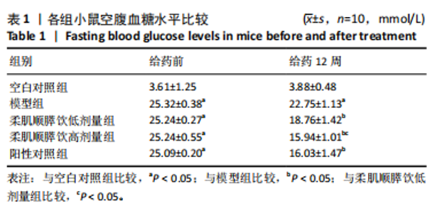
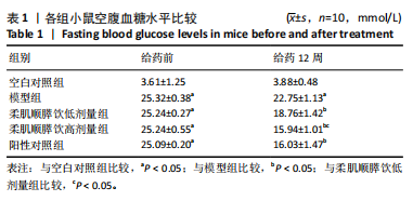
2.1 实验动物数量分析 实验过程中,各组小鼠营养状况良好,均可自由活动,进食进水,未发生脱落,均进入结果分析。 2.2 各组小鼠葡萄糖相关代谢指标 给药前,模型组、柔肌顺膵饮低、高剂量组及阳性对照组小鼠空腹血糖水平显著高于空白对照组(P < 0.05)。给药12周后,模型组小鼠空腹血糖水平仍显著高于空白对照组(P < 0.05);柔肌顺膵饮低、高剂量组及阳性对照组小鼠空腹血糖水平显著低于模型组(P < 0.05),柔肌顺膵饮高剂量组小鼠空腹血糖水平显著低于柔肌顺膵饮低剂量组(P < 0.05),柔肌顺膵饮高剂量组小鼠空腹血糖水平与阳性对照组无显著差异(P > 0.05),见表1。"
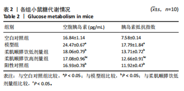
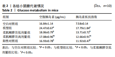
给药12周后,模型组小鼠空腹胰岛素和胰岛素抵抗指数均显著高于空白对照组(P < 0.05);柔肌顺膵饮低、高剂量组及阳性对照组小鼠空腹胰岛素和胰岛素抵抗指数显著低于模型组(P < 0.05);柔肌顺膵饮高剂量组小鼠空腹胰岛素和胰岛素抵抗指数显著低于柔肌顺膵饮低剂量组(P < 0.05);柔肌顺膵饮高剂量组小鼠空腹胰岛素和胰岛素抵抗指数与阳性对照组无显著差异(P > 0.05),见表2。"
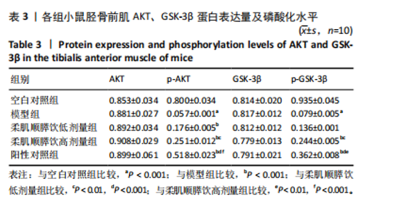
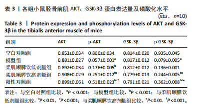
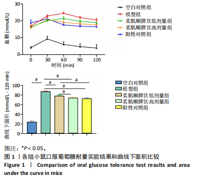
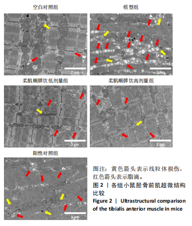
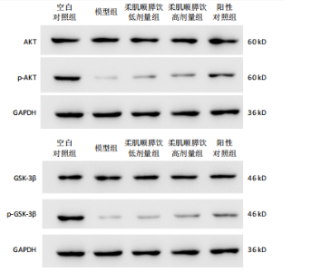
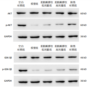
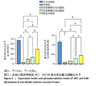
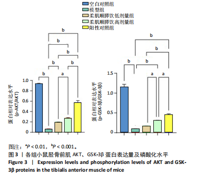
2.3 各组小鼠口服葡萄糖耐量实验结果 口服葡萄糖耐量实验结果显示:模型组小鼠各时间点血糖显著高于空白对照组,在60 min时达到血糖最高峰,曲线下面积显著增加(P < 0.05);柔肌顺膵饮低剂量组、高剂量组和阳性对照组小鼠各时间点血糖显著低于模型组,曲线下面积显著减少(P < 0.05),且柔肌顺膵饮高剂量组的血糖值在30 min时即达到最高峰,呈现与正常组相似的血糖曲线变化;柔肌顺膵饮高剂量组曲线下面积显著低于柔肌顺膵饮低剂量组(P < 0.05);柔肌顺膵饮高剂量组曲线下面积与阳性对照组无显著差异(P > 0.05),见图1。 2.4 各组小鼠胫骨前肌超微结构 透射电镜下观察可见:空白对照组小鼠骨骼肌组织各肌节Z线、M线、H带完整清晰,骨骼肌细丝排列整齐,线粒体边界较清楚,存在少量脂滴(红色箭头);模型组小鼠骨骼肌肌纤维排列不规则,肌纤维排列紊乱,各肌节Z线、M线中断、H带模糊,大量线粒体损伤(黄色箭头),肌间质存在大量脂滴;柔肌顺膵饮低剂量组小鼠骨骼肌间质内脂滴减少,线粒体损伤改善;柔肌顺膵饮高剂量组和阳性药物组小鼠骨骼肌间质内脂滴明显减少,线粒体结构较规则,线粒体损伤较治疗前显著改善,见图2。 2.5 各组小鼠胫骨前肌AKT、GSK-3β蛋白表达量及磷酸化水平 模型组小鼠胫骨前肌p-AKT、p-GSK-3β蛋白表达量显著低于空白对照组(P < 0.001);柔肌顺膵饮低、高剂量组及阳性对照组小鼠胫骨前肌p-AKT、p-GSK-3β蛋白表达量显著高于模型组(P < 0.001);柔肌顺膵饮高剂量组小鼠胫骨前肌p-AKT、p-GSK-3β蛋白表达量显著高于柔肌顺膵饮低剂量组(P < 0.01);柔肌顺膵饮高剂量组小鼠胫骨前肌p-AKT、p-GSK-3β蛋白表达量显著低于阳性对照组(P < 0.001,P < 0.01),见表3,图3。"

"

"
"
"
| [1] The Prevention of Diabetes Mellitus. JAMA. 2021;325(2):190. [2] CHEISSON G, JACQUEMINET S, COSSON E, et al. Perioperative management of adult diabetic patients. Intraoperative period. Anaesth Crit Care Pain Med. 2018;37 Suppl 1:S21-S25. [3] YU CG, FU Y, FANG Y, et al. Fighting Type-2 Diabetes: Present and Future Perspectives. Curr Med Chem. 2019;26(10):1891-1907. [4] LEE SH, PARK SY, CHOI CS. Insulin Resistance: From Mechanisms to Therapeutic Strategies. Diabetes Metab J. 2022;46(1):15-37. [5] WANG CC, CHEN HJ, CHAN DC, et al. Low-Dose Acrolein, an Endogenous and Exogenous Toxic Molecule, Inhibits Glucose Transport via an Inhibition of Akt-Regulated GLUT4 Signaling in Skeletal Muscle Cells. Int J Mol Sci. 2021;22(13):7228. [6] 张六妹,刘菁菁,林晓烨,等.高强度间歇运动改善骨骼肌损伤和提高大鼠运动能力的机制[J].中国组织工程研究,2023,27(35):5603-5609. [7] CHAO CN, LO JF, KHAN FB, et al. Tid1-S attenuates LPS-induced cardiac hypertrophy and apoptosis through ER-a mediated modulation of p-PI3K/p-Akt signaling cascade. J Cell Biochem. 2019;120(10):16703-16710. [8] LUNDELL LS, MASSART J, ALTINTAŞ A, et al. Regulation of glucose uptake and inflammation markers by FOXO1 and FOXO3 in skeletal muscle. Mol Metab. 2019;20:79-88. [9] CHEN CC, LII CK, LIN YH, et al. Andrographis paniculata Improves Insulin Resistance in High-Fat Diet-Induced Obese Mice and TNFα-Treated 3T3-L1 Adipocytes. Am J Chin Med. 2020;48(5):1073-1090. [10] SALTIEL AR. Insulin signaling in health and disease. J Clin Invest. 2021; 131(1):e142241. [11] RYAN AS, LI G, MCMILLIN S, et al. Pathways in Skeletal Muscle: Protein Signaling and Insulin Sensitivity after Exercise Training and Weight Loss Interventions in Middle-Aged and Older Adults. Cells. 2021;10(12):3490. [12] MIAO L, ZHANG X, ZHANG H, et al. Baicalin ameliorates insulin resistance and regulates hepatic glucose metabolism via activating insulin signaling pathway in obese pre-diabetic mice. Phytomedicine. 2024;124:155296. [13] HUANG S, YU C, HU M, et al. Electroacupuncture ameliorates hepatic defects in a rat model of polycystic ovary syndrome induced by letrozole and a high-fat diet. Acupunct Med. 2024;42(2):87-99. [14] LANZILLOTTA C, TRAMUTOLA A, LANZILLOTTA S, et al. Biliverdin Reductase-A integrates insulin signaling with mitochondrial metabolism through phosphorylation of GSK3β. Redox Biol. 2024;73:103221. [15] 蔡之幸,陈越,朱丹阳,等.柔肌顺膵饮改善2型糖尿病骨骼肌胰岛素抵抗的实验研究[J].新疆医科大学学报,2024,47(7): 1024-1031. [16] 彭岚玉,李定祥,姚敬心,等.基于GPR43/β-arrestin-2/IκBα/NF-κB通路探讨左归降糖通脉方对糖尿病大鼠血管内皮炎性保护的影响[J].中国中西医结合杂志,2024,44(5):558-566. [17] JUÁREZ-REYES K, BRINDIS F, MEDINA-CAMPOS ON, et al. Hypoglycemic, antihyperglycemic, and antioxidant effects of the edible plant Anoda cristata. J Ethnopharmacol. 2015;161:36-45. [18] WANG T, LU J, SHI L, et al. Association of insulin resistance and β-cell dysfunction with incident diabetes among adults in China: a nationwide, population-based, prospective cohort study. Lancet Diabetes Endocrinol. 2020;8(2):115-124. [19] HUTCHISON AL, TAVAGLIONE F, ROMEO S, et al. Endocrine aspects of metabolic dysfunction-associated steatotic liver disease (MASLD): Beyond insulin resistance. J Hepatol. 2023;79(6):1524-1541. [20] SI X, TIAN J, SHU C, et al. Serum Ceramide Reduction by Blueberry Anthocyanin-Rich Extract Alleviates Insulin Resistance in Hyperlipidemia Mice. J Agric Food Chem. 2020;68(31):8185-8194. [21] MCCORMICK N, O’CONNOR MJ, YOKOSE C, et al. Assessing the Causal Relationships Between Insulin Resistance and Hyperuricemia and Gout Using Bidirectional Mendelian Randomization. Arthritis Rheumatol. 2021;73(11):2096-2104. [22] HOU XZ, LV YF, LI YS, et al. Association between different insulin resistance surrogates and all-cause mortality in patients with coronary heart disease and hypertension: NHANES longitudinal cohort study. Cardiovasc Diabetol. 2024;23(1):86.
[23] KELLAR D, CRAFT S. Brain insulin resistance in Alzheimer’s disease and related disorders: mechanisms and therapeutic approaches. Lancet Neurol. 2020;19(9):758-766. [24] ZHAO H, ZHANG J, CHENG X, et al. Insulin resistance in polycystic ovary syndrome across various tissues: an updated review of pathogenesis, evaluation, and treatment. J Ovarian Res. 2023;16(1):9. [25] HE J, WANG H, VIJG J. New Insights into Bioactive Compounds of Traditional Chinese Medicines for Insulin Resistance Based on Signaling Pathways. Chem Biodivers. 2019;16(9):e1900176. [26] ZHANG Y, YANG Y, DING L, et al. Emerging Applications of Metabolomics to Assess the Efficacy of Traditional Chinese Medicines for Treating Type 2 Diabetes Mellitus. Front Pharmacol. 2021;12:735410. [27] 夏翔,柳玉瑾,邰杏芳,等.回春饮治疗老年期痴呆的临床观察和实验研究[J].上海中医药大学上海市中医药研究院学报,1996(1): 22-26. [28] MA D, CHEN Z, WANG Y, et al. Effects of rapid growth on fasting insulin and insulin resistance: a system review and meta-analysis. Eur J Clin Nutr. 2021;75(8):1193-1204. [29] MATTHEWS DR, HOSKER JP, RUDENSKI AS, et al. Homeostasis model assessment: insulin resistance and beta-cell function from fasting plasma glucose and insulin concentrations in man. Diabetologia. 1985;28(7):412-419. [30] ZHANG X, YUAN Y, LI C, et al. Effect of a Salt Substitute on Incidence of Hypertension and Hypotension Among Normotensive Adults. J Am Coll Cardiol. 2024;83(7):711-722. [31] HANLEY AJ, WILLIAMS K, STERN MP, et al. Homeostasis model assessment of insulin resistance in relation to the incidence of cardiovascular disease: the San Antonio Heart Study. Diabetes Care. 2002;25(7):1177-1184. [32] MONNERET D, BONNEFONT-ROUSSELOT D. Homeostasis model assessment of insulin resistance and lobular inflammation in nondiabetic patients with nonalcoholic fatty liver disease: methodological considerations. Eur J Gastroenterol Hepatol. 2020; 32(4):542. [33] KIM JJ, HWANG KR, OH SH, et al. Prevalence of insulin resistance in Korean women with polycystic ovary syndrome according to various homeostasis model assessment for insulin resistance cutoff values. Fertil Steril. 2019;112(5):959-966.e1. [34] FASCIOLO G, NAPOLITANO G, APRILE M, et al. Muscle Oxidative Stress Plays a Role in Hyperthyroidism-Linked Insulin Resistance. Antioxidants (Basel). 2023;12(3):592. [35] COBELLI C, DALLA MAN C, TOFFOLO G, et al. The oral minimal model method. Diabetes. 2014;63(4):1203-1213. [36] LEE PD, LUSTIG RH, LENDERS C, et al. Insulin-like growth factor binding protein 1 predicts insulin sensitivity and insulin area-under-the-curve in obese, nondiabetic adolescents. Endocr Pract. 2016;22(2):136-142. [37] MITSUSHIO K, BADEN MY, KAGISAKI T, et al. Interrelationships Among Accumulations of Intra- and Periorgan Fats, Visceral Fat, and Subcutaneous Fat. Diabetes. 2024;73(7):1122-1126. [38] 张艳,何瑞波,王庆博,等. 不同负荷量有氧运动对肥胖大鼠骨骼肌炎症反应和胰岛素信号途径的影响及机制[J].中国组织工程研究,2023,27(8):1237-1244. [39] 许趁意,岳仁宋,吕雪莲. 岳仁宋从少阳阳明辨治2型糖尿病合并血脂异常经验[J].中华中医药杂志,2021,36(9):5322-5325. [40] 杨璐瑜,林安盈,杨正望,等.健脾化痰方通过调控 FOXO1/PDK4信号通路干预 PCOS-IR大鼠脂代谢的作用机制研究[J].中国实验动物学报,2024,32(4):451-460. [41] 朱梦梦,王立鑫,封若雨,等.脾虚湿阻型单纯性肥胖小鼠模型的构建及评价[J].中国中医基础医学杂志,2021,27(2):247-250+328. [42] KOBAYASHI Y, WATANABE N, KITAKAZE T, et al. Oleamide rescues tibialis anterior muscle atrophy of mice housed in small cages. Br J Nutr. 2021;126(4):481-491. [43] PALOMO-FERNÁNDEZ I, MARTÍN-CASADO L, MARCOS-TEJEDOR F, et al. Lateral wedge insoles and their use in ankle instability. Scand J Med Sci Sports. 2023;33(9):1716-1725. [44] MITSUYA H, NAKAZATO K, HAKKAKU T, et al. Hip flexion angle affects longitudinal muscle activity of the rectus femoris in leg extension exercise. Eur J Appl Physiol. 2023;123(6):1299-1309. [45] LI Y, SHEN Z, JIANG X, et al. Mouse mesenchymal stem cell-derived exosomal miR-466f-3p reverses EMT process through inhibiting AKT/GSK3β pathway via c-MET in radiation-induced lung injury. J Exp Clin Cancer Res. 2022;41(1):128. [46] HE Q, YANG J, ZHANG G, et al. Sanhuang Jiangtang tablet protects type 2 diabetes osteoporosis via AKT-GSK3β-NFATc1 signaling pathway by integrating bioinformatics analysis and experimental validation. J Ethnopharmacol. 2021;273:113946. [47] KAN H, WANG P, YANG Y, et al. Apigenin inhibits proliferation and differentiation of cardiac fibroblasts through AKT/GSK3β signaling pathway. J Ethnopharmacol. 2024;334:118518. [48] XIONG W, LIU Y, ZHOU H, et al. Human dental pulp stem cells mitigate the neuropathology and cognitive decline via AKT-GSK3β-Nrf2 pathways in Alzheimer’s disease. Int J Oral Sci. 2024;16(1):40. [49] RAMIRES JÚNIOR OV, SILVEIRA JS, et al. Homocysteine May Decrease Glucose Uptake and Alter the Akt/GSK3β/GLUT1 Signaling Pathway in Hippocampal Slices: Neuroprotective Effects of Rivastigmine and Ibuprofen. Mol Neurobiol. 2023;60(9):5468-5481. [50] ZHANG J, WANG X, WANG F, et al. Xiangsha Liujunzi Decoction improves gastrointestinal motility in functional dyspepsia with spleen deficiency syndrome by restoring mitochondrial quality control homeostasis. Phytomedicine. 2022;105:154374. [51] SONG M, ZHANG J, HUO S, et al. Mitophagy alleviates AIF-mediated spleen apoptosis induced by AlCl3 through Parkin stabilization in mice. Food Chem Toxicol. 2023;176:113762. [52] KONG BS, MIN SH, LEE C, et al. Mitochondrial-encoded MOTS-c prevents pancreatic islet destruction in autoimmune diabetes. Cell Rep. 2021;36(4):109447. |
| [1] | Li Kaiying, Wei Xiaoge, Song Fei, Yang Nan, Zhao Zhenning, Wang Yan, Mu Jing, Ma Huisheng. Mechanism of Lijin manipulation regulating scar formation in skeletal muscle injury repair in rabbits [J]. Chinese Journal of Tissue Engineering Research, 2025, 29(8): 1600-1608. |
| [2] | Li Huayuan, Li Chun, Liu Junwei, Wang Ting, Li Long, Wu Yongli. Effect of warm acupuncture on PINK1/Parkin pathway in the skeletal muscle of rats with chronic fatigue syndrome [J]. Chinese Journal of Tissue Engineering Research, 2025, 29(8): 1618-1625. |
| [3] | Wang Xuanqiang, Zhang Wenyang, Li Yang, Kong Weiqian, Li Wei, Wang Le, Li Zhongshan, Bai Shi. Effects of chronic exposure to low-frequency pulsed magnetic fields on contractility and morphology of the quadriceps muscle in healthy adults [J]. Chinese Journal of Tissue Engineering Research, 2025, 29(8): 1634-1642. |
| [4] | Yin Lu, Jiang Chuanfeng, Chen Junjie, Yi Ming, Wang Zihe, Shi Houyin, Wang Guoyou, Shen Huarui. Effect of Complanatoside A on the apoptosis of articular chondrocytes [J]. Chinese Journal of Tissue Engineering Research, 2025, 29(8): 1541-1547. |
| [5] | Aikepaer · Aierken, Chen Xiaotao, Wufanbieke · Baheti. Osteogenesis-induced exosomes derived from human periodontal ligament stem cells promote osteogenic differentiation of human periodontal ligament stem cells in an inflammatory microenvironment [J]. Chinese Journal of Tissue Engineering Research, 2025, 29(7): 1388-1394. |
| [6] | Zhang Haojun, Li Hongyi, Zhang Hui, Chen Haoran, Zhang Lizhong, Geng Jie, Hou Chuandong, Yu Qi, He Peifeng, Jia Jinpeng, Lu Xuechun. Identification and drug sensitivity analysis of key molecular markers in mesenchymal cell-derived osteosarcoma [J]. Chinese Journal of Tissue Engineering Research, 2025, 29(7): 1448-1456. |
| [7] | Sun Yuting, Wu Jiayuan, Zhang Jian. Physical factors and action mechanisms affecting osteogenic/odontogenic differentiation of dental pulp stem cells [J]. Chinese Journal of Tissue Engineering Research, 2025, 29(7): 1531-1540. |
| [8] | Yu Ting, Lyu Dongmei, Deng Hao, Sun Tao, Cheng Qian. Icariin pretreatment enhances effect of human periodontal stem cells on M1-type macrophages [J]. Chinese Journal of Tissue Engineering Research, 2025, 29(7): 1328-1335. |
| [9] | Zhao Ruihua, Chen Sixian, Guo Yang, Shi Lei, Wu Chengjie, Wu Mao, Yang Guanglu, Zhang Haoheng, Ma Yong. Wen-Shen-Tong-Du Decoction promoting spinal cord injury repair in mice [J]. Chinese Journal of Tissue Engineering Research, 2025, 29(6): 1118-1126. |
| [10] | Zheng Lin, Jin Wenjun, Luo Shanshan, Huang Rui, Wang Jie, Cheng Yuting, An Zheqing, Xiong Yue, Gong Zipeng, Liao Jian. Eucommia ulmoides promotes alveolar bone formation in ovariectomized rats [J]. Chinese Journal of Tissue Engineering Research, 2025, 29(6): 1159-1167. |
| [11] | Zhang Debao, Wang Peng, Li Kun, Zhang Shaojie, Li Zhijun, Li Shuwen, Wu Yimin. Epidural fibrous scar formation in rabbits following autologous ligamentum flavum intervention [J]. Chinese Journal of Tissue Engineering Research, 2025, 29(6): 1168-1175. |
| [12] | Ji Huihui, Jiang Xu, Zhang Zhimin, Xing Yunhong, Wang Liangliang, Li Na, Song Yuting, Luo Xuguang, Cui Huilin, Cao Ximei. SR9009 combined with indolepropionic acid alleviates inflammation in C2C12 myoblasts through the nuclear factor-kappa B signaling pathway [J]. Chinese Journal of Tissue Engineering Research, 2025, 29(6): 1220-1229. |
| [13] | He Bo, Chen Wen, Ma Suilu, He Zhijun, Song Yuan, Li Jinpeng, Liu Tao, Wei Xiaotao, Wang Weiwei, Xie Jing . Pathogenesis and treatment progress of flap ischemia-reperfusion injury [J]. Chinese Journal of Tissue Engineering Research, 2025, 29(6): 1230-1238. |
| [14] | Zhang Wenhua, Li Xun, Zhang Weichao, Li Xinying, Ma Guoao, Wang Xiaoqiang . Promoting myogenesis based on the SphK1/S1P/S1PR2 signaling pathway: a new perspective on improving skeletal muscle health through exercise [J]. Chinese Journal of Tissue Engineering Research, 2025, 29(6): 1265-1275. |
| [15] | Lan Shuangli, Xiang Feifan, Deng Guanghui, Xiao Yukun, Yang Yunkang, Liang Jie. Naringin inhibits iron deposition and cell apoptosis in bone tissue of osteoporotic rats [J]. Chinese Journal of Tissue Engineering Research, 2025, 29(5): 888-898. |
| Viewed | ||||||
|
Full text |
|
|||||
|
Abstract |
|
|||||