Chinese Journal of Tissue Engineering Research ›› 2023, Vol. 27 ›› Issue (24): 3818-3823.doi: 10.12307/2023.446
Previous Articles Next Articles
Effects of mycophenolate mofetil on the development of rat embryonic auricle
Chen Qian, Zhang Yang, Li Gaofeng
- The First Affiliated Hospital of Hunan Normal University, Hunan Provincial People’s Hospital, Changsha 410013, Hunan Province, China
-
Received:2022-05-18Accepted:2022-07-12Online:2023-08-28Published:2023-01-19 -
Contact:Li Gaofeng, MD, Chief physician, Professor, Master’s supervisor, The First Affiliated Hospital of Hunan Normal University, Hunan Provincial People’s Hospital, Changsha 410013, Hunan Province, China -
About author:Chen Qian, Master candidate, Physician, The First Affiliated Hospital of Hunan Normal University, Hunan Provincial People’s Hospital, Changsha 410013, Hunan Province, China -
Supported by:Natural Science Foundation of Hunan Province, No. 2021JJ30401 (to LGF)
CLC Number:
Cite this article
Chen Qian, Zhang Yang, Li Gaofeng. Effects of mycophenolate mofetil on the development of rat embryonic auricle[J]. Chinese Journal of Tissue Engineering Research, 2023, 27(24): 3818-3823.
share this article
Add to citation manager EndNote|Reference Manager|ProCite|BibTeX|RefWorks

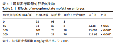
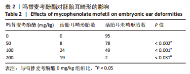
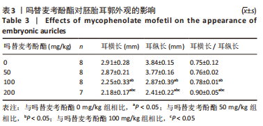
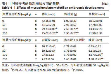
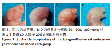
2.1 实验动物数量分析 实验所选用32只SD大鼠孕鼠,随机分为4组,最终纳入对照组8只,吗替麦考酚酯50,100,200 mg/kg组各8,8,7只。 2.2 大体指标 观察记录孕20.5 d时各组胚胎数量,测量各组活胎的发育指标,如表1所示,对照组孕鼠胚胎发育基本正常,随吗替麦考酚酯剂量增加,各实验组死胎数、吸收胎数增加,活胎数及活胎率降低,对照组与吗替麦考酚酯 50,100和200 mg/kg组相比,活胎数、死胎数、吸收胎数存在明显统计学差异(P < 0.05)。如表2所示,随吗替麦考酚酯剂量增加,吗替麦考酚酯组畸形胎数、耳畸形胎数增加,耳畸形胎率逐渐升高。如表3所示,对照组与吗替麦考酚酯100,200 mg/kg组相比,耳横长、耳纵长、耳纵长/耳横长存在明显统计学差异(P < 0.05)。如表4所示,对照组与吗替麦考酚酯 100,200 mg/kg组相比,体长、体质量、鼻枕距/双顶径、眼距等各指标存在明显统计学差异(P < 0.05)。吗替麦考酚酯 50 mg/kg组与吗替麦考酚酯 100,200 mg/kg组相比,各指标存在明显统计学差异(P < 0.05)。吗替麦考酚酯 100 mg/kg组与吗替麦考酚酯200 mg/kg组相比,耳纵长、体长、体质量、鼻枕距存在明显统计学差异(P < 0.05)。各组SD孕鼠胚胎第20.5天耳郭形态,见图1。"

"

"

"

"

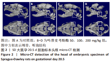
2.3 影像学检查结果 micro-CT检测孕20.5 d胚胎标本头颅,观察中耳及内耳发育情况。对照组颅骨、听泡结构发育完整,见图2A;吗替麦考酚酯50 mg/kg组听泡结构发育基本完整,见图2B;吗替麦考酚酯100 mg/kg组听泡结构不连续,见图2C;吗替麦考酚酯200 mg/kg组颅骨明显发育不良,听泡基本未发育,见图2D。"

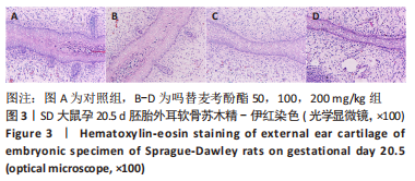
2.4 组织学检测结果 2.4.1 耳软骨苏木精-伊红染色观察结果 孕20.5 d胚胎外耳标本苏木精-伊红染色,对照组与吗替麦考酚酯50 mg/kg组靠近软骨膜处软骨细胞较小,密度较高,软骨基质淡红,软骨中部细胞逐渐增大、稀疏,软骨陷窝周围基质内含大量弹性纤维。对照组软骨细胞较吗替麦考酚酯50 mg/kg组密集,见图3A,B。吗替麦考酚酯 100 mg/kg组软骨发育不良,细胞移行不明显,软骨基质减少,见图3C。吗替麦考酚酯200 mg/kg组软骨发育迟缓,软骨薄,细胞排列不规则,软骨基质明显减少,见图3D。"

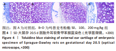
2.4.2 耳软骨甲苯胺蓝染色观察结果 孕20.5 d胚胎外耳标本甲胺蓝染色,对照组靠骨膜处可见软骨细胞较小,单个分布,中部软骨细胞较大、密集,软骨陷窝内同源细胞多,软骨细胞增生活跃,见图4A。吗替麦考酚酯50 mg/kg组软骨与对照组相近,见图4B。吗替麦考酚酯100 mg/kg组软骨细胞较前两组少,从近骨膜处至中部软骨细胞移行不明显,见图4C。吗替麦考酚酯200 mg/kg组软骨细胞稀疏,排列不规则,软骨陷窝内同源细胞少,软骨增生不活跃,见图4D。"

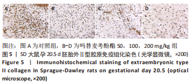
2.4.3 耳软骨Ⅱ型胶原免疫组化观察结果 孕20.5 d胚胎外耳标本Ⅱ型胶原免疫组化检测,对照组软骨膜、软骨囊及细胞间质呈褐黄色均质状或颗粒状,呈阳性表现,靠近软骨膜、软骨囊处呈强阳性,靠近软骨中部表达逐渐减弱,见图5A。吗替麦考酚酯50 mg/kg组可见棕黄色颗粒,呈中等阳性表达,见图5B。吗替麦考酚酯100,200 mg/kg组可见淡黄色颗粒,呈弱阳性表达,各组Ⅱ型胶原分布均匀,见图5C,D。"
| [1] 吗替麦考酚酯在自身免疫病治疗中应用的风湿病专家共识小组.吗替麦考酚酯在自身免疫病治疗中应用的风湿病专家共识[J].中华风湿病学杂志,2019,23(7):436-440. [2] CONTI F, CECCARELLI F, PERRICONE C, et al. Mycophenolate mofetil in systemic lupus erythematosus: results from a retrospective study in a large monocentric cohort and review of the literature. Immunol Res. 2014;60(2-3):270-276. [3] 岳瑞珍,李素领.自身免疫性肝炎中西医诊疗进展[J].中西医结合肝病杂志,2021,31(12):1146-1149. [4] BENATAR M, ROWLAND LP. The muddle of mycophenolate mofetil in myasthenia. Neurology. 2008;71(6):390-391. [5] SALZMANN J, LIGHTMAN S. The potential of newer immunomodulating drugs in the treatment of uveitis: a review. BioDrugs. 2000;13(6):397-408. [6] TULLUS K, MARKS SD. Vasculitis in children and adolescents: clinical presentation, etiopathogenesis, and treatment. Paediatr Drugs. 2009; 11(6):375-380. [7] RICCI G, DONDI A, PATRIZI A, et al. Systemic therapy of atopic dermatitis in children. Drugs. 2009;69(3):297-306. [8] 杨晓华,何乐人.先天性小耳畸形患者颅颌面发育异常的研究进展[J].中国医疗美容,2022,12(2):65-69. [9] 陆楠,李冬梅.面部畸形患儿心理健康状态的研究进展[J].国际眼科纵览,2016,40(3):206-212. [10] 郭蕊,王冰清,刘暾,等.小耳畸形及其伴发畸形的临床流行病学研究[J].中华耳科学杂志,2022,20(3):409-416. [11] DENG K, DAI L, YI L, et al. Epidemiologic characteristics and time trend in the prevalence of anotia and microtia in China. Birth Defects Res A Clin Mol Teratol. 2016;106(2):88-94. [12] 赵尚华,康深松.先天性小耳畸形耳廓支架的研究进展[J].中国美容医学,2019,28(10):170-173. [13] 王爽,何乐人,杨锦秀,等.耳后双蒂扩张皮瓣法耳廓再造术[J].中华整形外科杂志,2022,38(1):58-63. [14] BRENT B. The correction of mi-rotia with autogenous cartilage grafts: I. The classic deformity. Plast Reconstr Surg. 1980;66(1):1-12. [15] 刘铮,王喜梅,李志斌.自体肋软骨Nagata法耳廓再造术对再造耳廓皮肤敏感度的影响[J].中国美容医学,2021,30(8):49-53. [16] 庄洪兴,蒋海越,潘博,等.先天性小耳畸形的皮肤软组织扩张器法外耳再造术[J].中华整形外科杂志,2006,22(4):286-289. [17] 李高峰,谭军,罗滔,等.耳后延迟皮瓣自体肋软骨支架法外耳再造术[J].中华医学美学美容杂志,2014,20(4):241-244. [18] LI G, ZHANG F, DING W, et al. A New Microtia Reconstruction Method Using Delayed Postauricular Skin Flap. Plast Reconstr Surg. 2017;139(4):946-955. [19] SIEGERT R, MAGRITZ R. Otoplasty and Auricular Reconstruction. Facial Plast Surg. 2019;35(4):377-386. [20] 刘怡伶.先天性小耳畸形术后皮瓣破裂或坏死的危险因素及护理对策[J].青岛医药卫生,2021,53(5):392-394. [21] MARTÍN MC, CRISTIANO E, VILLANUEVA M, et al. Esophageal atresia and prenatal exposure to mycophenolate. Reprod Toxicol. 2014;50:117-121. [22] ALSEBAYEL MM, ABAALKHAIL FA, ALSEBAYEL FM, et al. Congenital Esophageal Atresia and Microtia in a Newborn Secondary to Mycophenolate Mofetil Exposure During Pregnancy: A Case Report and Review of the Literature. Am J Case Rep. 2018;19:523-526. [23] ANDERKA MT, LIN AE, ABUELO DN, et al. Reviewing the evidence for mycophenolate mofetil as a new teratogen: case report and review of the literature. Am J Med Genet A. 2009;149A(6):1241-1248. [24] PEREZ-AYTES A, MARIN-REINA P, BOSO V, et al. Mycophenolate mofetil embryopathy: A newly recognized teratogenic syndrome. Eur J Med Genet. 2017;60(1):16-21. [25] MASTRANGELO S, SOTTILE G, SUTERA AM, et al. Genome-wide association study reveals the locus responsible for microtia in Valle del Belice sheep breed. Anim Genet. 2018;49(6):636-640. [26] HE S, ZHANG Z, SUN Y, et al. Genome-wide association study shows that microtia in Altay sheep is caused by a 76 bp duplication of HMX1. Anim Genet. 2020;51(1):132-136. [27] DIMOPOULOU M, VERHOEF A, PENNINGS JLA, et al. Embryotoxic and pharmacologic potency ranking of six azoles in the rat whole embryo culture by morphological and transcriptomic analysis. Toxicol Appl Pharmacol. 2017;322:15-26. [28] 李学川.先天性小耳残耳软骨中Ⅱ型胶原和弹性蛋白的表达及生物力学研究[D].北京:中国协和医科大学,2006. [29] ROMERO MANTEOLA EJ, RAVETTA P, PATIÑO GONZÁLEZ CC, et al. Congenital esophageal stenosis: diagnosis and treatment. Cases review. Arch Argent Pediatr. 2018;116(1):e110-e114. [30] LIAN I, KIM J, OKAZAWA H, et al. The role of YAP transcription coactivator in regulating stem cell self-renewal and differentiation. Genes Dev. 2010;24(11):1106-1118. [31] CHEN X, ZHANG R. Microtia epigenetics: An overview of review and new viewpoint. Medicine (Baltimore). 2019;98(41):e17468. [32] 郭蕊,张玲,杨庆华.先天性小耳畸形遗传学研究进展[J].中华整形外科杂志,2019,35(5):507-512. [33] 郭佩佩. CYP26A1基因突变与先天性小耳畸形发病的相关性研究[D].北京:北京协和医学院,2021. [34] SCHMIDT F, ECKARDT K, SHAKIBAEI M, et al. Effects of mycophenolic acid alone and in combination with its metabolite mycophenolic acid glucuronide on rat embryos in vitro. Arch Toxicol. 2013;87(2):361-370. [35] LAKE JI, AVETISYAN M, ZIMMERMANN AG, et al. Neural crest requires Impdh2 for development of the enteric nervous system, great vessels, and craniofacial skeleton. Dev Biol. 2016;409(1):152-165. [36] 蒋卓远,查艳,石小峰,等.神经嵴细胞和神经嵴病及其致病机制的研究进展[J].遗传,2022,44(2):117-134. [37] SNIDER TN, MISHINA Y. Cranial neural crest cell contribution to craniofacial formation, pathology, and future directions in tissue engineering. Birth Defects Res C Embryo Today. 2014;102(3):324-332. [38] TRAINOR PA, TAM PP. Cranial paraxial mesoderm and neural crest cells of the mouse embryo: co-distribution in the craniofacial mesenchyme but distinct segregation in branchial arches. Development. 1995; 121(8):2569-2582. [39] 卢毅,孙飞,王志豪,等.组织工程气管软骨再生的新策略[J].国际外科学杂志,2022,49(4):273-278. [40] 李学川.先天性小耳残耳软骨中Ⅱ型胶原和弹性蛋白的表达及生物力学研究[D].北京:中国协和医科大学,2006. [41] 何清义,李起鸿,许建中.两种诱导方法对去分化软骨细胞II型胶原的诱导[J].中国矫形外科杂志,2004(Z4):95-98. |
| [1] | Sun Kexin, Zeng Jinshi, Li Jia, Jiang Haiyue, Liu Xia. Mechanical stimulation enhances matrix formation of three-dimensional bioprinted cartilage constructs [J]. Chinese Journal of Tissue Engineering Research, 2023, 27(在线): 1-7. |
| [2] | He Xi, Wan Yu, Tang Yuting, Yang Anning, Wu Kai, Jiao Yun, Bai Zhigang, Jiang Yideng, Shen Jiangyong. Erastin inhibits proliferation of hypertrophic scar fibroblasts [J]. Chinese Journal of Tissue Engineering Research, 2023, 27(在线): 1-. |
| [3] | Zhong Yizheng, Huang Peizhen, Cai Qunbin, Zheng Liqin, He Xingpeng, Dong Hang. Microstructural indexes that determine the trabecular bone maximum stress of micro-finite element models [J]. Chinese Journal of Tissue Engineering Research, 2023, 27(9): 1313-1318. |
| [4] | Cao Sheng, Kong Lingwei, Xu Kun, Sun Zhijie. Correlation of cervical sagittal force line parameters with degenerative segment and Pfirrmann classification in patients with cervical intervertebral disc degeneration [J]. Chinese Journal of Tissue Engineering Research, 2023, 27(9): 1319-1324. |
| [5] | Ke Yuqi, Chen Changjian, Wu Hao, Zheng Lianjie. Comparison of 12-month follow-up results of primary total hip arthroplasty between modified direct anterior approach and direct anterior approach [J]. Chinese Journal of Tissue Engineering Research, 2023, 27(9): 1377-1382. |
| [6] | Zhang Lichuang, Gao Huali, Wang Jingchao, Lin Huijun, Wu Chonggui, Ma Yinghui, Huang Yunfei, Fang Xue, Zhai Weitao. Effect of tendon manipulation with equal emphasis on muscles and bones on accelerating the functional rehabilitation of quadriceps femoris after total knee arthroplasty [J]. Chinese Journal of Tissue Engineering Research, 2023, 27(9): 1383-1389. |
| [7] | Du Xueting, Zhang Xiaodong, Chen Yanjun, Wang Mei, Chen Wubiao, Huang Wenhua. Application of compressed sensing technology in two-dimensional magnetic resonance imaging of the ankle joint [J]. Chinese Journal of Tissue Engineering Research, 2023, 27(9): 1396-1402. |
| [8] | You Zhengqiu, Zhang Zhongzu, Wang Qunbo. Early symptomatic intervertebral disc pseudocysts after discectomy detected on MRI [J]. Chinese Journal of Tissue Engineering Research, 2023, 27(9): 1403-1409. |
| [9] | Li Chao, Zhang Peipei, Xu Mengting, Li Linlin, Ding Jiangtao, Liu Xihua, Bi Hongyan. Respiratory training improves morphological changes of the multifidus muscle in patients with chronic nonspecific lower back pain assessed by musculoskeletal ultrasound [J]. Chinese Journal of Tissue Engineering Research, 2023, 27(9): 1417-1421. |
| [10] | He Yinhao, Li Xiaosheng, Chen Hongwen, Chen Tiezhu. 3D printed porous tantalum metal in the treatment of developmental dysplasia of the hip: current status and application prospect [J]. Chinese Journal of Tissue Engineering Research, 2023, 27(9): 1455-1461. |
| [11] | Jiang Xiaocheng, Shi Lu, Wang Yinbin, Li Qiujiang, Xi Chuangzhen, Ma Zefeng, Cai Lijun. Systematical evaluation of bone fusion rate after interbody fusion in patients with osteoporosis and lumbar degenerative disease treated with teriparatide [J]. Chinese Journal of Tissue Engineering Research, 2023, 27(9): 1427-1433. |
| [12] | Wu Dongzhe, Gao Xiaolin, Li Chuangtao, Wang Hao. Constructing the prediction model of maximal oxygen uptake by back-propagation neural network based on the cardiorespiratory optimal point [J]. Chinese Journal of Tissue Engineering Research, 2023, 27(8): 1224-1231. |
| [13] | Yang Jiujie, Li Zhi, Wang Shujie, Tian Ye, Zhao Wei. Intraoperative neurophysiological monitoring of functional changes following durotomy with decompression for acute spinal cord injury [J]. Chinese Journal of Tissue Engineering Research, 2023, 27(8): 1232-1236. |
| [14] | Li Mengfei, Zhang Hong, Zhao Shaojian, Yin Guanghao, Wang Qibao. Expression of forkhead box protein 3 in refractory periapical periodontitis in rats with Enterococcus faecalis infection [J]. Chinese Journal of Tissue Engineering Research, 2023, 27(8): 1187-1192. |
| [15] | Lian Shilin, Zhang Yan, Jiang Qiang, Zhang Hanshuo, Li Tusheng, Ding Yu. Interventional effects of whole blood and platelet-rich plasma with different preparation methods on nucleus pulposus cells [J]. Chinese Journal of Tissue Engineering Research, 2023, 27(8): 1199-1204. |
| Viewed | ||||||
|
Full text |
|
|||||
|
Abstract |
|
|||||